Review
. 2013 Jan:32:181-95.
doi: 10.1016/j.preteyeres.2012.10.004. Epub 2012 Nov 2.

A contemporary concept of the blood-aqueous barrier

Affiliations
Review

A contemporary concept of the blood-aqueous barrier

Thomas F Freddo. Prog Retin Eye Res. 2013 Jan.

Abstract

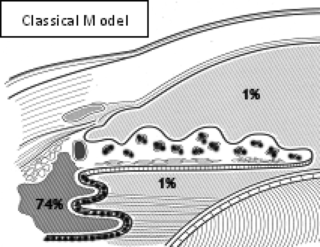
This review traces the evolution of the concept of the blood-aqueous barrier (BAB) during the past 20 years. The Classical model simply stipulated that the tight junctions of the iris vasculature and ciliary epithelium excluded plasma proteins from the aqueous humor (AH). It failed to reconcile the presence of AH protein levels equal to 1% of that found in plasma. Moreover, models of barrier kinetics assumed that the processes of AH secretion and plasma protein entry were directly linked. Thus, elevations of AH protein levels could only be explained by a pathological breakdown of the BAB. Over the last 20 years it has been shown that the plasma proteins in normal AH by-pass the posterior chamber entirely. Instead, these proteins diffuse from the capillaries of ciliary body stroma, into the iris stroma and then into the anterior chamber. This creates a reservoir of plasma-proteins in the iris stroma that is not derived from the iris vessels. This reservoir is prevented from diffusing posteriorly by tight junctions in the posterior iris epithelium. The one-way valve created by the pupil resting on the anterior lens capsule, combined with the continuous, forward flow of AH through the pupil, prevents protein reflux into the posterior chamber. Importantly, in the new paradigm, secretion of AH and the entry of plasma proteins into AH, are semi-independent events. This opens the possibility that AH protein levels could increase in the absence of breakdown of the BAB. Clinical consequences of this new paradigm of the BAB are discussed.


Conflict of interest statement

No conflict of interest exists in the work of the author summarized in this review.

Figures

Figure #1
Figure #1
TOP- Transmission electron micrograph. Black, HRP reaction product fills the ciliary body stroma (*). The tracer has diffused between adjacent pigmented ciliary epithelial cells (PCE) and into the cleft between the apical surfaces of the adjoined pigmented and non-pigmented ciliary epithelium (NPCE). Attempting to diffuse between adjacent non-pigmented ciliary epithelial cells, the HRP is stopped abruptly by the presence of a tight junction joining the apico-lateral surfaces of these cells. (curved arrows) (X20,000). BOTTOM – Freeze-fracture electron micrograph shows branching and anastomosing strands of continuous tight junction joining the apico-lateral surfaces of non-pigmented ciliary epithelial cells.(X57,500). INSET – Transmission electron micrograph shows fusion points of tight junction joining iris vascular endothelial cells. L – lumen. (X130,000). With permission from Morrison, J.C., VanBuskirk, E.M., Freddo, T., 1989. Anatomy, microcirculation and ultrastructure of the ciliary body. In: Ritch, R., Shields, M.B. and Krupin, T., eds. The Glaucomas. C.V. Mosby Co., St. Louis.
Figure #2
Figure #2
In the classical model of the blood-aqueous barrier, key tight junctions (•••••) are present joining iris vascular endothelial cells and non-pigmented ciliary epithelial cells. The concentration of plasma protein in the ciliary body stroma is assumed to be approximately 74% and that within the aqueous humor, at all points, is assumed to be 1%.
Figure #3
Figure #3
Traditional two-compartment model of the anterior segment in which tracers diffuse directly from the plasma into the anterior chamber, with secondary diffusional losses to the corneal stroma. Va- Volume of anterior chamber, Vc-Volume of cornea, kd-blood-aqueous barrier transfer coefficient, kfoutflow transfer coefficient, kc.ca – aqueous to cornea transfer coefficient. With permission from Academic Press. McLaren, J., Ziai, N. and Brubaker, R.F., 1993. A simple three-compartment model of anterior segment kinetics. Exp. Eye Res. 56: 355–66.
Figure #4
Figure #4
(a) One minute after HRP injection, the tracer fills the vessels of the iris, ciliary body and conjunctiva. Early leakage is present in the latter two locations. C, cornea; AC, anterior chamber; PC, posterior chamber. (b) Three min after HRP injection, filling of the ciliary body stroma is more evident and migration of HRP can be followed toward the iris. (c) Eight min after HRP injection, the ciliary body stroma is filled and HRP has reached the anterior chamber at the root of the iris (arrow). With permission from The Assoc. for Research in Vision and Ophthalmology. Freddo, T.F., Bartels, S.P., Barsotti, M.F. and Kamm, R.D., 1990. The source of proteins in the aqueous humor of the normal rabbit. Invest. Ophthalmol. Vis Sci. 31: 125–137.
Figure #4
Figure #4
(a) One minute after HRP injection, the tracer fills the vessels of the iris, ciliary body and conjunctiva. Early leakage is present in the latter two locations. C, cornea; AC, anterior chamber; PC, posterior chamber. (b) Three min after HRP injection, filling of the ciliary body stroma is more evident and migration of HRP can be followed toward the iris. (c) Eight min after HRP injection, the ciliary body stroma is filled and HRP has reached the anterior chamber at the root of the iris (arrow). With permission from The Assoc. for Research in Vision and Ophthalmology. Freddo, T.F., Bartels, S.P., Barsotti, M.F. and Kamm, R.D., 1990. The source of proteins in the aqueous humor of the normal rabbit. Invest. Ophthalmol. Vis Sci. 31: 125–137.
Figure #5
Figure #5
Fluorescein concentration in a rabbit anterior chamber after intravenous fluorescein injection. The three-compartment model (solid line) matched the measured concentrations (•) considerably better than the two-compartment model. With permission from Academic Press. McLaren, J., Ziai, N. and Brubaker, R.F., 1993. A simple three-compartment model of anterior segment kinetics. Exp. Eye Res. 56: 355–66.
Figure #6
Figure #6
Three-compartment model of the anterior segment. Tracers must pass through the iris enroute from the plasma to the anterior chamber. Va- Volume of anterior chamber, Vc-Volume of cornea, Vi – Volume of the iris, ki.ip - iris to plasma transfer coefficient referred to the volume of the iris, ki.ia – iris to anterior chamber transfer coefficient referred to the volume of the iris, kf - outflow transfer coefficient, kc.ca – aqueous to cornea transfer coefficient. With permission from Academic Press. McLaren, J., Ziai, N. and Brubaker, R.F., 1993. A simple three-compartment model of anterior segment kinetics. Exp. Eye Res. 56: 355–66.
Figure #7
Figure #7
Mean percent signal enhancement as a function of time after administration of gadolinium contrast material for the ciliary body and the anterior and posterior chambers. With permission from The Assoc. for Research in Vision and Ophthalmology. Kolodny, N., Freddo, T.F., Lawrence, B., Suarez, C., and Bartels, S.P., 1996. Contrast-enhanced MRI confirmation of an anterior protein pathway in the normal rabbit eye. Invest. Ophthalmol. Vis Sci. 37: 1602–1607.
Figure #8
Figure #8
A: Pre-contrast image shows details of tissues in the anterior segment of the eye, including iris, ciliary body and both the anterior and posterior chambers. B: Within two minutes of contrast infusion, there is clear enhancement of the ciliary body and the choroid, but the anterior chamber, posterior chamber and vitreous body show no enhancement. C: After 90 minutes, the enhancement in the ciliary body and choroid has begun to diminish. There is clear enhancement in the anterior chamber but the posterior chamber and the vitreous remain unchanged from Figures A and B.
Figure #9
Figure #9
T1- weighted MRI images (TR/TE=1400/15ms, acquisition time 4 min 45 sec) of 56 year-old male with pigment dispersion syndrome and no glaucoma. A: baseline pre-constrast image clearly shows the iris, ciliary body and anterior and posterior chambers. B: Image shows same eye 49 minutes after intravenous infusion of 0.4 mg/kg Gadolinium dimeglumine i.v (Magnevist, Berlex Imaging, Wayne, NJ). In this subject with mid-peripheral transillumination defects of the nasal and inferior iris, gadolinium has enhanced the AC as expected, but has secondarily entered the PC on the nasal side through the iris epithelial defects (arrow). T= temporal.
Figure #9
Figure #9
T1- weighted MRI images (TR/TE=1400/15ms, acquisition time 4 min 45 sec) of 56 year-old male with pigment dispersion syndrome and no glaucoma. A: baseline pre-constrast image clearly shows the iris, ciliary body and anterior and posterior chambers. B: Image shows same eye 49 minutes after intravenous infusion of 0.4 mg/kg Gadolinium dimeglumine i.v (Magnevist, Berlex Imaging, Wayne, NJ). In this subject with mid-peripheral transillumination defects of the nasal and inferior iris, gadolinium has enhanced the AC as expected, but has secondarily entered the PC on the nasal side through the iris epithelial defects (arrow). T= temporal.
Figure #10
Figure #10
A: In the Classical model, the tight junctions of the non-pigmented ciliary epithelium and of the iris vascular endothelium are the key elements. The iris stroma is presumed to be free of plasma proteins and the concentration of plasma proteins in the aqueous is uniform throughout the anterior and posterior chambers. Elevation of aqueous humor plasma protein concentrations can be explained only by an increase in blood-aqueous barrier permeability. B: In the new paradigm, the small amount of plasmaderived protein present in aqueous humor diffuses from the ciliary body stroma, to the root of the iris, accumulates in the iris stroma and is then released into the aqueous humor of the anterior chamber (arrows). The posterior chamber is essentially plasma-protein-free. Some of the protein delivered to the iris root immediately enters the trabecular outflow pathways (arrows). The tight junctions of the nonpigmented ciliary epithelium and of the iris vasculature endothelium remain key elements (•••••). An additional key element becomes the tight junctions of the posterior iris epithelium (•••••), which prevent the protein in the iris stroma from diffusing posteriorly, when combined with the one way valve created by the pupil resting on the anterior lens capsule, and the continuous forward flow of aqueous humor through the pupil. The entry of aqueous humor and the plasma proteins contained within it are semi-independent variables. As such, plasma protein concentrations in the aqueous humor can increase in the absence of an increase in blood-aqueous barrier permeability.
Figure #11
Figure #11
Diagram of the ciliary body, iris and anterior chamber angle demonstrates pathway taken by plasma proteins entering the aqueous of the normal eye (large arrow). Additional protein, beyond that measurable in the circulating aqueous humor, is likely shunted directly into the trabecular outflow pathway (small arrow). With permission from The Mosby Publishers. Morrison, J and Freddo, T.F. Anatomy, microcirculation and ultrastructure of the ciliary body. In: The Glaucomas, 2nd ed, Vol. I, Ritch, R., Shields, M.B. and Krupin, T., 1996. Cptr 6, pp. 125–138.
Figure #12
Figure #12
Oxygen distribution in the nonsurgical eye and the proposed effect of vitrectomy and/or cataract surgery on oxygen delivery to the anterior chamber angle. (A) In the nonsurgical eye, oxygen enters from the retinal vasculature through the vitreous, across the ciliary epithelium from the ciliary body vasculature, and into the anterior chamber across the cornea. Oxygen is consumed by the lens and the ciliary epithelium. A small amount of oxygen enters the anterior chamber angle by diffusing across the ciliary body and iris stroma (curved red arrow). This is the same pathway as that of the plasma proteins (gold arrow). (B) After vitrectomy, more oxygen reaches the posterior chamber. This supplies more oxygen to the “aqueous surface” of the ciliary epithelium, reducing the amount of oxygen that the ciliary epithelium removes from the blood and slightly increasing the amount of oxygen available to enter the anterior chamber angle from the ciliary body stroma. (C) Cataract surgery reduces oxygen consumption by the lens, thereby increasing the pO2 anterior to the lens and in the posterior chamber. The increased oxygen on the aqueous surface of the ciliary epithelium reduces the amount of oxygen that the ciliary epithelium removes from the blood, thereby slightly increasing the amount of oxygen available to enter the anterior chamber angle from the ciliary body stroma. (D) After vitrectomy and cataract surgery, significantly more oxygen is available on the aqueous surface of the ciliary epithelium, resulting in the removal of significantly less oxygen from the blood. This process increases the amount of oxygen available to diffuse from the ciliary body stroma, across the iris stroma, and into the anterior chamber angle, exposing the outflow system to a large excess of oxygen and/or oxygen metabolites. With permission from The Assoc. for Research in Vision and Ophthalmology. Siegfried, C.J., Shui, Y-B., Holekamp, N.M., Bai, F. and Beebe, D.C., 2010. Oxygen distribution in the human eye: Relevance to the etiology of open-angle glaucoma after vitrectomy. Invest. Ophthalmol. Vis. Sci.51, 5731–5738.
Figure #13
Figure #13
T1-weighted spin-echo MR images (TR/TE = 1400/15 msec-image, each with an acquisition time of 4 min, 45 sec. LEFT: Baseline image acquired 45 min after instillation of 3% pilocarpine (note miotic iris) but prior to administration of intravenous gadolinium. RIGHT: Image obtained 60 min after gadolinium infusion shows enhancement in the anterior chamber but not in the posterior chamber or vitreous body. Insert compares split images of the anterior segments from the two time points shown in the main figure. With permission from Elsevier. Freddo, T.F., Patz, S and Arshanskiy, Y., 2006. Pilocarpine’s effects on the blood-aqueous barrier of the human eye as assessed by high-resolution, contrast magnetic resonance imaging. Exp. Eye Res. 82, 458–464.

References

    1. Abraham SV. Miotic iridocyclitis: Its role in the surgical treatment of glaucoma. Am. J. Ophthalmol. 1959;48:634–643. - PubMed
    1. Anjou CI. Physiological variations of the aqueous flare density in normal human eyes. Acta Ophthalmol. (Copenh.) 1961a;39:525–539. - PubMed
    1. Anjou CI. Influence of light on the 24-hour variation in aqueous flare density and intra-ocular pressure in normal rabbits' eyes. Acta Ophthalmol. (Copenh.) 1961b;39:852–873. - PubMed
    1. Babizhayev MA. Biomarkers and special features of oxidative stress in the anterior segment of the eye linked to lens cataract and the trabecular meshwork injury in primary open-angle glaucoma: Challenges of dual combination therapy with N-acetylcarnosine lubricant eye drops and oral formulation of nonhydrolyzed carnosine. Fundamental & Clin. Pharmacol. 2012;26:86–117. - PubMed
    1. Bárány EH. Simultaneous measurement of changing intraocular pressure and outflow facility in the vervet monkey by constant pressure infusion. Invest. Ophthalmol. Vis. Sci. 1964;3:135–143. - PubMed

Publication types