

Advanced glycation end-products stimulate basic fibroblast growth factor expression in cultured Müller cells
- PMID: 23129015
- PMCID: PMC3572729
- DOI: 10.3892/mmr.2012.1152
Advanced glycation end-products stimulate basic fibroblast growth factor expression in cultured Müller cells
Abstract
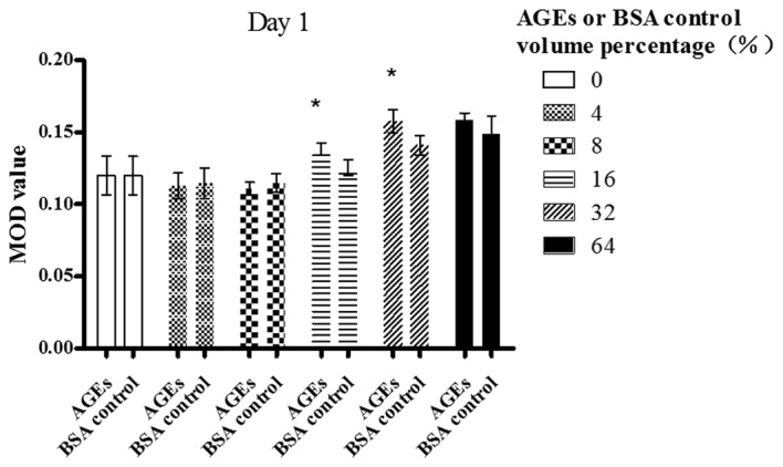
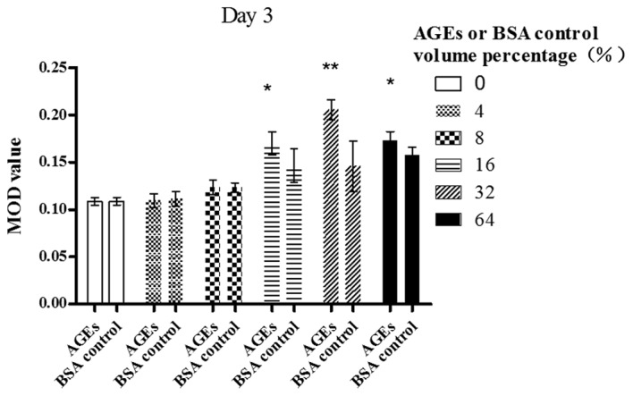
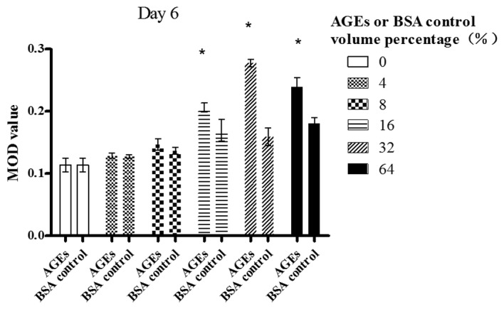
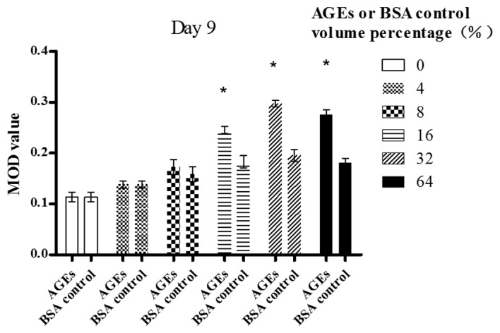
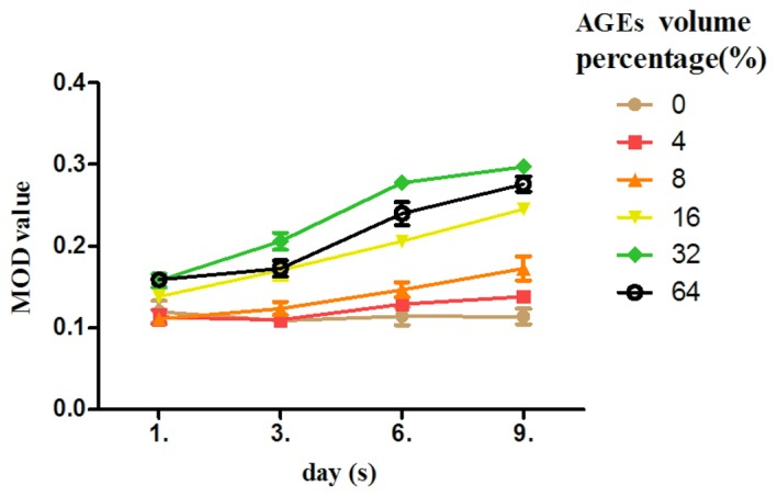
Accumulating evidence points to a causal role for advanced glycation end-products (AGEs) in the development of diabetic vascular complications, including diabetic retinopathy (DR). To assess the reciprocal correlation between AGEs and basic fibroblast growth factor (bFGF), the effects of AGEs on the production of bFGF by Müller cells were investigated. Müller cells were cultured from adult rabbit retinas. The AGEs were prepared with highly glycated bovine serum albumin (BSA) and the control non‑glycated BSA (BSA control) was incubated under the same conditions without glucose. Cultured Müller cells were exposed to AGEs or BSA control (volume percentages were 4, 8, 16, 32 and 64%) for a time course of 1, 3, 6 and 9 days in their desired medium. The expression of bFGF in Müller cells was evaluated by immunocytochemistry. Quantification was performed by densitometry using computerized image analysis with dedicated software. AGEs in a volume percentage of 16 and 32% on day 1 and in a volume percentage of 16, 32 and 64% on days 3, 6 and 9 increased the bFGF expression in Müller cells (P<0.05). Additionally, AGEs upregulated bFGF expression in Müller cells in a time‑dependent manner. In conclusion, the treatment of Müller cells with AGEs resulted in a dose- and time‑dependent elevation of bFGF in the culture medium. The results from this study suggest that the increased formation of AGEs in the vitreous may be involved in the development of DR by inducing the production of bFGF by retinal Müller cells.
Figures
References
-
- Daroux M, Prévost G, Maillard-Lefebvre H, Gaxatte C, D’Agati VD, Schmidt AM, Boulanger E. Advanced glycation end-products: implications for diabetic and non-diabetic nephropathies. Diabetes Metab. 2010;36:1–10. - PubMed
-
- Berrou J, Tostivint I, Verrecchia F, Berthier C, Boulanger E, Mauviel A, Marti HP, Wautier MP, Wautier JL, Rondeau E, Hertig A. Advanced glycation end products regulate extracellular matrix protein and protease expression by human glomerular mesangial cells. Int J Mol Med. 2009;23:513–520. - PubMed
-
- Wolffenbuttel BH, Giordano D, Founds HW, Bucala R. Long-term assessment of glucose control by haemoglobin-AGE measurement. Lancet. 1996;347:513–515. - PubMed
-
- Yamagishi S. Role of advanced glycation end products (AGEs) and receptor for AGEs (RAGE) in vascular damage in diabetes. Exp Gerontol. 2011;46:217–224. - PubMed
Publication types
MeSH terms
Substances
LinkOut - more resources
Full Text Sources
