. 2012 Dec;3(6):561-71.
doi: 10.1007/s13244-012-0195-7. Epub 2012 Nov 6.

A pictorial review of acute aortic syndrome: discriminating and overlapping features as revealed by ECG-gated multidetector-row CT angiography

Affiliations

A pictorial review of acute aortic syndrome: discriminating and overlapping features as revealed by ECG-gated multidetector-row CT angiography

Takuya Ueda et al. Insights Imaging. 2012 Dec.

Abstract

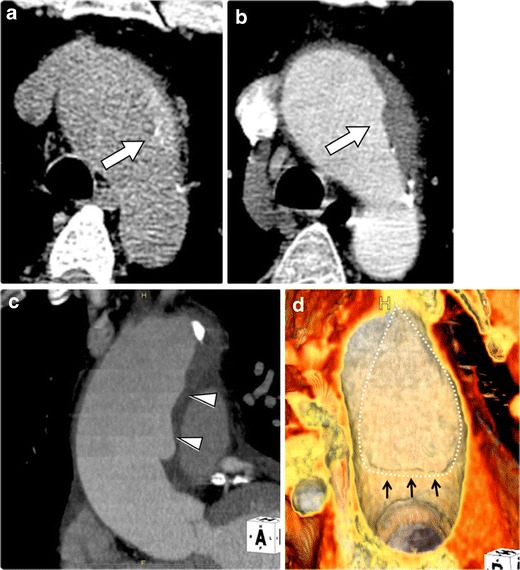
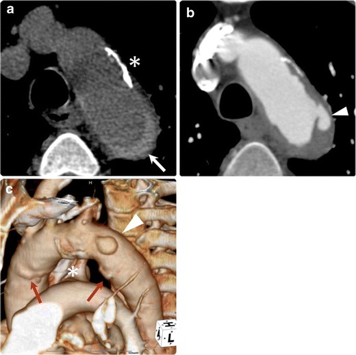
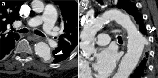
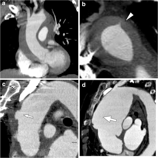
Background: The term "acute aortic syndrome" (AAS) encompasses a spectrum of life-threatening conditions characterized by acute aortic pain. AAS traditionally embraces three abnormalities including classic aortic dissection, intramural haematoma, and penetrating atherosclerotic ulcer. Although the underlying etiologies and conditions predisposing to AAS are diverse, the clinical features are indistinguishable.

Methods: Multidetector-row computed tomography (CT) with electrocardiographic gating (ECG-gated MDCT) has greatly improved imaging of acute thoracic aortic diseases by virtually eliminating pulsation artifacts transmitted from cardiac motion and reveals subtle aortic abnormalities, which have been difficult to recognize by conventional non-gated CT.

Results: While these advances in imaging technology provide additional discriminating features of acute aortic diseases, they also reveal a range of overlapping features of these life-threatening conditions that not uncommonly are dynamic and evolving. These overlapping and transitional features may be a major source of misunderstanding, confusion, and controversy for diseases that cause AAS.

Conclusion: In this pictorial review, we describe the discriminating and typical imaging features as revealed by modern ECG-gated MDCT angiography. In addition to the discriminating features, recognition of the overlapping and transitional features in AAS will allow a more comprehensive understanding of their underlying pathophysiologic conditions and their natural history, and may improve therapeutic management.

Main messages: • The superior visualization of ECG-gated CTA improves the diagnostic accuracy of acute aortic syndrome. • ECG-gated CTA provides discriminating features of underlying pathophysiologic conditions of AAS. • Also, recognition of the overlapping features in AAS will allow a more comprehensive understanding.


Figures

Fig. 1
Fig. 1
Impact of ECG-gated CT angiography (CTA). Non-gated (a, b) and ECG-gated (c, d) CTA in a 66-year-old man with limited intimal tear. a Non-gated axial CT image shows a ‘dissection-like’ motion artifact (arrow) on the contour of the aorta because of pulsatile motion of the aorta. b Oblique sagittal slab maximum intensity projection (slab-MIP) image shows an undulating contour of the aorta caused by misregistration from the pulsatile motion. c ECG-gated CTA, performed 1 day after the non-gated CT, eliminates the pulsatile motion artifact. The image clearly reveals a hidden focal intimal tear (arrowhead). d Slab MIP image of ECG-gated CTA demonstrates visualization of the ascending aorta without misregistration artifacts. A focal bulge with a tiny intimal tear is revealed (curved arrow)
Fig. 2
Fig. 2
Discriminating and overlapping features of acute aortic syndrome. In addition to the certain discriminating features as revealed by recent advances in imaging, each disease in acute aortic syndrome (AAS) may show overlapping features and transition from one to another. In such overlapping and transitional features of aortic diseases, the diagnosis may be affected by the time point at which the imaging has been examined and also by the detectability of tiny pathology by the imaging modality. Awareness of some related and distinguishing radiologic features in AAS is helpful in understanding these diseases and providing new insight into the pathophysiology
Fig. 3
Fig. 3
Acute Stanford type A classic aortic dissection in a 71-year-old woman with abrupt, severe retrosternal chest pain. ac Transverse images from an ECG-gated CTA demonstrate an extensive intimo-medial flap involving the entire thoracic aorta. The true lumen (*) appears small in the ascending aorta where it is nearly circumferentially separated from the rest of the aortic wall (b). Extension of the flap down to the aortic root to involve the aortic valve apparatus is clearly demonstrated in this patient who had aortic regurgitation confirmed on echocardiography, a significant complication of acute dissection (c). d Multiplanar reformations in a coronal plane show the involvement of the brachiocephalic artery to better advantage (arrow), the near circumferential separation of the aortic media at the level of the ascending aorta and its extension down to the aortic valve apparatus (arrowhead)
Fig. 4
Fig. 4
Dissection variant intramural haematoma (IMH) in a 54-year-old man who presented with acute chest pain. a Non-contrast CT image shows a crescent-shaped high-density area in the ascending aorta and descending aorta (arrowhead). b On the axial contrast-enhanced CT image, neither the flow channel nor intimal flap is detected along the entire aorta, consistent with IMH. c Oblique sagittal MPR image demonstrates widespread extension of type A IMH throughout the whole thoracic aorta. An ‘ulcer-like projection,’ the radiological finding of a focal disruption of the surface of the aortic wall from the true lumen into the thrombosed false lumen, is identified at the mid ascending aorta (arrow). During surgery, a small thrombosed focal primary intimal tear (PIT) was identified at the corresponding site
Fig. 5
Fig. 5
Resolved tiny primary intimal tear of dissection variant IMH in a 68-year-old woman with hypertension who presented with unstable thoracoabdominal pain. a Non-contrast CT image shows a crescent-shaped high-density area in the descending aorta (arrowhead). b Slab MIP image demonstrates haematoma extending through the whole thoracic descending aorta, which consists of dissection variant IMH. A tiny PIT is detected at the distal end of the haematoma (arrow). c The patient was followed with stable limited chest pain. After 7-day follow-up, the PIT resolved spontaneously, leaving a slightly enlarged haematoma (arrow). The patient was treated conservatively, and the haematoma eventually resolved
Fig. 6
Fig. 6
Limited intimal tear with intramural haemorrhagic content in a 48-year-old male. a Non-contrast CT demonstrates the aortic wall thickening with haemorrhagic content (arrow) at the proximal aortic arch. b Contrast-enhanced CTA demonstrates the aortic wall thickening (arrow) without clear intimal-medial flap or false lumen visualized; this was initially diagnosed as traditional IMH. c Slab-MIP image demonstrates the ‘eccentric one-sided bulge’ of the aortic wall along the ascending aorta (arrowheads). d 3D-VR luminal image right in front of the bulge shows a teardrop-shaped intimal tear (dotted line) with a localized intimal flap (black arrows) at the inferior border of the intimal tear
Fig. 7
Fig. 7
Penetrating atherosclerotic ulcer with haemorrhagic content in a 74-year-old man. a Non-contrast CT shows intramural haemorrhagic content (arrow) in the descending aorta. Note the thick calcification and plaque on the aortic wall (*), suggesting an atherosclerotic background of the patient. b Axial ECG-gated CTA demonstrates the ulcerative lesion at the proximal ascending aorta (arrowhead) into the aortic media, which shows associated haemorrhage. c 3D-VR image depicts the crater-like ulceration (arrowhead) and atherosclerotic calcification (*) on the aortic arch. Some non-penetrating atherosclerotic ulcers are also identified (arrows). The condition is considered as an overlapping feature between IMH (in the broad sense) and penetrating atherosclerotic ulcer
Fig. 8
Fig. 8
Penetrating atherosclerotic ulcer in an 81-year-old man. a Axial CTA images demonstrate an ulcerative lesion (arrowhead) on the thickened aortic wall with haemorrhagic content in the descending aorta. b Oblique sagittal MPR image clearly visualizes a crater-like feature of the ulcerative lesion (arrowhead) deeply penetrating into the atherosclerotic aortic wall. Entire aorta depicts severe atherosclerosis with plaque burden and calcifications
Fig. 9
Fig. 9
Impending rupture of an atherosclerotic aortic aneurysm in an 83-year-old man. The patient had been followed for stable aortic aneurysm for many years and suddenly presented with acute chest pain. a Non-contrast CT demonstrates ballooning of the aorta at the aortic arch. The aortic wall shows haemorrhagic content within the aortic wall (arrow), which suggests an acute process of this event. b Axial CTA depicts bulged dilatation of the aorta on the right side of the aortic arch (arrowheads), which was not depicted on the previous follow-up CT images. c Oblique coronal MPR image demonstrates dilatation of the aorta (arrowheads) with a thickened aortic wall with haemorrhagic content. The aortic lumen shows an eccentric bulge, a sign of impending rupture of an atherosclerotic aortic aneurysm
Fig. 10
Fig. 10
Aneurysmal dilatation of the aorta secondary to dissection variant IMH in a 66-year-old woman. a Oblique sagittal contrast-enhanced CT demonstrates IMH extending from the ascending aorta through the proximal descending aorta. b Axial image depicts a streak-form ulcer-like projection (primary intimal tear) in the descending aorta (arrowhead). c Two days after onset, an ulcer-like projection was enlarged to form an outpouching cavity into the thrombosed false lumen (small arrow). d One week later, the haemorrhagic content is resolved and the ulcer-like projection not been detected. On the other hand, the aorta shows aneurysmal dilatation. Note that the dilatation starts from the location where the haematoma was present (large arrow). Although the pathological features and clinical course are different, such aneurysmal dilatation of dissection variant IMH might be confused with an atherosclerotic aortic aneurysm if an ulcer-like projection is not detected on the initial CT examination

References

    1. Vilacosta I, Roman JA. Acute aortic syndrome. Heart. 2001;85:365–368. doi: 10.1136/heart.85.4.365. - DOI - PMC - PubMed
    1. Hiratzka LF, Bakris GL, Beckman JA, et al. ACCF/AHA/AATS/ACR/ASA/SCA/SCAI/ SIR/STS/SVM guidelines for the diagnosis and management of patients with Thoracic Aortic Disease: a report of the American College of Cardiology Foundation/American Heart Association Task Force on Practice Guidelines, American Association for Thoracic Surgery, American College of Radiology, American Stroke Association, Society of Cardiovascular Anesthesiologists, Society for Cardiovascular Angiography and Interventions, Society of Interventional Radiology. Circulation. 2010;121:e266–369. doi: 10.1161/CIR.0b013e3181d4739e. - DOI - PubMed
    1. Vilacosta I, Aragoncillo P, Canadas V, San Roman JA, Ferreiros J, Rodriguez E. Acute aortic syndrome: a new look at an old conundrum. Heart. 2009;95:1130–1139. - PubMed
    1. Hayter RG, Rhea JT, Small A, Tafazoli FS, Novelline RA. Suspected aortic dissection and other aortic disorders: multi-detector row CT in 373 cases in the emergency setting. Radiology. 2006;238:841–852. doi: 10.1148/radiol.2383041528. - DOI - PubMed
    1. Shiga T, Wajima Z, Apfel CC, Inoue T, Ohe Y. Diagnostic accuracy of transesophageal echocardiography, helical computed tomography, and magnetic resonance imaging for suspected thoracic aortic dissection: systematic review and meta-analysis. Arch Intern Med. 2006;166:1350–1356. doi: 10.1001/archinte.166.13.1350. - DOI - PubMed

LinkOut - more resources