. 2013 Jun;228(6):1174-88.
doi: 10.1002/jcp.24271.

Regulation of human Cripto-1 expression by nuclear receptors and DNA promoter methylation in human embryonal and breast cancer cells

Affiliations

Regulation of human Cripto-1 expression by nuclear receptors and DNA promoter methylation in human embryonal and breast cancer cells

Caterina Bianco et al. J Cell Physiol. 2013 Jun.

Abstract

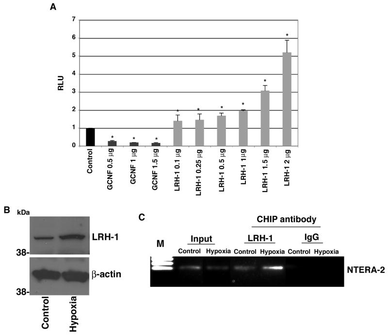
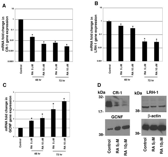
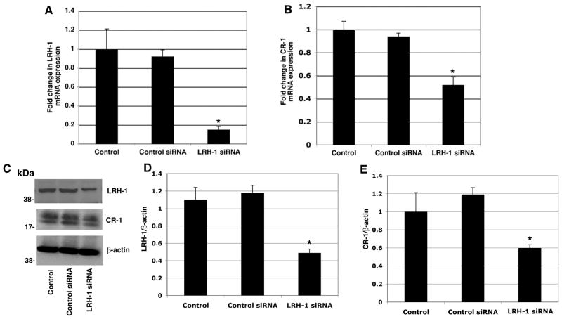
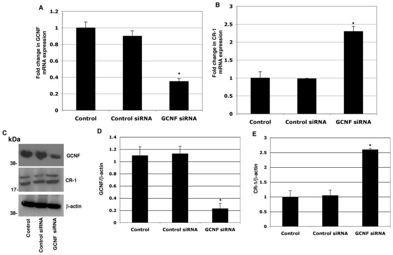
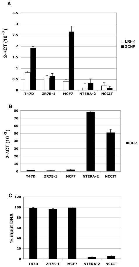
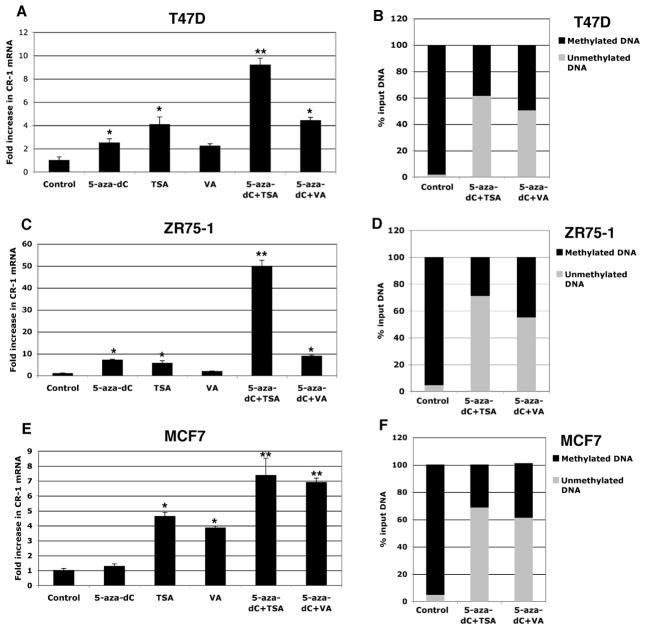
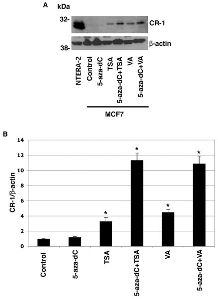
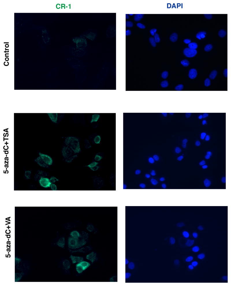
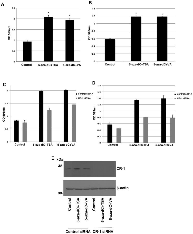
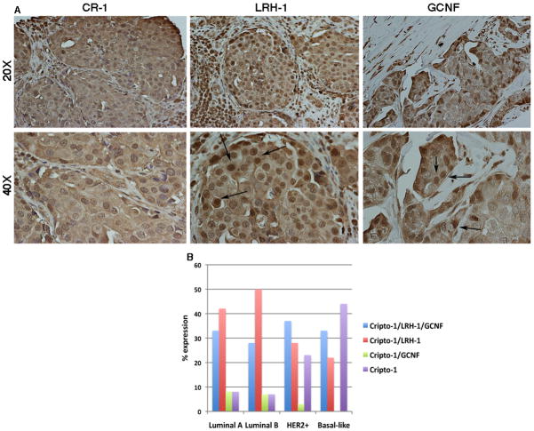
Human Cripto-1 (CR-1) plays an important role in regulating embryonic development while also regulating various stages of tumor progression. However, mechanisms that regulate CR-1 expression during embryogenesis and tumorigenesis are still not well defined. In the present study, we investigated the effects of two nuclear receptors, liver receptor homolog (LRH)-1 and germ cell nuclear factor receptor (GCNF) and epigenetic modifications on CR-1 gene expression in NTERA-2 human embryonal carcinoma cells and in breast cancer cells. CR-1 expression in NTERA-2 cells was positively regulated by LRH-1 through direct binding to a DR0 element within the CR-1 promoter, while GCNF strongly suppressed CR-1 expression in these cells. In addition, the CR-1 promoter was unmethylated in NTERA-2 cells, while T47D, ZR75-1, and MCF7 breast cancer cells showed high levels of CR-1 promoter methylation and low CR-1 mRNA and protein expression. Treatment of breast cancer cells with a demethylating agent and histone deacetylase inhibitors reduced methylation of the CR-1 promoter and reactivated CR-1 mRNA and protein expression in these cells, promoting migration and invasion of breast cancer cells. Analysis of a breast cancer tissue array revealed that CR-1 was highly expressed in the majority of human breast tumors, suggesting that CR-1 expression in breast cancer cell lines might not be representative of in vivo expression. Collectively, these findings offer some insight into the transcriptional regulation of CR-1 gene expression and its critical role in the pathogenesis of human cancer.


Figures

Figure 1
Figure 1. LRH-1 binds to the CR-1 promoter and enhances CR-1 promoter luciferase activity in NTERA-2 cells
A: Dual luciferase assay of transiently co-transfected NTERA-2 cells with a full-length CR-1 promoter luciferase reporter vector and GCNF or LRH-1 expression plasmids. These results are the mean ± SD of triplicates of three separate experiments (*P<0.01). B: Western blot analysis for LRH-1 and β-actin in NTERA-2 cells grown in normoxic (20% oxygen) or hypoxic (0.5% oxygen) conditions for 24 hours. C: ChIP assay in NTETRA-2 cells following 24 hours of incubation in a hypoxic chamber. The cross-linked protein-DNA complexes were immunoprecipitated with anti-LRH-1 antibody or with an isotype control IgG as negative control, and the purified DNA was amplified by PCR using specific primers spanning the DR0 region within the CR-1 promoter.
Figure 2
Figure 2. RA treatment downregulates CR-1 and LRH-1 and upregulates GCNF expression in NTERA-2 cells
Quantitative real time PCR for CR-1 (A), LRH-1 (B) and GCNF (C) in NTERA-2 cells treated with RA for 48 and 72 hours. Control cells were treated with vehicle DMSO. mRNA levels were normalized to GAPDH expression. The values shown are averages ± SD for duplicate samples from one of three experiments. *P<0.05, as compared to control DMSO treated NTERA-2 cells. D: Western blot analysis for CR-1, LRH-1, GCNF and βactin in NTERA-2 cells treated for 72 hours with 5 or 10 μM RA. Control cells were treated with vehicle DMSO.
Figure 3
Figure 3. Knockdown of LRH-1 expression by siRNA downregulates CR-1 mRNA and protein expression in NTERA-2 cells
Real time PCR for LRH-1 (A) and CR-1 (B) in NTERA-2 cells transfected with control or LRH-1 siRNAs. Data are representative of three experiments with duplicate samples. *P<0.05, as compared to control non-transfected samples. C: Western blot analysis for LRH-1, CR-1 and β-actin in siRNA transfected cells. D, E: Fold difference in LRH-1 and CR-1 expression by densitometric analysis of LRH-1 or CR-1 in NTERA-2 cell lysates normalized to β-actin content. *P<0.05, as compared to control non-transfected samples.
Figure 4
Figure 4. Knockdown of GCNF expression by siRNA upregulates CR-1 mRNA and protein expression in NTERA-2 cells
Real time PCR for GCNF (A) and CR-1 (B) in NTERA-2 cells transfected with control or GCNF siRNAs. Data are representative of three experiments with duplicate samples. *P<0.05, as compared to control non-transfected samples. C Western blot analysis for GCNF, CR-1 and β-actin in siRNA transfected cells. D, E: Fold difference in GCNF and CR-1 expression by densitometric analysis of GCNF or CR-1 in NTERA-2 cell lysates normalized to β-actin content. *P<0.05, as compared to control non-transfected samples.
Fig. 5
Fig. 5. GCNF, LRH-1 and CR-1 mRNA expression and methylation status of the CR-1 promoter in human breast and embryonal carcinoma cells
A, B: Real time PCR for GCNF, LRH-1 and CR-1 in breast T47D, MCF7 and ZR75-1 and embryonal NCCIT and NTERA-2 carcinoma cells. Data were normalized to GAPDH expression and are representative of three experiments with duplicate samples. C: Methylation of the CR-1 promoter in breast and embryonal carcinoma cells after enzymatic digestion of genomic DNA with methylation sensitive or methylation dependent restriction enzymes (see Materials and Methods). DNA was subsequently amplified using specific primers spanning CpG islands in the CR-1 promoter. Experiment was repeated three times on duplicate samples.
Fig. 6
Fig. 6. DNMT and HDAC inhibitors reactivate CR-1 mRNA expression in breast cancer cell lines
A, C, E: Real time PCR for CR-1 in T47D, ZR75-1 and MCF7 cells treated with 5-aza-dC, TSA or VA alone or in combination. Real time data were normalized to GAPDH expression. Data are representative of three experiments with duplicate samples. *P<0.05. **P<0.01. B, D, F: Methylation of the CR-1 promoter in drug-treated and vehicle-treated T47D, ZR-75-1 and MCF7 cells. A representative example of two independent experiments with duplicate samples is presented.
Fig. 7
Fig. 7. DNMT and HDAC inhibitors reactivate CR-1 protein expression in MCF7 cells
A: Western blot analysis for CR-1 in drug-treated and vehicle-treated MCF-7 cells. NTERA-2 cells were used as positive control. β-actin was used as a protein loading control. B: Fold difference in CR-1 protein expression by densitometric analysis in MCF7 cell lysates normalized to β-actin content. *P<0.01, as compared to control vehicle-treated cells.
Fig. 8
Fig. 8. Immunofluorescence staining for CR-1 in vehicle-treated or DNMT and HDAC inhibitors-treated MCF7 cells
DAPI staining of nuclei is shown in blue.
Fig. 9
Fig. 9. DNMT and HDAC inhibitors enhance migration and invasion of MCF7 cells in a CR-1-dependent manner
Migration (A) and invasion (B) assays of 5-aza-dC, TSA and VA treated MCF7 cells. MCF7 cells were also transfected with control or CR-1 siRNAs and their migratory (C) and invasive (D) behavior was assessed. E: Western blot analysis for CR-1 and β-actin in MCF7 control and treated cells that have been transfected with control or CR-1 siRNA pools.
Fig. 10
Fig. 10. CR-1 overexpression in MCF7 tumor spheres is associated with increased expression of embryonic stem cell markers
Representative images of MCF7 Neo (A) and MCF7 CR-1 (B) spheres after 7 days in culture in ultra-low attachment plates. The number (C) and the size (D) of spheres derived from MCF7 Neo and MCF7 CR-1 cells were assessed. Quantitative real time PCR for CR-1 (E), Nanog (F), Oct-4 (G) and CD133 (H) in MCF7 Neo and MCF7 CR-1 spheres. Data are representative of two independent experiments performed in a 24 well plate for each cell line. *P<0.0001.
Fig. 11
Fig. 11. Expression of CR-1, LRH-1 and GCNF in human invasive ductal breast carcinomas
A: Immunohistochemical analysis shows positive staining for CR-1, LRH-1 and GCNF. LRH-1 and GCNF show cytoplasmic and nuclear staining. Arrows are pointing to some cells with LRH-1 and GCNF nuclear staining. Original magnifications are 20X and 40X. B: Expression of CR-1, LRH-1 and GCNF in breast cancer molecular subtypes as assessed by immunohistochemical staining.

Similar articles

Cited by

References

    1. Aasen T, Raya A, Barrero MJ, Garreta E, Consiglio A, Gonzalez F, Vassena R, Bilic J, Pekarik V, Tiscornia G, Edel M, Boue S, Izpisua Belmonte JC. Efficient and rapid generation of induced pluripotent stem cells from human keratinocytes. Nat Biotechnol. 2008;26(11):1276–1284. - PubMed
    1. Annicotte JS, Chavey C, Servant N, Teyssier J, Bardin A, Licznar A, Badia E, Pujol P, Vignon F, Maudelonde T, Lazennec G, Cavailles V, Fajas L. The nuclear receptor liver receptor homolog-1 is an estrogen receptor target gene. Oncogene. 2005;24(55):8167–8175. - PMC - PubMed
    1. Aoi T, Yae K, Nakagawa M, Ichisaka T, Okita K, Takahashi K, Chiba T, Yamanaka S. Generation of pluripotent stem cells from adult mouse liver and stomach cells. Science. 2008;321(5889):699–702. - PubMed
    1. Assou S, Lecarrour T, Tondeur S, Strom S, Gabelle A, Marty S, Nadal L, Pantesco V, Reme T, Hugnot JP, Gasca S, Hovatta O, Hamamah S, Klein B, De Vos J. A meta-analysis of human embryonic stem cells transcriptome integrated into a web-based expression atlas. Stem Cells. 2007;25:961–973. - PMC - PubMed
    1. Bianco C, Adkins HB, Wechselberger C, Seno M, Normanno N, De Luca A, Sun Y, Khan N, Kenney N, Ebert A, Williams KP, Sanicola M, Salomon DS. Cripto-1 activates nodal- and ALK4-dependent and -independent signaling pathways in mammary epithelial Cells. Mol Cell Biol. 2002;22(8):2586–2597. - PMC - PubMed

Publication types

MeSH terms

LinkOut - more resources