. 2012 Nov 20;109(47):19202-7.
doi: 10.1073/pnas.1217338109. Epub 2012 Nov 5.

Formation of triple-helical structures by the 3'-end sequences of MALAT1 and MENβ noncoding RNAs

Affiliations

Formation of triple-helical structures by the 3'-end sequences of MALAT1 and MENβ noncoding RNAs

Jessica A Brown et al. Proc Natl Acad Sci U S A. .

Abstract

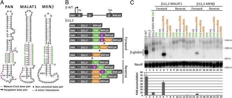
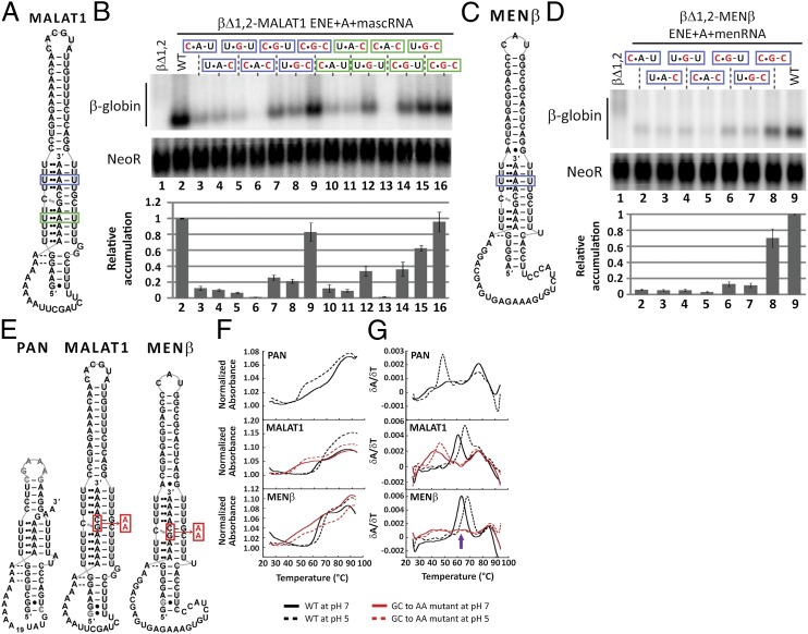
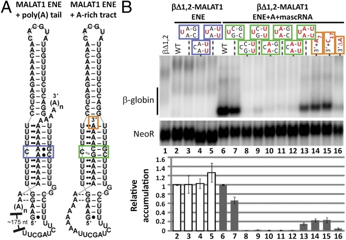
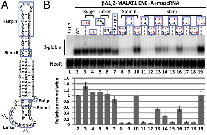
Stability of the long noncoding-polyadenylated nuclear (PAN) RNA from Kaposi's sarcoma-associated herpesvirus is conferred by an expression and nuclear retention element (ENE). The ENE protects PAN RNA from a rapid deadenylation-dependent decay pathway via formation of a triple helix between the U-rich internal loop of the ENE and the 3'-poly(A) tail. Because viruses borrow molecular mechanisms from their hosts, we searched highly abundant human long-noncoding RNAs and identified putative ENE-like structures in metastasis-associated lung adenocarcinoma transcript 1 (MALAT1) and multiple endocrine neoplasia-β (MENβ) RNAs. Unlike the PAN ENE, the U-rich internal loops of both predicted cellular ENEs are interrupted by G and C nucleotides and reside upstream of genomically encoded A-rich tracts. We confirmed the ability of MALAT1 and MENβ sequences containing the predicted ENE and A-rich tract to increase the levels of an intronless β-globin reporter RNA. UV thermal denaturation profiles at different pH values support formation of a triple-helical structure composed of multiple U•A-U base triples and a single C•G-C base triple. Additional analyses of the MALAT1 ENE revealed that robust stabilization activity requires an intact triple helix, strong stems at the duplex-triplex junctions, a G-C base pair flanking the triplex to mediate potential A-minor interactions, and the 3'-terminal A of the A-rich tract to form a blunt-ended triplex lacking unpaired nucleotides at the duplex-triplex junction. These examples of triple-helical, ENE-like structures in cellular noncoding RNAs, are unique.


Conflict of interest statement

The authors declare no conflict of interest.

Figures

Fig. 1.
Fig. 1.
Models and stabilization activity of the predicted cellular ENE-like structures. (A) Schematic diagrams for the predicted ENE-like structures from MALAT1 (nucleotides 8263–8355) and MENβ (nucleotides 22651–22743) RNAs are based upon the known secondary and tertiary contacts of the PAN ENE (nucleotides 895–959) (14, 15). Nucleotides in the U-rich internal loop are in green and nucleotides in the poly(A) tail and A-rich tract are in purple. (B) Schematic diagrams of the β-globin plasmid constructs with different combinations of the predicted cellular ENE (green), A-rich tract (purple), and tRNA-like sequence (orange, representing mascRNA or menRNA) from MALAT1 or MENβ in either the forward or reverse orientation (see Fig. S2 for construct details). β-Globin expression is driven by the CMV promoter; the RNase P cleavage site is indicated by an arrowhead; and there is a bovine growth hormone polyadenylation signal (BGH pA) downstream of the 3′ UTR. (C) Northern blots (Upper) were probed for β-globin and Neomycin resistance (NeoR) RNAs, with the βΔ1,2-KSHV PAN ENE (βΔ1,2–79F in ref. 12) included as a control. RNA sizes are indicated to the right of the blot. Results were quantitated (Lower) by normalizing the β-globin signal to the NeoR signal, which served as a loading and transfection control. The empty βΔ1,2 reporter level was set at an arbitrary value of 1. Fold-accumulation is the average of at least three independent experiments; error bars represent SD.
Fig. 2.
Fig. 2.
In vivo and in vitro biochemical evidence for an RNA triple-helical structure. (A and C) The same schematics as in Fig. 1A indicate the putative U•A-U base triples replaced with C•G-C in a blue (upper) or green (lower) box. (B and D) Northern blot analysis of β-globin and NeoR mRNAs (Upper) and quantitations (Lower) were performed as in Fig. 1C. Black nucleotides are WT sequence; mutated nucleotides are red. The WT βΔ1,2 reporter level was set at an arbitrary value of 1. Relative accumulation is the average of at least three independent experiments; error bars represent SD. (E) Schematic diagrams show the RNAs used in UV thermal denaturation experiments. The alignment of the PAN ENE core with A32GA2 is arbitrary. The red box and arrow denote the GC dinucleotide that was substituted with AA in MALAT1 ENE+A and MENβ ENE+A. (F) Plots of normalized absorbance at 260 nm versus temperature and (G) first derivative plots (δA/δT) versus temperature are shown for the PAN ENE core + A32GA2, the MALAT1 ENE+A WT and GC-to-AA, and the MENβ ENE+A WT and GC-to-AA mutant. Melting profiles of WT sequences are in black and the GC-to-AA mutant RNAs are in red. A solid line denotes pH 7; a dashed line denotes pH 5. Note that the MENβ ENE+A GC-to-AA mutant exhibits three transitions or peaks. The peak at 55–70 °C (purple arrow) shifted by several degrees in different experiments and once split into two defined peaks, making it difficult to explain its origin.
Fig. 3.
Fig. 3.
Functional significance of the intervening C and G nucleotides in the U-rich loop and A-rich tract. (A) A schematic diagram is shown for the ENE-like structures predicted to form by the putative MALAT1 ENE with a 3′-poly(A) tail (nucleotides 8263–8336) or its adjacent A-rich tract (nucleotodes 8263–8355). The blue, green, and orange boxes outline the nucleotides targeted for mutagenesis. (B) Northern blot analysis of β-globin and NeoR mRNAs (Upper) and quantitation (Lower) were performed as in Fig. 1C. Black denotes WT sequence; mutated nucleotides are red. The plus (+) and delta (Δ) symbols represent a nucleotide insertion and deletion, respectively. The WT βΔ1,2 reporter levels were set at an arbitrary value of 1 for the βΔ1,2-MALAT1 ENE WT (white bar, lane 2) and the βΔ1,2-MALAT1 ENE+A+mascRNA WT (gray bar, lane 6). Each mutant was normalized relative to its WT counterpart. Relative accumulation is the average of at least three independent experiments; error bars represent SD.
Fig. 4.
Fig. 4.
Other structural features contribute to stabilization. (A) The same MALAT1 schematic as in Fig. 1A has blue lines designating mutated regions. (B) Northern blot analysis of β-globin and NeoR RNAs (Upper) and quantitations (Lower) were performed as in Fig. 1C. WT nucleotides are black; mutated nucleotides are red. The delta (Δ) symbol represents a nucleotide deletion. In Δhairpin, a GCAA tetraloop was substituted to maintain a predicted stem-loop structure. The WT βΔ1,2 reporter level was set at an arbitrary value of 1. Relative accumulation is the average of at least three independent experiments; error bars represent SD.

References

    1. Carninci P. Tagging mammalian transcription complexity. Trends Genet. 2006;22(9):501–510. - PubMed
    1. Wang KC, Chang HY. Molecular mechanisms of long noncoding RNAs. Mol Cell. 2011;43(6):904–914. - PMC - PubMed
    1. Clark MB, et al. Genome-wide analysis of long noncoding RNA stability. Genome Res. 2012;22(5):885–898. - PMC - PubMed
    1. Tani H, et al. Genome-wide determination of RNA stability reveals hundreds of short-lived noncoding transcripts in mammals. Genome Res. 2012;22(5):947–956. - PMC - PubMed
    1. Hutchinson JN, et al. A screen for nuclear transcripts identifies two linked noncoding RNAs associated with SC35 splicing domains. BMC Genomics. 2007;8:39. - PMC - PubMed

Publication types

LinkOut - more resources