. 2012 Nov 20;109(47):19103-7.
doi: 10.1073/pnas.1211384109. Epub 2012 Nov 5.

Energy-dispersive X-ray emission spectroscopy using an X-ray free-electron laser in a shot-by-shot mode

Affiliations

Energy-dispersive X-ray emission spectroscopy using an X-ray free-electron laser in a shot-by-shot mode

Roberto Alonso-Mori et al. Proc Natl Acad Sci U S A. .

Abstract

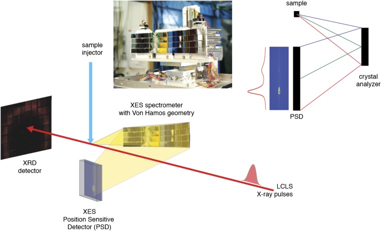
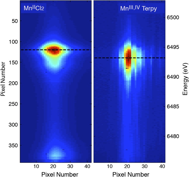
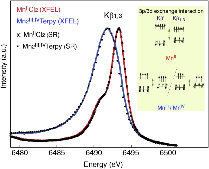
The ultrabright femtosecond X-ray pulses provided by X-ray free-electron lasers open capabilities for studying the structure and dynamics of a wide variety of systems beyond what is possible with synchrotron sources. Recently, this "probe-before-destroy" approach has been demonstrated for atomic structure determination by serial X-ray diffraction of microcrystals. There has been the question whether a similar approach can be extended to probe the local electronic structure by X-ray spectroscopy. To address this, we have carried out femtosecond X-ray emission spectroscopy (XES) at the Linac Coherent Light Source using redox-active Mn complexes. XES probes the charge and spin states as well as the ligand environment, critical for understanding the functional role of redox-active metal sites. Kβ(1,3) XES spectra of Mn(II) and Mn(2)(III,IV) complexes at room temperature were collected using a wavelength dispersive spectrometer and femtosecond X-ray pulses with an individual dose of up to >100 MGy. The spectra were found in agreement with undamaged spectra collected at low dose using synchrotron radiation. Our results demonstrate that the intact electronic structure of redox active transition metal compounds in different oxidation states can be characterized with this shot-by-shot method. This opens the door for studying the chemical dynamics of metal catalytic sites by following reactions under functional conditions. The technique can be combined with X-ray diffraction to simultaneously obtain the geometric structure of the overall protein and the local chemistry of active metal sites and is expected to prove valuable for understanding the mechanism of important metalloproteins, such as photosystem II.


Conflict of interest statement

The authors declare no conflict of interest.

Figures

Fig. 1.
Fig. 1.
(Lower) The energy-dispersive XES experimental setup. The setup allows simultaneous detection of X-ray diffraction (XRD) and X-ray emission spectra (XES) from a stream of crystals that intersects the XFEL beam. The XRD detector is downstream of the X-ray laser beam. The XES spectrometer is at 90° to the direction of the X-ray beam, focusing the emission spectrum on a custom-built 2D detector placed below the intersection point. (Upper Right) Vertical cut of the von Hamos geometry with a crystal analyzer and a position-sensitive detector; Bragg scattering from a point source is analyzed by the spectrometer array, resulting in an energy-dispersed spectrum on the detector. (Upper Left) A picture of the von Hamos spectrometer used in this work showing the array of 16 crystal analyzers.
Fig. 2.
Fig. 2.
The focused Kβ1,3 XES spectra from all of the crystal analyzers. Two-dimensional images showing the Mn Kβ1,3 X-ray emission spectra of (Left) MnIICl2 and (Right) Mn2III,IVTerpy collected at the LCLS. The images show the clear differences in the emission spectrum between the MnII and the Mn2III,IV complexes. The vertical axis reflects the energy dispersion. Each of the crystals is tuned to focus the spectrum on the horizontal axis such that all 16 spectra coincide, thereby improving the signal-to-noise ratio.
Fig. 3.
Fig. 3.
The integrated Kβ1,3 spectra from the MnII and Mn2III,IV complexes. The integrated RT Mn Kβ1,3 X-ray emission spectra of MnIICl2 (red, 500 mM Mn) and Mn2III,IVTerpy (blue, 180 mM Mn) were collected at the LCLS with 50-fs X-ray pulses. The symbols show MnIICl2 and Mn2III,IVTerpy spectra collected with a similar setup at a synchrotron for comparison purposes. The Kβ1,3 spectrum arises from the 3d–3p exchange coupling (Inset) that makes the spectrum sensitive to the number of unpaired electrons in the 3d orbitals, thereby providing information about the spin state of the complex. For high-spin complexes the position of the peak, therefore, is an indicator of the oxidation state or charge density on the metal.

References

    1. Emma P, et al. First lasing and operation of an angstrom-wavelength free-electron laser. Nat Photonics. 2010;4:641–647.
    1. Chapman HN, et al. Femtosecond X-ray protein nanocrystallography. Nature. 2011;470(7332):73–77. - PMC - PubMed
    1. Aquila A, et al. Time-resolved protein nanocrystallography using an X-ray free-electron laser. Opt Express. 2012;20(3):2706–2716. - PMC - PubMed
    1. Koopmann R, et al. In vivo protein crystallization opens new routes in structural biology. Nat Methods. 2012;9(3):259–262. - PMC - PubMed
    1. Kern J, et al. Room temperature femtosecond X-ray diffraction of photosystem II microcrystals. Proc Natl Acad Sci USA. 2012;109(25):9721–9726. - PMC - PubMed

Publication types

LinkOut - more resources