. 2012 Dec 15;189(12):5849-59.
doi: 10.4049/jimmunol.1200585. Epub 2012 Nov 5.

Intrapulmonary G-CSF rescues neutrophil recruitment to the lung and neutrophil release to blood in Gram-negative bacterial infection in MCP-1-/- mice

Affiliations

Intrapulmonary G-CSF rescues neutrophil recruitment to the lung and neutrophil release to blood in Gram-negative bacterial infection in MCP-1-/- mice

Gayathriy Balamayooran et al. J Immunol. .

Abstract

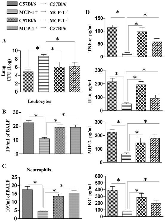
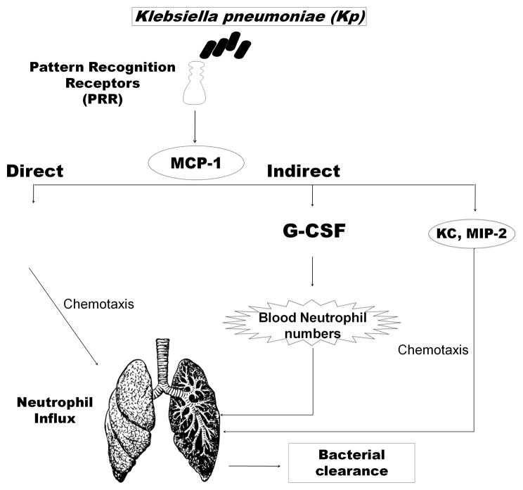
We previously demonstrated that MCP-1 is important for E. coli-induced neutrophil migration to the lungs. However, E. coli neither disseminates nor induces death in mice. Furthermore, the cell types and the host defense mechanisms that contribute to MCP-1-dependent neutrophil trafficking have not been defined. In this study, we sought to explore the cell types and the mechanisms associated with Klebsiella pneumoniae-mediated MCP-1-dependent neutrophil influx. MCP-1(-/-) mice are more susceptible to pulmonary K. pneumoniae infection and show higher bacterial burden in the lungs and dissemination. MCP-1(-/-) mice also display attenuated neutrophil influx, cytokine/chemokine production, and activation of NF-κB and MAPKs following intratracheal K. pneumoniae infection. rMCP-1 treatment in MCP-1(-/-) mice following K. pneumoniae infection rescued impairment in survival, bacterial clearance, and neutrophil accumulation in the lung. Neutrophil numbers in the blood of MCP-1(-/-) mice were associated with G-CSF concentrations in bronchoalveolar lavage fluid and blood. Bone marrow or resident cell-derived MCP-1 contributed to bacterial clearance, neutrophil accumulation, and cytokine/chemokine production in the lungs following infection. Furthermore, exogenous MCP-1 dose dependently increased neutrophil counts and G-CSF concentrations in the blood. Intriguingly, administration of intratracheal rG-CSF to MCP-1(-/-) mice after K. pneumoniae infection rescued survival, bacterial clearance and dissemination, and neutrophil influx in MCP-1(-/-) mice. Collectively, these novel findings unveil an unrecognized role of MCP-1 in neutrophil-mediated host immunity during K. pneumoniae pneumonia and illustrate that G-CSF could be used to rescue impairment in host immunity in individuals with absent or malfunctional MCP-1.


Figures

Figure 1
Figure 1. Importance of MCP-1 in host defense against pulmonary K. pneumoniae infection
A. Reduced survival in MCP-1−/− mice following i.t. K. pneumoniae infection. MCP-1−/− and WT (C57Bl/6) mice were i.t. inoculated with 103/mouse of K. pneumoniae, and survival was monitored up to 10 days. n=20 mice in each group. *, p<0.05 determined by Wilcoxon Rank Sign Test. B-E. Impaired bacterial clearance in the in MCP-1−/− mice after i.t. K. pneumoniae infection (103/mouse). Bacterial CFUs in mice were determined in lungs, spleens, livers and blood of MCP-1−/− and WT mice after K. pneumoniae infection. (n=4–6 mice/time point/group; *, Significant differences between MCP-1−/− and WT mice). F–H. Total leukocyte, neutrophil and macrophage numbers in the lungs of MCP-1−/− mice after K. pneumoniae inoculation. Both MCP-1−/− and WT animals underwent BAL fluid and lung collection after challenge with K. pneumoniae (n=4–6 mice/time point/group). I. MPO activity in homogenized (unlavaged) whole lungs of WT and MCP-1−/− mice infected with K. pneumoniae (103 CFU/mouse) for 24 and 36 h.(n=6–8 mice/group; * indicates p<0.05 compared with MCP-1−/− mice). J. Lung histology in MCP-1−/− mice following K. pneumoniae inoculation. Lung sections were made 36 h after bacterial or saline challenge and stained with H&E. Score for leukocyte numbers in the lesion and bacterial numbers were calculated as described in Materials and Methods. These are representative sections of 4 mice in each experiment with similar results (Magnification x200).
Figure 2
Figure 2. Impairment of airspace cytokine responses in MCP-1−/− mice after infection with K. pneumoniae
A. Activation of NF-κB in the lung following infection with K. pneumoniae. Lung homogenates from MCP-1−/− mice and their controls were prepared at 24 and 36 h after infection with K. pneumoniae. NF-κB expression and phosphorylation of NF-κB and IκBα were determined using western blots of lung homogenates. The blot is a representative of 3 independent blots with similar results. B. Relative densities normalized against GAPDH are representatives of 3 independent blots. C. Activation of MAPKs in the lung following K. pneumoniae infection. Total proteins in the lung were isolated from MCP-1−/− and control mice at 24 and 36 h after infection with K. pneumoniae, resolved on an SDS-PAGE and the membrane was blotted with the Abs against activated/phosphorylated form of MAPKs as described in Materials and Methods. This is a representative blot of 3 separate blots with similar results. D. Densitometric analysis of MAPK activation was performed from 3 separate blots. *denotes the difference between MCP-1−/− mice and their WT controls (p<0.05). E–H. Cytokine (TNF-α, IL-6)and chemokine (MIP-2 and KC) concentrations in BAL fluid were measured by sandwich ELISA after infection with K. pneumoniae. (n=4–6 mice were used at each time point in each group. Significant differences between MCP-1−/− and WT mice are indicated by asterisks.)
Figure 3
Figure 3. Impaired survival, bacterial clearance and neutrophil influx in the lungs of MCP-1−/− mice are restored by rMCP-1
Mortality (A) in WT and MCP-1−/− mice infected with 103CFUs of K. pneumoniae and administered with rMCP-1(5 μg/mouse) or vehicle (BSA) 1 h later, and survival was assessed up to 10 days. Data are presented as % survival (n=20 mice/group) and analyzed using Wilcoxon signed-rank test. * indicates the difference between rMCP-1 or vehicle (BSA) control treated MCP-1−/− mice (p<0.05) and ** indicates the difference between WT and MCP-1−/− mice. Bacterial clearance (B, C) in the lungs and spleen were examined following exogenous MCP-1 administration in MCP-1−/− mice at 24 h post-K. pneumoniae challenge (103CFUs/mouse). Data are presented as mean ± SE (n=6–8 mice/group/time point). * indicates p<0.05 compared with BSA (vehicle) administered mice. Cellular infiltration (D–E) in airspaces at 24 h after i.t. treatment with rMCP-1(5 μg/mouse) or vehicle (BSA) control (n=6–8 mice/group/time point).
Figure 4
Figure 4. Total leukocyte numbers, neutrophil numbers, bacterial burden and cytokine chemokine levels in bone marrow chimeras following K. pneumoniae infection
Bone marrow transplantation was performed between WT and MCP-1−/− mice in four combinations as is described in the materials and methods section and infected with K. pneumoniae i.t and at 36 h post-infection unlavaged lung bacterial counts were determined (A) while BALF was collected to determine the WBC (B) and neutrophil counts (C) in lungs, and TNF-α, IL-6, KC and MIP-2 levels (D). *indicates significant difference between different chimeras; (p<0.05; n=4–6/group).
Figure 5
Figure 5. CCR2 expression in blood neutrophils and G-CSF production in the lungs of WT and MCP-1−/− mice after K. pneumoniae infection
Flow cytometric analysis of blood from WT mice at 24 and 36 h after i.t K. pneumoniae (1X10³ CFU/mouse) infection using tagged antibodies against CCR2 and Gr1(Ly6G) (A–B) (n=5–6 mice/group). C. Neutrophil numbers in blood of both WT and MCP-1−/− mice following K. pneumoniae infection. Neutrophils were gated and the percentage of Gr-1/Ly6G positive cells were determined. G-CSF concentrations in BALF and in blood of WT and MCP-1−/− mice following i.t K. pneumoniae infection (E-F) determined by using sandwich ELISA. n=4–6; *p<0.05 between WT and MCP-1−/− mice.
Figure 6
Figure 6. Increased neutrophil numbers and cytokine/chemokine levels in BALF and blood following i.t treatment with rMCP-1 (A–C)
A–C. Blood neutrophil numbers were determined 24 h after i.t treatment with rMCP-1 in two different doses (5 and 10μg/mouse) using Gr-1/Ly6G antibodies. Absolute blood neutrophil numbers were determined in total blood volume of mice as described in Materials and Methods. D. Protein concentrations of TNF-α, IL-6 and G-CSF in BALF and in serum were determined after treatment with 5 and 10 μg rMCP-1/mouse using sandwich ELISA. n=4–6/group/time point; *p<0.05.
Figure 7
Figure 7. Higher blood neutrophil numbers and G-CSF concentrations in BALF and in serum after K. pneumoniae infection of the lung
A–B. Blood neutrophil percentage and absolute blood neutrophil numbers were determined following i.t K. pneumoniae infection and rMCP-1 (5μg/mouse). C–D. G-CSF protein concentrations in BALF and in serum were determined by sandwich ELISA in mice after infection followed by rMCP-1 treatment. Data n=4–6 mice/group, and *p<0.05 between BSA and rMCP-1 treated mice.
Figure 8
Figure 8. Attenuated survival, bacterial clearance, neutrophil influx, and cytokine/chemokine production in the lungs of MCP-1−/− mice are restored by exogenous rG-CSF
A. Survival of WT and MCP-1−/− mice after infection with 103 CFUs of K. pneumoniae and administered rG-CSF(1 μg/mouse) or vehicle (BSA) 1 h later, and survival was assessed up to 10 days. Data are presented as % survival (n=20 mice/group) and analyzed using Wilcoxon signed-rank test. * indicates the difference between rG-CSF or vehicle (BSA) control treated MCP-1−/− mice (p<0.05) and ** indicates the difference between BSA and rG-CSF administered mice. Bacterial load (B) in the lungs and bacterial dissemination (C) were examined in lung homogenates of rG-CSF or BSA administered WT and MCP-1−/− mice at 24 h post-K. pneumoniae challenge (103 CFUs/mouse). (n=5–6 mice/group). * indicates p<0.05 rG-CSF compared with BSA (vehicle) administered mice. Cellular infiltration (D–E) in airspaces at 24 h after i.t. treatment with rG-CSF (1μg/mouse) or vehicle (BSA) control. (n=6–8 mice/group; * indicates p<0.05 as compared to BSA administered mice).
Figure 9
Figure 9. Model representing MCP-1 mediated neutrophil recruitment to the lungs during Klebsiella pneumonia
K. pneumoniae infection in lungs stimulates MCP-1 production through interaction with pattern recognition receptors. MCP-1 regulates neutrophil recruitment indirectly via controlling G-CSF, KC and MIP-2 levels and directly through the expression of CCR2 on neutrophils.

References

    1. Rubenfeld GD, Caldwell E, Peabody E, Weaver J, Martin DP, Neff M, Stern EJ, Hudson LD. Incidence and outcomes of acute lung injury. N Engl J Med. 2005;353:1685–1693. - PubMed
    1. Ho J, Tambyah PA, Paterson DL. Multiresistant Gram-negative infections: a global perspective. Curr Opin Infect Dis. 2010;23:546–553. - PubMed
    1. Foglia E, Meier MD, Elward A. Ventilator-associated pneumonia in neonatal and pediatric intensive care unit patients. Clin Microbiol Rev. 2007;20:409–425. table of contents. - PMC - PubMed
    1. Baughman RP. Antibiotic resistance in the intensive care unit. Curr Opin Crit Care. 2002;8:430–434. - PubMed
    1. Balamayooran G, Batra S, Balamayooran T, Cai S, Jeyaseelan S. Monocyte Chemoattractant Protein 1 Regulates Pulmonary Host Defense via Neutrophil Recruitment during Escherichia coli Infection. Infect Immun. 79:2567–2577. - PMC - PubMed

Publication types

MeSH terms

Substances