. 2013 Jan 15;591(2):571-92.
doi: 10.1113/jphysiol.2012.241760. Epub 2012 Nov 5.

Disturbed energy metabolism and muscular dystrophy caused by pure creatine deficiency are reversible by creatine intake

Affiliations

Disturbed energy metabolism and muscular dystrophy caused by pure creatine deficiency are reversible by creatine intake

C I Nabuurs et al. J Physiol. .

Abstract

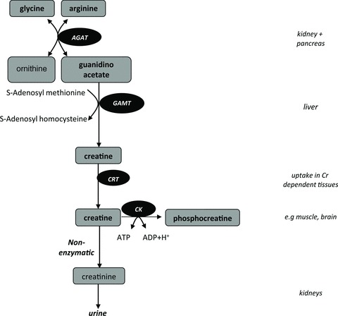
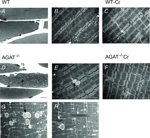
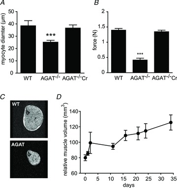
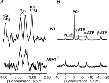
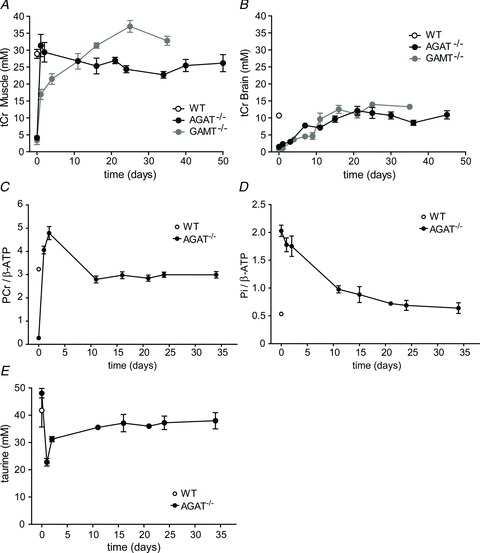
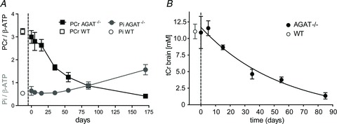
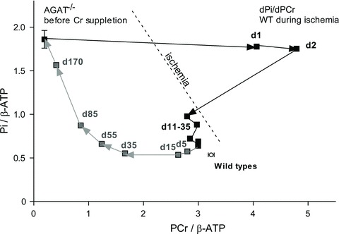
Creatine (Cr) plays an important role in muscle energy homeostasis by its participation in the ATP-phosphocreatine phosphoryl exchange reaction mediated by creatine kinase. Given that the consequences of Cr depletion are incompletely understood, we assessed the morphological, metabolic and functional consequences of systemic depletion on skeletal muscle in a mouse model with deficiency of l-arginine:glycine amidinotransferase (AGAT(-/-)), which catalyses the first step of Cr biosynthesis. In vivo magnetic resonance spectroscopy showed a near-complete absence of Cr and phosphocreatine in resting hindlimb muscle of AGAT(-/-) mice. Compared with wild-type, the inorganic phosphate/β-ATP ratio was increased fourfold, while ATP levels were reduced by nearly half. Activities of proton-pumping respiratory chain enzymes were reduced, whereas F(1)F(0)-ATPase activity and overall mitochondrial content were increased. The Cr-deficient AGAT(-/-) mice had a reduced grip strength and suffered from severe muscle atrophy. Electron microscopy revealed increased amounts of intramyocellular lipid droplets and crystal formation within mitochondria of AGAT(-/-) muscle fibres. Ischaemia resulted in exacerbation of the decrease of pH and increased glycolytic ATP synthesis. Oral Cr administration led to rapid accumulation in skeletal muscle (faster than in brain) and reversed all the muscle abnormalities, revealing that the condition of the AGAT(-/-) mice can be switched between Cr deficient and normal simply by dietary manipulation. Systemic creatine depletion results in mitochondrial dysfunction and intracellular energy deficiency, as well as structural and physiological abnormalities. The consequences of AGAT deficiency are more pronounced than those of muscle-specific creatine kinase deficiency, which suggests a multifaceted involvement of creatine in muscle energy homeostasis in addition to its role in the phosphocreatine-creatine kinase system.


Figures

Figure 1
Figure 1. Biosynthesis of creatine
De novo synthesis of creatine mainly takes place in the kidneys, pancreas and liver. The first step of the biosynthesis of creatine (Cr) is rate limiting and is catalysed by l-arginine:glycine amidinotransferase (AGAT). The second step is catalysed by guanidinoacetate methyltransferase (GAMT). The produced Cr is transported by Cr transporters (CRT) towards tissues that have a high energy demand, such as muscle or brain, where it is phosphorylated in the creatine kinase (CK) reaction, which plays an important role in maintaining ATP levels. A proportion (∼1.5%) of the total Cr is converted non-enzymatically into creatinine (Crn), which is excreted by the kidneys.
Figure 2
Figure 2. Photomicrographs of skeletal muscle sections
A and D, light microscopic images show Toluidine Blue-stained semi-thin longitudinal sections from hindlimb muscle of wild-type (WT; A) and AGAT−/− mice (D) on a normal Cr-free diet. D, AGAT−/− muscle shows an increased number of lipid droplets (small white dots) when compared with the muscles of the WT control animals. E, G and H, electron microscopic images of hindlimb skeletal muscle of AGAT−/− muscle demonstrate that the lipid droplets were mainly present in close proximity to the mitochondria (G). H, in AGAT−/− muscle multiple mitochondria contain electron-dense bodies between the mitochondrial cristae membranes. B, electron microscopic images of WT mice on a creatine-free diet show normal skeletal muscle for comparison. F, after 12 weeks of Cr supplementation, the number of lipid droplets in the electron microscopic images of the AGAT−/− mice decreases to normal amounts, and the abnormalities in the mitochondria are no longer observed. Creatine supplementation did not reveal any changes in the WT mice. Magnifications: A and D, ×440; and B, C and E–H, ×12,000. Scale bars: (A, D) = 20μm, (B, C, E–H) = 1μm.
Figure 3
Figure 3. Reduced muscle volume, myocyte diameter and grip strength in AGAT−/− mice
A, myocyte diameters determined from neuropathological analysis. B, muscle force determined by grip strength tests in WT (+/+), and AGAT-deficient knockout (−/−) mice on a Cr-free diet and after 12 weeks of Cr supplementation. C, cross-sectional gradient echo images of hindlimb (TR/TE = 250/5 ms, FOV = 20 mm × 20 mm, 256 × 256 matrix). D, increasing muscle volume of AGAT−/− mice upon Cr supplementation determined from cross-sectional MR images. Values are means ± SEM, n = 5–10 per group. Significant differences compared with all other groups: ***P < 0.001 (Student's unpaired t test).
Figure 4
Figure 4. Magnetic resonance spectra of muscle obtained from WT mice (top) and AGAT−/− mice on a Cr-free diet
A 1H MR spectrum (A) obtained from a 16 μl voxel and a non-localized 31P MR spectrum (B) obtained in hindlimb tissue of WT (upper spectra) and AGAT−/− mice (lower spectra) on a Cr-free diet. The unlocalized 31P AGAT−/− spectrum was multiplied by four to increase visibility, which was a direct consequence of the severe reduction in muscle volume. Note the absence of total creatine (tCr) and phosphocreatine (PCr) and the relatively large inorganic phosphate (Pi) signal in the AGAT−/− muscle.
Figure 5
Figure 5. Metabolic changes in AGAT−/− and GAMT−/− mice during Cr supplementation
A and B, changes in total creatine (tCr) concentrations were obtained from 1H spectra of muscle from a 16 μl voxel (STEAM) in the lower limb tibialis anterior/extensor digitorum longus (A) and an 8.8 μl voxel in the hypothalamic/hippocampal region of the brain (B). Total Cr levels in AGAT−/− mouse muscle (filled circles) were compared with tCr levels obtained in triceps surae in GAMT−/− mice (grey circles; the GAMT−/− data were used with permission from Kan et al. 2007). CE, changes in PCr/β-ATP (C) and Pi/β-ATP signal ratios (D) and taurine concentration (E) in hindleg muscle of AGAT−/− mice during Cr administration. The ratios were determined from unlocalized 31P MR spectra. Data are means ± SEM, n ≥ 3 per time point.
Figure 6
Figure 6. Changes in PCr and Pi levels in hindleg muscle and tCr levels in brain during Cr restriction
A, PCr/β-ATP follows an exponential decay in AGAT−/− mice upon Cr restriction, whereas Pi/β-ATP ratios gradually increase when Cr intake is restricted. The Pi levels start to increase when PCr/β-ATP ratios decrease below ∼1.5. Metabolite levels in muscle were determined from unlocalized 31P MR spectra. B, tCr concentrations in the thalamic/hippocampal region (8.8 μl volume) were determined by 1H MRS after 35 days of supplementation using water as a reference signal. Day 0 indicates the last day of the Cr administration. The WT reference values are given to the left of the dashed line. Data are means ± SD, n = 3–4 per time point.
Figure 7
Figure 7. Changes in PCr and Pi levels in hindleg muscle of AGAT−/− mice during Cr administration and restriction
Ratios of PCr/β-ATP and Pi/β-ATP during Cr supplementation (filled squares) and Cr restriction (grey squares). Note that the fast accumulation of PCr and slow adaptation of Pi levels during Cr treatment results in an immediate increase of total phosphate content in muscle. During Cr restriction, Pi levels start to increase when PCr/β-ATP ratios decrease below ∼1.5. Metabolite levels in muscle were determined from unlocalized 31P MR spectra. Data are means, n = 3–4 per time point. Note that the overshoot in PCr/β-ATP during the first 2 days of Cr supplementation can be explained by adaptations in gene expression, resulting in decreases in [ATP] that are likely still to be present after only 2 days of Cr supplementation.
Figure 8
Figure 8. Metabolic responses to ischaemia and calculated ATPase flux
AC, changes of PCr (A) and Pi signal intensities (B), as well as pH (C), in skeletal muscle before and during an ischaemic period. Signal intensities and pH levels were determined from 31P MR spectra in WT mice (black), AGAT−/− mice on a Cr-free diet (day 0; blue), and after 2 (red) and 21 days (grey) of Cr administration. The PCr and Pi signal intensities were normalized to the β-ATP signal intensity before ischaemia (β-ATP0). Data are means ± SEM, n = 5–7 per group. DF, rates of ATP generation by net breakdown of PCr (D), by glycolysis (E), and their sum (total ATPase rate; F), determined from the decreases in PCr and pH during the first 7 min of the ischaemic period, assuming normal ATP levels (7.8 mm) in WT and AGAT−/− mice on day 21 and reduced levels (60%) in AGAT−/− mice on day 0 and day 2. Data are means ± SD, n = 5–7 per group. Significant differences were determined by ANOVA. # Value obtained from the summed spectrum of the entire AGAT−/− group, because of low individual signal intensities.

Comment in

References

    1. Acosta ML, Kalloniatis M, Christie DL. Creatine transporter localization in developing and adult retina: importance of creatine to retinal function. Am J Physiol Cell Physiol. 2005;289:C1015–C1023. - PubMed
    1. Akima H, Lott D, Senesac C, Deol J, Germain S, Arpan I, Bendixen R, Lee Sweeney H, Walter G, Vandenborne K. Relationships of thigh muscle contractile and non-contractile tissue with function, strength, and age in boys with Duchenne muscular dystrophy. Neuromuscul Disord. 2012;22:16–25. - PMC - PubMed
    1. Battini R, Leuzzi V, Carducci C, Tosetti M, Bianchi MC, Item CB, Stöckler-Ipsiroglu S, Cioni G. Creatine depletion in a new case with AGAT deficiency: clinical and genetic study in a large pedigree. Mol Genet Metab. 2002;77:326–331. - PubMed
    1. Bergeron R, Previs SF, Cline GW, Perret P, Russell RR, 3rd, Young LH, Shulman GI. Effect of 5-aminoimidazole-4-carboxamide-1-β-D-ribofuranoside infusion on in vivo glucose and lipid metabolism in lean and obese Zucker rats. Diabetes. 2001;50:1076–1082. - PubMed
    1. Bianchi MC, Tosetti M, Battini R, Leuzzi V, Alessandri' MG, Carducci C, Antonozzi I, Cioni G. Treatment monitoring of brain creatine deficiency syndromes: a 1H- and 31P-MR spectroscopy study. AJNR Am J Neuroradiol. 2007;28:548–554. - PMC - PubMed

Publication types

MeSH terms

Supplementary concepts

LinkOut - more resources