. 2013 Jan;33(2):265-80.
doi: 10.1128/MCB.01008-12. Epub 2012 Nov 5.

ARID1a-DNA interactions are required for promoter occupancy by SWI/SNF

Affiliations

ARID1a-DNA interactions are required for promoter occupancy by SWI/SNF

Ronald L Chandler et al. Mol Cell Biol. 2013 Jan.

Abstract

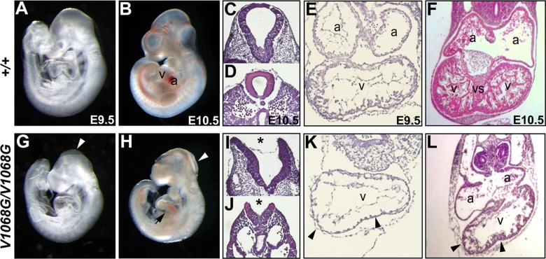
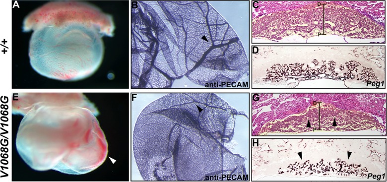
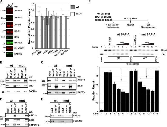
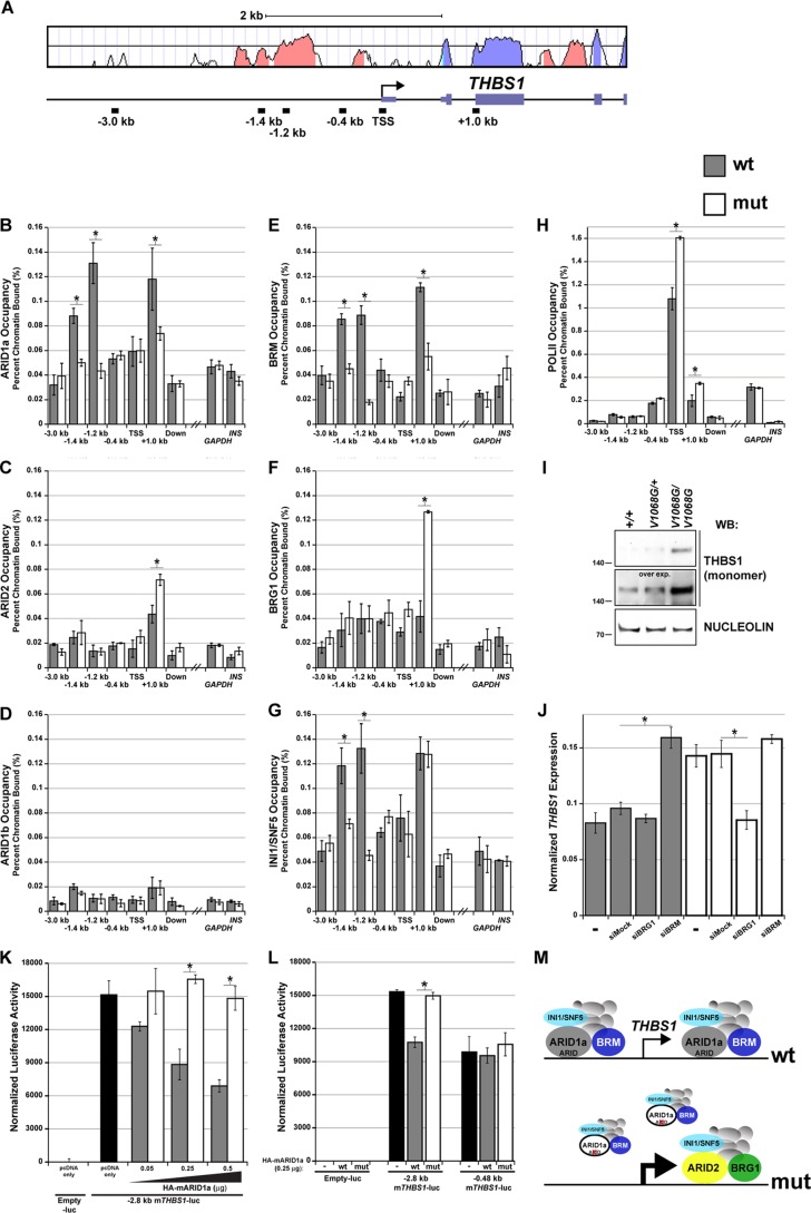
Every known SWI/SNF chromatin-remodeling complex incorporates an ARID DNA binding domain-containing subunit. Despite being a ubiquitous component of the complex, physiological roles for this domain remain undefined. Here, we show that disruption of ARID1a-DNA binding in mice results in embryonic lethality, with mutant embryos manifesting prominent defects in the heart and extraembryonic vasculature. The DNA binding-defective mutant ARID1a subunit is stably expressed and capable of assembling into a SWI/SNF complex with core catalytic properties, but nucleosome substrate binding and promoter occupancy by ARID1a-containing SWI/SNF complexes (BAF-A) are impaired. Depletion of ARID domain-dependent, BAF-A associations at THROMBOSPONDIN 1 (THBS1) led to the concomitant upregulation of this SWI/SNF target gene. Using a THBS1 promoter-reporter gene, we further show that BAF-A directly regulates THBS1 promoter activity in an ARID domain-dependent manner. Our data not only demonstrate that ARID1a-DNA interactions are physiologically relevant in higher eukaryotes but also indicate that these interactions facilitate SWI/SNF binding to target sites in vivo. These findings support the model wherein cooperative interactions among intrinsic subunit-chromatin interaction domains and sequence-specific transcription factors drive SWI/SNF recruitment.


Figures

Fig 1
Fig 1
Arid1aV1068G/V1068G embryos manifest neural tube and heart defects. Whole-mount images of E9.5 and 10.5 Arid1a+/+ (A and B) or Arid1aV1068G/V1068G (G and H) littermate embryos are shown. E9.5 and E10.5 Arid1aV1068G/V1068G embryos display open head folds (G and H, arrowheads). E10.5 Arid1aV1068G/V1068G hearts are grossly abnormal and appeared developmentally delayed (H, arrows). Transverse histological sections through the hindbrain region (I, asterisk) or base of tail (J, asterisk) of E9.5 Arid1aV1068G/V1068G embryos indicate neural tube closure defects. Transverse paraffin sections of E9.5 (E and K) and E10.5 (F and L) Arid1a+/+ and Arid1aV1068G/V1068G hearts are shown. Trabeculation defects are prominent in E9.5 Arid1aV1068G/V1068G hearts (K and L, arrowheads), and these defects are present at E10.5. Mutant E10.5 hearts (L) have hypoplastic myocardial walls and severe ventricular septal defects. v, ventricles; a, atria; vs, ventricular septum.
Fig 2
Fig 2
Prominent extraembryonic vascular abnormalities are present in Arid1aV1068G/V1068G concepti. Whole-mount images of E9.5 Arid1a+/+ (A) and Arid1aV1068G/V1068G (E) yolk sacs are shown. Blood pools in the exocoelomic space of Arid1aV1068G/V1068G (E, arrowhead) are indicative of hemorrhaging. Whole-mount PECAM staining of E10.5 Arid1a+/+ (B) and Arid1aV1068G/V1068G (F) yolk sacs was performed. Hierarchical vascular branching patterns are evident in wild-type yolk sacs (B, arrowhead), whereas highly branched large vessels are scarce in mutant yolk sacs (F, arrowhead). Hematoxylin- and eosin-stained paraffin sections of E10.5 Arid1a+/+ (C) and Arid1aV1068G/V1068G (G) placentas are shown. False-colored, yellow shading identifies the placenta. Proximal (P) to distal (D) placental axes are demarcated by brackets. Mutant placentas display thick spongiotrophoblast layers (G, arrowheads). Near-adjacent paraffin sections of E10.5 Arid1a+/+ (D) and Arid1aV1068G/V1068G (H) placentas hybridized with the pan-mesodermal marker Peg1 are shown. Vascular branching is reduced in mutant placentas (H, arrowheads).
Fig 3
Fig 3
The V1068G mutation does not affect the stability or the catalytic activity of BAF-A complexes. (A) Quantitative fluorescent Western blots (WB) of Arid1a+/+ and Arid1aV1068G/V1068G E9.5 whole-embryo lysates probed with SWI/SNF subunit-specific antibodies. Graph represents the average ± SEM of the normalized band intensity from three age- or littermate-matched whole-embryo lysates. Band intensity measurements are plotted as a ratio to β-actin signals, and wild-type (wt) measurements were set at 1. mut, mutant. (B to E) Coimmunoprecipitation of SWI/SNF complex subunits or cullin-2 from wild-type or Arid1aV1068G/V1068G MEFs. Shown are Western blot panels containing input protein, mock-precipitated (protein A/G-agarose bead only) protein, or protein precipitated with antibodies specific for ARID1a, BRG1, or BRM. Western blots were probed with antibodies specific for ARID1a, BRG1, BRM, INI1/SNF5, BAF60a, and cullin-2. (F) Mononucleosome disruption by wild-type and mutant ARID1a-containing complexes increases PstI restriction site exposure. Fixed amounts of anti-ARID1a immunopurified fractions from either wild-type or Arid1aV1068G/V1068G MEFs were incubated with limiting amounts of gel-purified, radiolabeled mononucleosomes over a 40-min time course in the presence of ATP. Reaction mixtures containing wild-type or mutant anti-ARID1a fractions were quenched at 10-min intervals. Reaction mixtures containing ATPγS or mock-purified (protein A/G-agarose bead only) fractions and incubated over the entire time course served as controls. Naked DNA (lane 1) or mononucleosomes (lane 2) incubated without immunopurified material are shown. Uncut fractions were plotted as a ratio of the total DNA. Error bars in panel F represent the standard deviations, and significant differences were calculated using a two-tailed Student t test (*, P < 0.05).
Fig 4
Fig 4
Structural implications of the conserved valine 1068 residue. (A) Clustal alignment of minimal ARID domains from M. musculus (Mm) ARID1a/BAF250a aligned with similar regions from D. melanogaster (Dm) Osa and S. cerevisiae (Sc) SWI1 ARID subunits, as well as D. melanogaster Retn/Dri/Bright and M. musculus ARID3a/BRIGHT proteins. The mutated valine 1068 residue (red) is highlighted in yellow. (B, left) PyMol rendition of all eight ARID domain NMR conformers (PDB code 1RYU) for the ARID domain structure of H. sapiens ARID1a. α-Helices are depicted in red. V1068 is located in α-helix 3. (B, right) Close-up view of the V1068 side chain contact neighborhood. Favorable, nearest-neighbor V1068, V1059, Y1097, L1077, and K1094 (blue boldface in panel A) side chain interactions form a hydrophobic pocket in the center of a multi-α-helical bundle (H2 to H5).
Fig 5
Fig 5
The V1068G mutation leads to loss of DNA binding. (A and B) Single-component saturating EMSAs (ssEMSAs) using titrated amounts of wild-type (wt) or mutant (mut; V → G) recombinant protein fragments containing the non-sequence-specific ARID domain of H. sapiens (Hs) ARID1a. (D and E) Similar ssEMSAs using titrated amounts of wild-type or mutant (V → G) recombinant protein fragments containing the sequence-specific ARID domain of D. melanogaster Retn/Dri/Bright. Subsaturating amounts of the dri.16 probe (50 pM) were used in each DNA binding reaction. (C and F) Fractional occupancies were calculated from band densitometry measurements and plotted versus log protein concentrations. Blue dotted lines in panels C and F represent curves fitted to EMSA results using Kaleidagraph software.
Fig 6
Fig 6
Loss of sequence-nonspecific ARID1a-DNA interactions reduces BAF-A affinity for nucleosomes. (A) Saturating amounts of immobilized wild-type or mutant BAF-A complexes were incubated with limiting amounts of radiolabeled mononucleosomes in reaction mixtures containing increasing amounts of cold competitor DNA, and then the beads were subjected to a pulldown and washing. Mock-purified fractions from wild-type or mutant cells were used as negative controls. (B) Graph depicts the average scintillation counts measured from independent binding reactions. The numbers above the graphical columns denote the fold difference in average scintillation counts of the wild type compared to those of the mutant. Significant differences were calculated using a two-tailed Student t test (*, P < 0.05).
Fig 7
Fig 7
Loss of DNA binding correlates with reduced promoter occupancy and concurrent changes in SWI/SNF target gene expression. (A) Formaldehyde-cross-linked chromatin from wild-type and Arid1aV1068G/V1068G MEFs was immunoprecipitated using an ARID1a-specific antibody. DNA was amplified by quantitative PCR to determine if mutant ARID1a occupancy was reduced at known SWI/SNF target gene promoters (THBS1, ADAMTS1, and CRABP1). Promoter control elements for a constitutive (GAPDH) or silent (INS-1) gene were used as negative genomic controls. Data were plotted as the percentage of total input or chromatin bound. (B) cDNA synthesized from RNA isolated from wild-type or Arid1aV1068G/V1068G MEFs was used in a quantitative PCR to examine target gene expression differences. Error bars in both panels represent the SEMs, and significant differences between the wild type and mutant were calculated using a two-tailed Student t test (*, P < 0.05).
Fig 8
Fig 8
The ARID domain of ARID1a is required for BAF-A occupancy at THBS1. (A) Mouse/human VISTA alignment of the THBS1 promoter and 5′ transcribed region. Salmon-colored peaks denote evolutionarily conserved regions, whereas lavender peaks denote exons. The locations of ChIP amplicons within this interval are plotted. (B to H) Formaldehyde-cross-linked chromatin from wild-type and Arid1aV1068G/V1068G MEFs was immunoprecipitated with ARID1a, ARID1b, ARID2, BRG1, BRM, INI1/SNF5, or Pol II antibodies. DNA was amplified by quantitative PCR to determine if loss of ARID-DNA binding leads to changes in BAF-A occupancy across THBS1. An intergenic, nonconserved downstream region (located at approximately kb +20) and two unlinked promoter control elements (GAPDH and INS-1) were used as negative genomic controls. Data were plotted as the percentage of total input or chromatin bound. (I) Whole-embryo protein lysates from triplicate pooled samples were used to examine THBS1 protein (reduced, monomeric form) expression differences by Western blotting. An overexposed image of the Western blot was also included to further emphasize these expression differences. The constitutive nuclear matrix protein, nucleolin, was used as a loading control. (J) cDNA synthesized from RNA isolated from wild-type or Arid1aV1068G/V1068G MEFs was used in a quantitative PCR to examine THBS1 expression differences following transfection with mock (nontargeting control), BRG1, or BRM siRNA. (K) Normalized luciferase activity of the THBS1 −2.8 kb promoter-luciferase fragment cotransfected with 0.05 to 0.5 μg of wild-type or mutant HA-mARID1a expression plasmids into NIH 3T3 cells. Cells cotransfected with the empty luciferase vector (−luc) or THBS1 −2.8 kb promoter-luciferase fragment and with empty pcDNA expression plasmids served as negative and positive controls, respectively. (L) Normalized luciferase activity of THBS1 −2.8 kb and −0.48 kb promoter-luciferase reporter plasmids cotransfected with 0.25 μg of pcDNA only (−) or wild-type or mutant HA-mARID1a expression plasmids. Empty luciferase reporter plasmid was used as a negative control. (M) Summary model of ChIP and expression data. Error bars in panels B to H and in panel J represent the SEMs. Error bars in panels K and L represent the standard deviations. Significant differences were calculated using a two-tailed Student t test (*, P < 0.05).

References

    1. Weintraub H, Groudine M. 1976. Chromosomal subunits in active genes have an altered conformation. Science 193: 848–856 - PubMed
    1. Neigeborn L, Carlson M. 1984. Genes affecting the regulation of SUC2 gene expression by glucose repression in Saccharomyces cerevisiae. Genetics 108: 845–858 - PMC - PubMed
    1. Stern M, Jensen R, Herskowitz I. 1984. Five SWI genes are required for expression of the HO gene in yeast. J. Mol. Biol. 178: 853–868 - PubMed
    1. Cairns BR, Kim YJ, Sayre MH, Laurent BC, Kornberg RD. 1994. A multisubunit complex containing the SWI1/ADR6, SWI2/SNF2, SWI3, SNF5, and SNF6 gene products isolated from yeast. Proc. Natl. Acad. Sci. U. S. A. 91: 1950–1954 - PMC - PubMed
    1. Cote J, Quinn J, Workman JL, Peterson CL. 1994. Stimulation of GAL4 derivative binding to nucleosomal DNA by the yeast SWI/SNF complex. Science 265: 53–60 - PubMed

Publication types

MeSH terms