. 2012 Jun;1(3):e001511.
doi: 10.1161/JAHA.112.001511. Epub 2012 Jun 22.

Vulnerable myocardial interstitium in patients with isolated left ventricular hypertrophy and sudden cardiac death: a postmortem histological evaluation

Affiliations

Vulnerable myocardial interstitium in patients with isolated left ventricular hypertrophy and sudden cardiac death: a postmortem histological evaluation

Balaji K Tamarappoo et al. J Am Heart Assoc. 2012 Jun.

Abstract

Background: Concentric left ventricular hypertrophy (LVH) is independently associated with increased risk of sudden cardiac death (SCD). Some animal models of LVH display specific alterations of the myocardial interstitium that could increase myocardial vulnerability to ventricular arrhythmias, but these merit evaluation in humans with LVH and SCD.

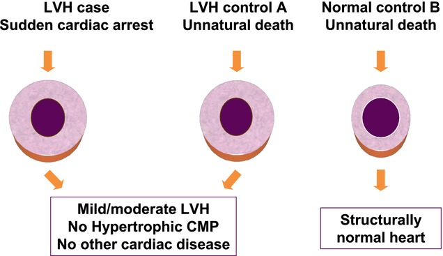
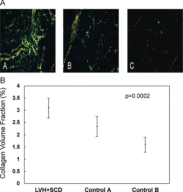
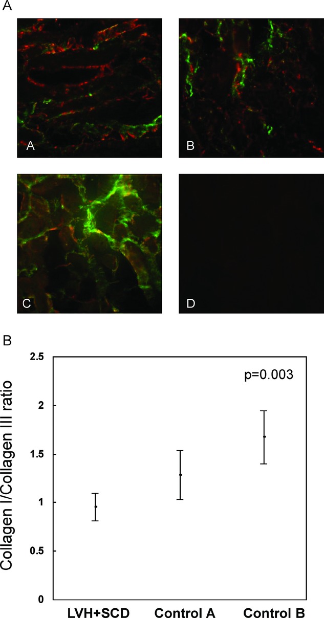
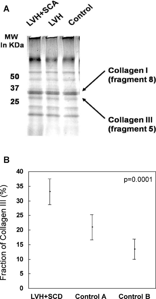
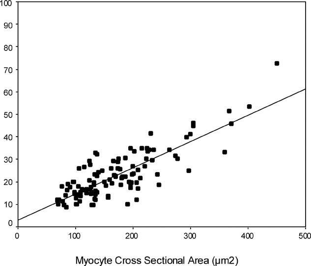
Methods and results: Twelve consecutive patients with isolated LVH and SCD (LVH+SCD) in the absence of hypertrophic cardiomyopathy, coronary disease, or other cardiac structural abnormality were ascertained in the Oregon Sudden Unexpected Death Study. Detailed postmortem comparisons were conducted with 18 controls who had isolated LVH and unnatural deaths (Control Group A) and 6 controls who had structurally normal hearts and unnatural deaths (Control Group B). Postmortem left ventricular myocardial sections were obtained for measurement of collagen volume fraction, characterization of gap junctions, and quantification of collagen subtypes. Heart weight normalized to body weight was higher in LVH+SCD cases (6.9±1.2 g/kg) than in Control Group A (5.3±1.4 g/kg) and Control Group B (4.2±0.3 g/kg); P=0.001. Collagen volume fraction was also higher in LVH+SCD cases (3.1±0.4) than in Control Group A (2.3±0.4) and Control Group B (1.6±0.3); P=0.0002. The relative amount of collagen III was significantly higher in LVH+SCD cases (33.0±4.4%) than in Control Group A (20.9±4.3%) and Control Group B (13.4±3.5%); P=0.0001. There was an overall increase in the number of connexin 43-labeled gap junctions with increasing myocyte size. No subject was found to have high-risk hypertrophic cardiomyopathy mutations.

Conclusions: In addition to the expected increase in myocardial mass and overall collagen content, SCD with isolated LVH was associated with relative abundance of type III collagen, a novel finding that warrants further mechanistic evaluation. (J Am Heart Assoc. 2012;1:e001511 doi: 10.1161/JAHA.111.001511.).

Keywords: collagen; death, sudden; hypertrophy; myocardium; remodeling.


Figures

Figure 1.
Figure 1.
Illustration of the postmortem human model with myocardial sections compared among cases (LVH and sudden cardiac arrest), Control Group A (LVH and unnatural death), and Control Group B (normal controls with unnatural death). CMP indicates cardiomyopathy.
Figure 2.
Figure 2.
A, Myocardial sections showing collagen fibrils stained with Picrosirius Red and viewed under polarized light (40×). Stain A shows a representative section from an individual with LVH+SCD. Stains B and C show representative sections from Control Groups A (LVH + non-SCD) and B (normal control), respectively, with significantly fewer collagen fibrils detectable in the interstitium. B, CVF was quantified as percentage of myocardium comprised of collagen fibrils per high-power field. CVF was expressed as mean±SD for individuals with LVH+SCD and controls. Subjects with LVH+SCD had significantly greater CVF (3.1±0.4%) than Control Group A (LVH + non-SCD) and Control Group B (normal control) (P=0.006 and P=0.007, respectively). There was also a significant difference in mean CVF between the 2 control groups (Control Group A 2.3±0.4% vs Control Group B 1.6±0.3%; P=0.02).
Figure 3.
Figure 3.
A, Comparisons of immunohistochemical staining for collagen type I (Anti-Type I, fluorescein-green) and type III (anti-Type III, Alexa 594-red) in representative cases (LVH+SCD), Control Group A (LVH + non-SCD), and Control Group B (non-LVH controls). Greatest intensity of staining for type III collagen (green) is seen for a case subject in stain A; significantly less intensity of staining is seen for Control Group A subject in stain B, and the least for Control Group B subject in stain C (predominant staining for collagen type I). Stain D represents a negative control with absence of autofluorescence. B, Semiquantitative measurement of the relative amounts of collagen type III and type I revealed a significant difference in the relative expression of collagen III and collagen I between cases and controls. The type I : type III collagen ratio was expressed as mean±SD for individuals with LVH+SCD and controls. Cases had a lower mean type I : type III collagen ratio (0.95±0.14) than those of Control Group A (1.28±0.25) and Control Group B (1.67±0.27); P=0.003.
Figure 4.
Figure 4.
A, Collagen III and collagen I were extracted by digestion of 1 g myocardium, separated by SDS-PAGE, and stained with Coomassie Blue-250. Representative gel with collagen I–band H (CNBr digest fragment 8) and collagen III–band M (CNBr fragments 5 and 9) is shown. SCA indicates sudden cardiac arrest; MW, molecular weight. B, Intensities of the Coomassie-stained protein bands were measured by densitometry. Relative amounts of type III : type I collagen in the extract were given by the ratio of the intensity of collagen III–band M (CNBr fragments 5 and 9) and collagen I–band H (CNBr digest fragment 8). Percent of collagen III compared to the sum of collagen I and collagen III extracted was expressed as mean±SD for individuals with LVH+SCD and controls. Percent myocardial collagen type III was significantly higher in cases (LVH+SCD; 33±4%) than in Control Group A (LVH + non-SCD; 21±4%; P=0.0001) and Control Group B (non-LVH, non-SCD; 13±3%; P=0.004).
Figure 5.
Figure 5.
Intercalated discs were identified by microscopy, and gap junctions were identified within intercalated discs by immunostaining of connexin 43. CSAs of intercalated discs and gap junctions were quantified from digitized microscopic images. Gap junction plaque area was compared to myocyte CSA. Increasing myocyte size as measured by CSA is associated with increase in gap junction plaque surface area (r=0.821, P<0.001).

Similar articles

Cited by

References

    1. Haider AW, Larson MG, Benjamin EJ, Levy D. Increased left ventricular mass and hypertrophy are associated with increased risk for sudden death. J Am Coll Cardiol. 1998;32:1454-1459 - PubMed
    1. Reinier K, Dervan C, Singh T, Uy-Evanado A, Lai S, Gunson K, Jui J, Chugh SS. Increased left ventricular mass and decreased left ventricular systolic function have independent pathways to ventricular arrhythmogenesis in coronary artery disease. Heart Rhythm. 2011;8:1177-1182 - PMC - PubMed
    1. Levy D, Anderson KM, Savage DD, Balkus SA, Kannel WB, Castelli WP. Risk of ventricular arrhythmias in left ventricular hypertrophy: the Framingham Heart Study. Am J Cardiol. 1987;60:560-565 - PubMed
    1. Chugh SS, Reinier K, Teodorescu C, Evanado A, Kehr E, Al Samara M, Mariani R, Gunson K, Jui J. Epidemiology of sudden cardiac death: clinical and research implications. Prog Cardiovasc Dis. 2008;51:213-228 - PMC - PubMed
    1. Fishman GI, Chugh SS, Dimarco JP, Albert CM, Anderson ME, Bonow RO, Buxton AE, Chen PS, Estes M, Jouven X, Kwong R, Lathrop DA, Mascette AM, Nerbonne JM, O'Rourke B, Page RL, Roden DM, Rosenbaum DS, Sotoodehnia N, Trayanova NA, Zheng ZJ. Sudden cardiac death prediction and prevention: report from a National Heart, Lung, and Blood Institute and Heart Rhythm Society Workshop. Circulation. 2010;122:2335-2348 - PMC - PubMed

LinkOut - more resources