. 2012 Aug;1(4):e001081.
doi: 10.1161/JAHA.112.001081. Epub 2012 Aug 24.

CaMK4 Gene Deletion Induces Hypertension

Affiliations

CaMK4 Gene Deletion Induces Hypertension

Gaetano Santulli et al. J Am Heart Assoc. 2012 Aug.

Abstract

Background: The expression of calcium/calmodulin-dependent kinase IV (CaMKIV) was hitherto thought to be confined to the nervous system. However, a recent genome-wide analysis indicated an association between hypertension and a single-nucleotide polymorphism (rs10491334) of the human CaMKIV gene (CaMK4), which suggests a role for this kinase in the regulation of vascular tone.

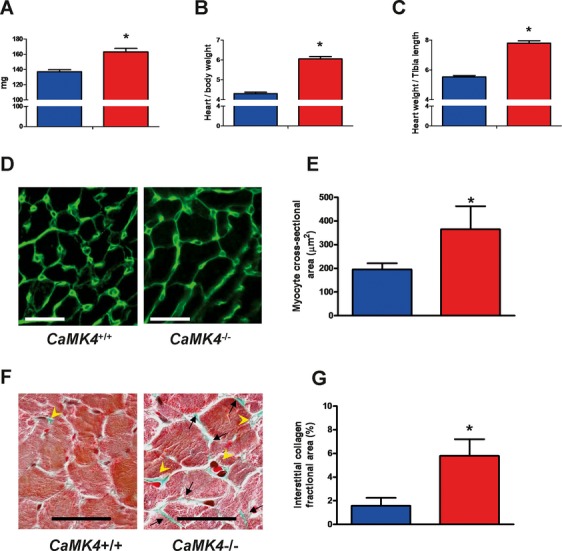
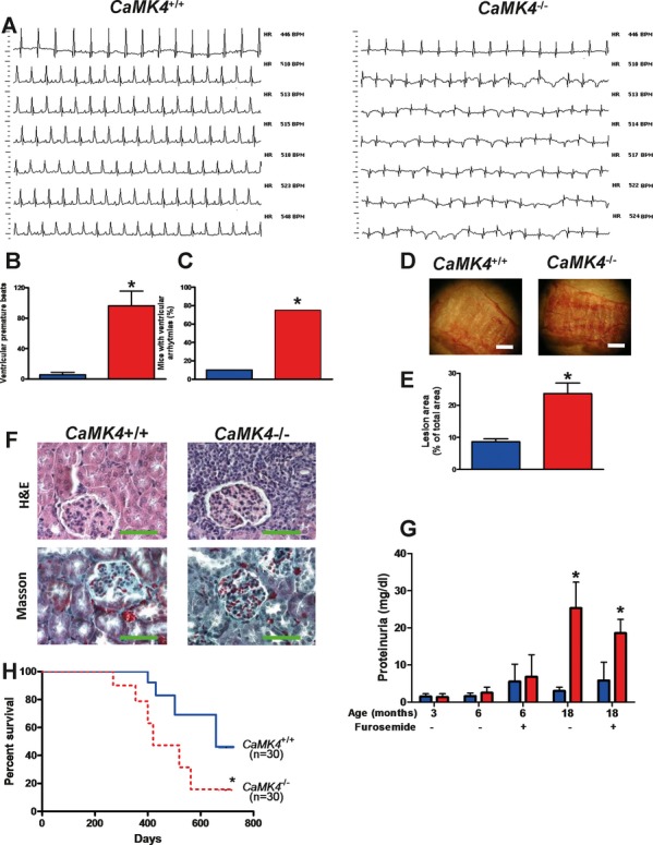
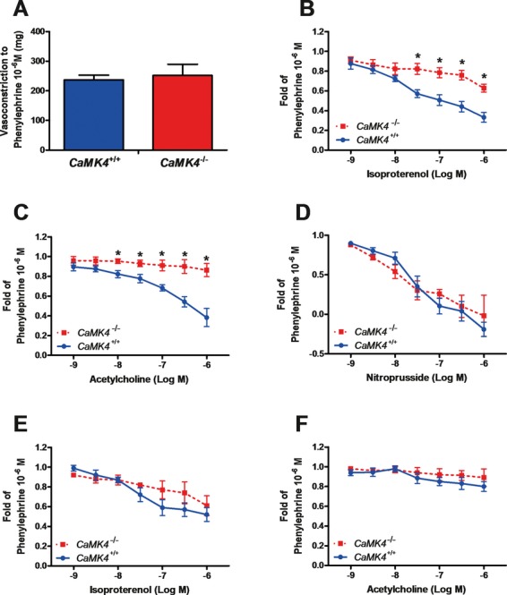
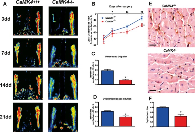
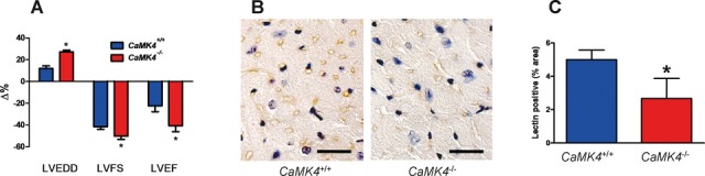
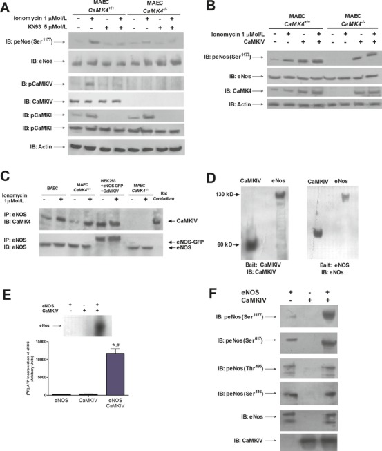
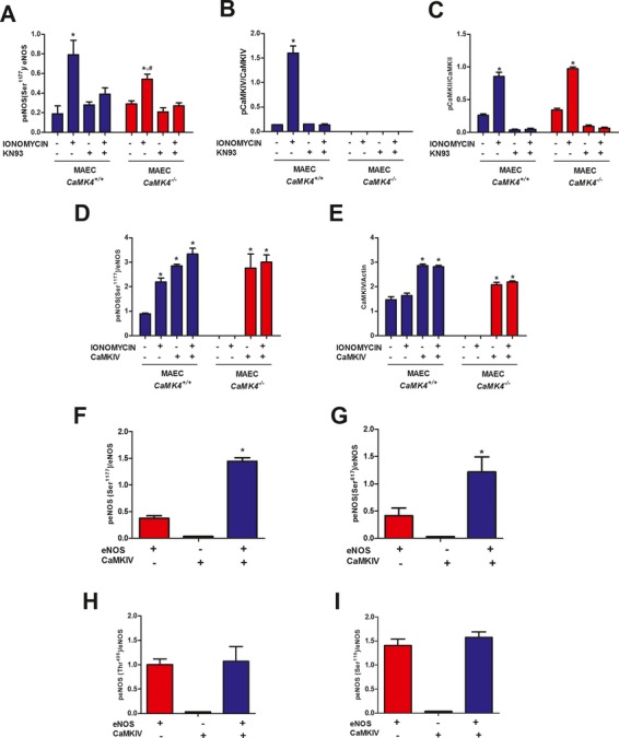
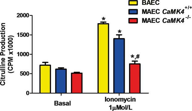
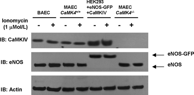
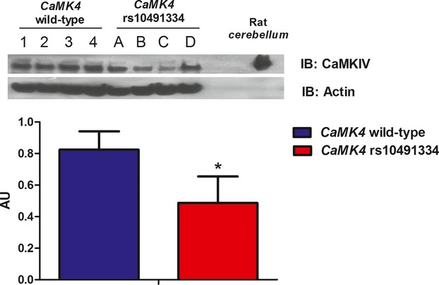
Methods and results: To directly assess the role of CaMKIV in hypertension, we characterized the cardiovascular phenotype of CaMK4(-/-) mice. They displayed a typical hypertensive phenotype, including high blood pressure levels, cardiac hypertrophy, vascular and kidney damage, and reduced tolerance to chronic ischemia and myocardial infarction compared with wild-type littermates. Interestingly, in vitro experiments showed the ability of this kinase to activate endothelial nitric oxide synthase. Eventually, in a population study, we found that the rs10491334 variant associates with a reduction in the expression levels of CaMKIV in lymphocytes from hypertensive patients.

Conclusions: Taken together, our results provide evidence that CaMKIV plays a pivotal role in blood pressure regulation through the control of endothelial nitric oxide synthase activity. (J Am Heart Assoc. 2012;1:e001081 doi: 10.1161/JAHA.112.001081.).

Keywords: angiogenesis; arrhythmia; endothelium; hypertension; hypertrophy.


Figures

Figure 1.
Figure 1.
LVH in CaMK4−/− mice. CaMK4−/− 6-month-old mice present cardiac hypertrophy, as determined by an increase in heart weight (A), normalized by body weight (B) or tibial length (C). Fluorescence-tagged wheat germ agglutinin, which binds to saccharides of cellular membranes, showed increased cardiac myocyte cross-sectional area in CaMK4−/− mice (D and E; magnification ×200, scale bar = 15 μm). Masson's trichrome staining (F; magnification ×300, scale bar = 15 μm; interstitial and perivascular collagen deposition indicated by black arrows and yellow arrowheads, respectively) also revealed an increase in fibrosis, quantified as described in Methods (G). Blue bars are CaMK4+/+ mice; red bars, CaMK4−/− mice. *P<0.05 vs CaMK4+/+.
Figure 2.
Figure 2.
Organ damage in 18-month-old mice. Old CaMK4−/− mice displayed a dilatation of the LV (see also Table 1) and exhibited several spontaneous ventricular arrhythmias (A; representative ECG records at different heart rates). These data are quantified in B and C (n=10 per group; *P<0.05). Of note, heart rate among mutated animals did not significantly differ (see Table 1). D and E, Lipid deposition in vessels of CaMK4+/+ and CaMK4−/− mice (n=12 per group). Representative pictures of oil red O–stained ascending aortas (D; white bar = 600 μm); areas of atherosclerotic lesions were quantified using the free software Fiji and are represented as percentage of lesion area to total aortic area (E; *P<0.05 vs CaMK4+/+). Kidneys (F) from the CaMK4−/− group exhibited increased glomerulosclerosis, inflammatory cell infiltration, and tubulointerstitial fibrosis compared with CaMK4+/+ mice (n=8 per group; representative pictures of hematoxylin and eosin or Masson's trichrome staining; magnification ×60; green bar = 100 μm). Moreover, 18-month-old CaMK4−/− mice presented another typical feature of the hypertensive phenotype (G), showing greater proteinuria than CaMK4+/+ mice (n=12 per group; *P<0.05 vs CaMK4+/+). In all histograms, blue bars are CaMK4+/+ mice; red bars, CaMK4−/− mice. Notably, compared with CaMK4+/+ mice, CaMK4-null mice displayed significantly (*P<0.05) impaired survival, as well (Kaplan-Meier curves; H). Blue line indicates CaMK4+/+ mice; red dotted line, CaMK4−/− mice.
Figure 3.
Figure 3.
Vascular responses on isolated aortic rings from 6-month-old mice. Vasoconstriction to α1-adrenergic agonist phenylephrine was similar in CaMK4+/+ and CaMK4−/− mice (A). Endothelium-dependent vasorelaxation induced by the β-adrenergic agonist isoproterenol (B) or by the muscarinic agonist acetylcholine (C) was blunted in CaMK4−/− vessels, whereas endothelium-independent vasodilation to nitroprusside was not different between CaMK4+/+ and CaMK4−/− (D). To better explore the role of nitric oxide in endothelial responses, we also evaluated vascular responses in the presence (10 μmol/L) of the specific eNOS inhibitor NG-nitro-l-arginine methyl ester (E and F). *P<0.05 vs CaMK4+/+.
Figure 4.
Figure 4.
Decreased neoangiogenic responses in CaMK4−/− mice during chronic ischemia in vivo (n=6 per group). Determination of laser Doppler blood flow (A and B) on postoperative days 3, 7, 14, and 21 showed a deficit in ischemic hindlimb perfusion, compared with the contralateral hindlimb, that was significantly increased in CaMK4−/− vs CaMK4+/+ mice (*P<0.05, repeated measurements, ANOVA; laser Doppler blood flow data are expressed as percent of ischemic to nonischemic limb). Ultrasound Doppler flowmetry of posterior tibial artery (C), performed 3 weeks after femoral artery removal, confirmed the attenuated blood flow restoration in CaMK4−/− mice (*P<0.05 vs CaMK4+/+). This result was mirrored by the dyed-bead perfusion analysis (D; *P<0.05 vs CaMK4+/+). Lectin staining of capillaries in the ischemic hindlimb (E; magnification ×20, black bar = 100 μm) showed that chronic ischemia produced a greater rarefaction of the capillary density, evaluated as number of capillaries corrected for the number of muscle fibers (F), in CaMK4−/− compared with CaMK4+/+ mice (*P<0.05). In all histograms, blue bars are CaMK4+/+ mice; red bars, CaMK4−/− mice.
Figure 5.
Figure 5.
Cardiac evaluation after cryoinjury. Changes in echocardiographic parameters (A) 8 weeks after myocardial infarction. (LVEDD indicates LV end-diastolic diameter; LVFS, LV fractional shortening; and LVEF, LV ejection fraction. n=12 per group. *P<0.05 vs CaMK4+/+). Immunohistochemical analysis (B; lectin staining, magnification ×60, black bar = 30 μm) of myocardium in mice 8 weeks after infarction. The peri-infarct area demonstrated lower capillary density, as confirmed by quantitative analysis (C); blue bars are CaMK4+/+ mice; red bars, CaMK4−/− mice. *P<0.05 vs CaMK4+/+.
Figure 6.
Figure 6.
Interaction between CaMKIV and eNOS. eNOS phosphorylation (Ser1177) is enhanced by ionomycin, echoing the phosphorylation of CaMKIV, and is prevented by the CaMK inhibitor KN93 (A). Notably, eNOS activation was less evident in CaMK4−/− MAEC, where CaMK4 was not expressed (A). Transgenic restoration of CaMKIV expression in CaMK4−/− ECs corrected calcium-induced eNOS activation (B). The interaction between CaMKIV and eNOS was demonstrated by performing immunoprecipitation (IP) experiments in different cellular settings, both in basal conditions and after stimulation with ionomycin (C). Such interaction is shown in BAEC and CaMK4+/+ MAEC but not in CaMK4−/− MAEC. In a nonendothelial cell type, HEK293, we confirmed the interaction after reconstituting the system by using a plasmid encoding CaMKIV and a plasmid encoding eNOS linked to GFP (C; rat cerebellum was used as CaMKIV-positive control). The input protein levels are shown in Figure 8. Overlay assay with purified CaMKIV (left blot) or eNOS (right blot) as bait (D). CaMKIV induced eNOS [32P]-γATP incorporation (E). Purified CaMKIV induced eNOS phosphorylation on Ser1177 and Ser615 but not on Ser114 and Thr495 (F). *P<0.05 vs eNOS, *P<0.05 vs CaMKIV; representative images from triplicate experiments are shown. Densitometric analyses are reported in Figure 7. MAEC indicates murine aortic ECs; BAEC, bovine aortic ECs.
Figure 7.
Figure 7.
Quantification of blot results presented in Figure 6. Relative protein quantification levels for Figure 6A (A–C) and Figure 6B (D–E). Blue bars are CaMK4+/+; red bars, CaMK4−/−. *P<0.05 vs untreated cells; #P<0.05 vs MAEC CaMK4+/+. MAEC indicates murine aortic ECs. Densitometric analyses for Figure 6F (F–H) showing CaMKIV-mediated eNOS phosphorylation on Ser1177 and Ser615 but not on Ser114 and Thr495. Red bars indicate eNOS; black bars, CaMKIV; and blue bars, eNOS and CaMKIV. *P<0.05 vs eNOS.
Figure 8.
Figure 8.
eNOS activity in ECs. eNOS activity, assessed by arginine–citrulline conversion, after stimulation by ionomycin (1 μmol/L) was blunted in CaMK4−/− MAEC. *P<0.05 vs basal; #P<0.05 vs CaMK4+/+. MAEC indicates murine aortic ECs; CPM, counts per minute.
Figure 9.
Figure 9.
Input Western blots of immunoprecipitation assay represented in Figure 6C. To confirm that equal amounts of proteins were present in the cell lysates used for immunoprecipitation as depicted in Figure 6C, we performed Western blotting on 30 μg of proteins of corresponding cell lysates with the same antibodies used in the experiment represented in Figure 6C, raised respectively against CaMKIV and eNOS. Furthermore, actin was detected to confirm equal amount of proteins.
Figure 10.
Figure 10.
Expression levels of CaMKIV in circulating lymphocytes of hypertensive patients. Western blot analysis of CaMKIV on peripheral blood lymphocytes showed that CaMKIV levels were higher in subjects with the CaMK4 wild-type genotype (1, 2, 3, and 4 represent samples from 4 different individuals) than in subjects homozygous for the polymorphic CaMK4 rs10491334 variant (A, B, C, and D represent samples from 4 different individuals). Rat cerebellum was used as CaMKIV-positive control. Data from immunoblots (IB; representative images from 5 experiments are shown) were quantified by densitometric analysis. CaMKIV levels were normalized to actin densitometry. *P<0.05 vs CaMK4+/+. AU indicates arbitrary units.

References

    1. Kushnir A, Shan J, Betzenhauser MJ, Reiken S, Marks AR. Role of CaMKIIdelta phosphorylation of the cardiac ryanodine receptor in the force frequency relationship and heart failure. Proc Natl Acad Sci USA. 2010;107:10274-10279 - PMC - PubMed
    1. Wagner S, Ruff HM, Weber SL, Bellmann S, Sowa T, Schulte T, Anderson ME, Grandi E, Bers DM, Backs J, Belardinelli L, Maier LS. Reactive oxygen species–activated Ca/calmodulin kinase IIdelta is required for late I(Na) augmentation leading to cellular Na and Ca overload. Circ Res. 2011;108:555-565 - PMC - PubMed
    1. Zhang T, Brown JH. Role of Ca2+/calmodulin-dependent protein kinase II in cardiac hypertrophy and heart failure. Cardiovasc Res. 2004;63:476-486 - PubMed
    1. Colomer JM, Mao L, Rockman HA, Means AR. Pressure overload selectively up-regulates Ca2+/calmodulin-dependent protein kinase II in vivo. Mol Endocrinol. 2003;17:183-192 - PubMed
    1. Wayman GA, Tokumitsu H, Davare MA, Soderling TR. Analysis of CaM-kinase signaling in cells. Cell Calcium. 2011;50:1-8 - PMC - PubMed

LinkOut - more resources