. 2012 Nov 6:13:31.
doi: 10.1186/1471-2121-13-31.

Yeast importin-β is required for nuclear import of the Mig2 repressor

Affiliations

Yeast importin-β is required for nuclear import of the Mig2 repressor

Alejandra Fernández-Cid et al. BMC Cell Biol. .

Abstract

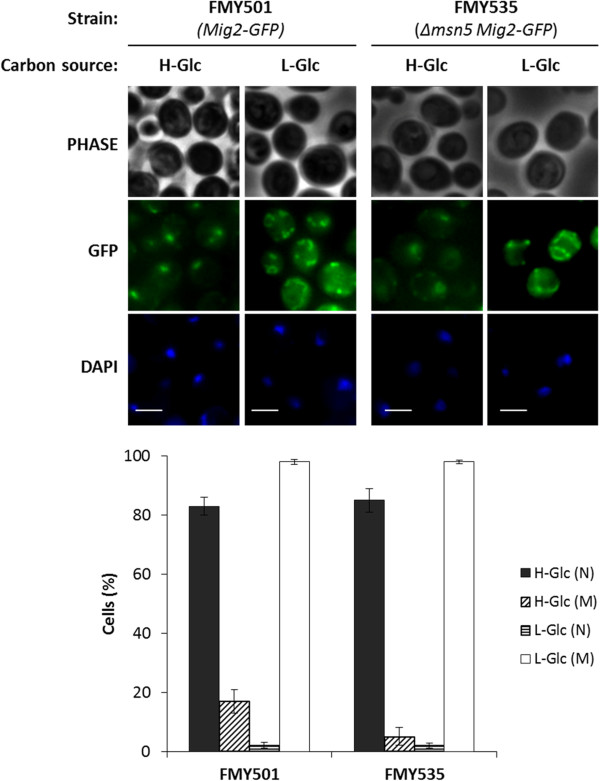
Background: Mig2 has been described as a transcriptional factor that in the absence of Mig1 protein is required for glucose repression of the SUC2 gene. Recently it has been reported that Mig2 has two different subcellular localizations. In high-glucose conditions it is a nuclear modulator of several Mig1-regulated genes, but in low-glucose most of the Mig2 protein accumulates in mitochondria. Thus, the Mig2 protein enters and leaves the nucleus in a glucose regulated manner. However, the mechanism by which Mig2 enters into the nucleus was unknown until now.

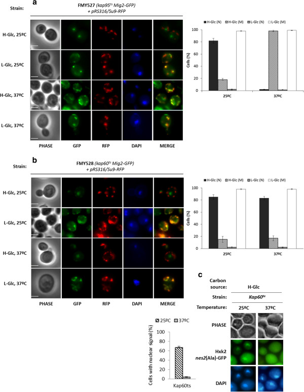
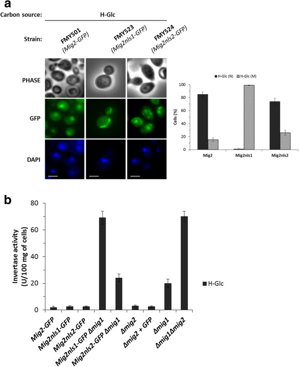
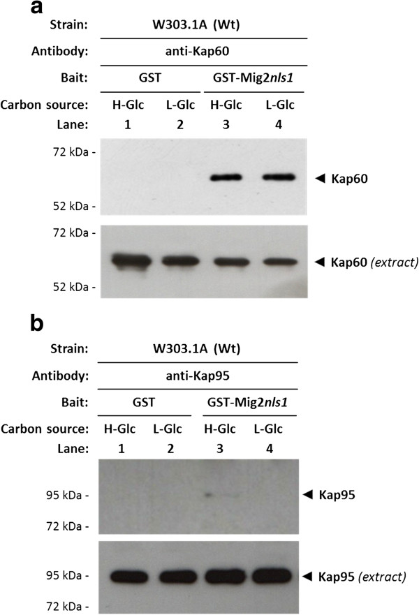
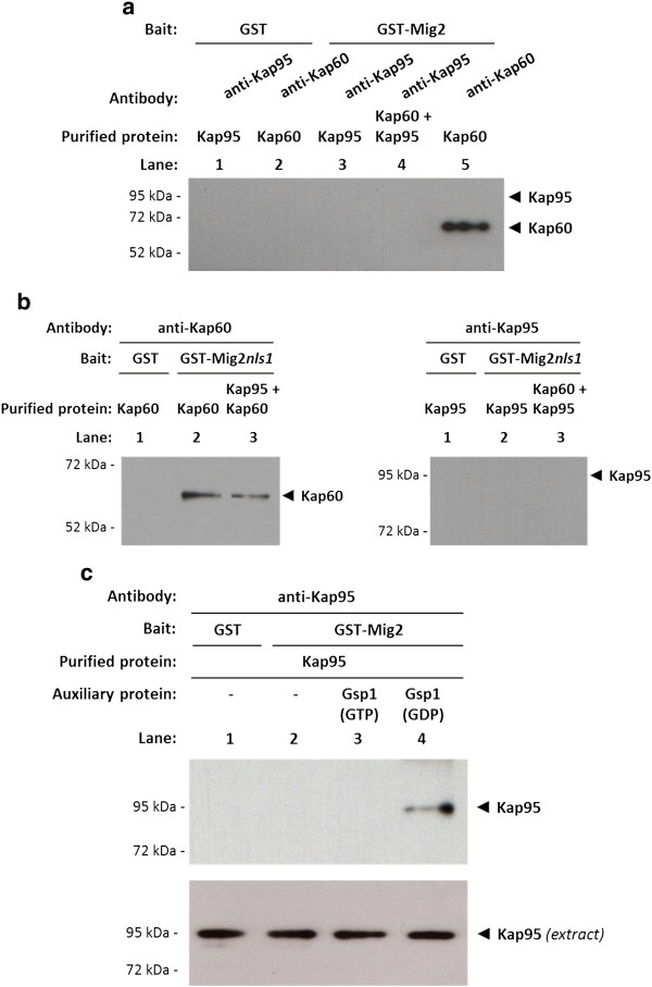
Results: Here, we report that the Mig2 protein is an import substrate of the carrier Kap95 (importin-β). The Mig2 nuclear import mechanism bypasses the requirement for Kap60 (importin-α) as an adaptor protein, since Mig2 directly binds to Kap95 in the presence of Gsp1(GDP). We also show that the Mig2 nuclear import and the binding of Mig2 with Kap95 are not glucose-dependent processes and require a basic NLS motif, located between lysine-32 and arginine-37. Mig2 interaction with Kap95 was assessed in vitro using purified proteins, demonstrating that importin-β, together with the GTP-binding protein Gsp1, is able to mediate efficient Mig2-Kap95 interaction in the absence of the importin-α (Kap60). It was also demonstrated, that the directionality of Mig2 transport is regulated by association with the small GTPase Gsp1 in the GDP- or GTP-bound forms, which promote cargo recognition and release, respectively.

Conclusions: The Mig2 protein accumulates in the nucleus through a Kap95 and NLS-dependent nuclear import pathway, which is independent of importin-α in Saccharomyces cerevisiae.


Figures

Figure 1
Figure 1
Localization of Mig2-GFP in Δmsn5 yeast cells. The FMY501(Mig2-GFP) and FMY535 (Δmsn5 Mig2-GFP) yeast strains, were grown in YEPD high-glucose medium (H-Glc) until an A600nm of 1.0 was reached and then transferred to YEPGly low glucose medium (L-Glc) for 5 min. Cells were stained with DAPI and imaged for GFP and DAPI fluorescence. Scale bar is 10 μm. The nuclear and mitochondrial localization of Mig2-GFP protein was determined in at least 100 cells per growth condition. No statistically significant differences were detected between the wild-type and the mutant strains. N, denotes a nuclear fluorescence signal and M, mitochondrial fluorescence signal.
Figure 2
Figure 2
Localization of Mig2-GFP in kap95tsand kap60tsyeast cells. (a) The FMY527 (kap95ts Mig2-GFP) and (b) FMY528 (kap60ts Mig2-GFP) strains were transformed with plasmid pRS316/Su9-RFP. Transformed cells were grown in high-glucose synthetic medium (H-Glc) until an A600nm of 1.0 was reached and then transferred to low glucose synthetic medium (L-Glc) for 5 min. (c) The JCY1410 (kap95ts) strain transformed with plasmid YEp352/Hxk2nes2(Ala)-GFP was used as positive control. Transformed cells were grown in high-glucose synthetic medium (H-Glc) until an A600nm of 1.0 was reached. The cells were grown at 25°C (permissive temperature) and then shifted to 37°C (not permissive temperature) for 1 h. Cells were stained with DAPI and imaged for GFP, RFP and DAPI fluorescence. Scale bar is 10 μm. The nuclear or mitochondrial localization of Mig2-GFP and the nuclear or cytoplasmic localization of Hxk2nes2(Ala) proteins was determined in at least 100 cells per growth condition. Error bars represent standard deviations for three independent experiments. N denotes a nuclear fluorescence signal and M mitochondrial fluorescence signal.
Figure 3
Figure 3
Identification of Mig2 NLSs. (a) The FMY501 (Mig2-GFP), FMY523 (Mig2nls1-GFP) and FMY524 (Mig2nls2-GFP) were grown in YEPD high-glucose medium (H-Glc) until an A600nm of 1.0 was reached and then transferred to YEPGly low glucose medium (L-Glc) for 5 min. The cells were visualized by fluorescence microscopy, DAPI staining revealed nuclear DNA. Scale bar is 10 μm. The nuclear localization of fluorescent reporter proteins was determined in at least 100 cells in three independent experiments. Means and standard deviations are shown for at least three independent experiments. N denotes a nuclear fluorescence signal and M mitochondrial fluorescence signal. (b) The FMY501 (Mig2-GFP), FMY523 (Mig2nls1-GFP), FMY524 (Mig2nls2-GFP), FMY525 (Mig2nls1-GFP Δmig1), FMY526 (Mig2nls2-GFP Δmig1), Y14575 (Δmig2), FMY536 (Δmig2 + GFP), H174 (Δmig1) and MAP24 (Δmig1 Δmig2) strains were grown in YEPD high-glucose medium (H-Glc) until an A600nm of 0.8 was reached. Invertase activity was assayed in whole cells. Values are the averages of results obtained in four independent experiments.
Figure 4
Figure 4
Interaction of Kap60 and Kap95 with Mig2. In vivo co-immunoprecipitation of Kap60 and Kap95 with Mig2. The wild-type, FMY501 (Mig2-GFP) (a) or a control strain, (TetR-GFP) synthetizing GFP only (b) were grown in YEPD-media until an A600nm of 0.8 was reached and then shifted to low glucose (L-Glc) conditions for 1 h. The cell extracts were immunoprecipitated with a polyclonal anti-Kap60 or anti-Kap95 antibody (lanes 3-6) or a polyclonal antibody against Pho4 (lanes 1 and 2). Immunoprecipitates were separated by 12% SDS-PAGE, and co-precipitated Mig2-GFP was visualized by Western blot with polyclonal anti-GFP antibodies. The level of immunoprecipitated Kap60 or Kap95 in the blotted samples was determined by using anti-Kap60 and anti-Kap95 antibodies, respectively. The level of Mig2-GFP and GFP present in the different extracts was determined by Western blot using anti-GFP antibody. The Western blots shown are representative of results obtained from four independent experiments. The GST-Mig2 fusion protein was purified on glutathione-Sepharose columns. Equal amounts of GST-Mig2 were incubated with cell extracts from the wild-type strain W303-1A. The yeasts were grown in YEPD media until an A600nm of 0.8 was reached and then shifted to low (L-Glc) glucose conditions for 1 h. After exhaustive washing the proteins were separated by 12% SDS-PAGE, and retained Kap60 and Kap95 proteins were visualized by Western blot using polyclonal anti-Kap60 (c) and anti-Kap95 (d) antibodies respectively. For the control samples, GST protein was also incubated with high- (H-Glc) and low-glucose (L-Glc) cell extracts, but no signals were detected. The level of Kap60 and Kap95 proteins present in the different extracts used in Figure  4c and 4d was determined by Western blot using anti-Kap60 and anti-Kap95 antibodies respectively. The Western blots shown are representative of results obtained from four independent experiments.
Figure 5
Figure 5
Interaction of Kap60 and Kap95 with Mig2 nls1. The GST-Mig2nls1fusion protein was purified on glutathione-Sepharose columns. Equal amounts of GST-Mig2nls1 were incubated with cell extracts from the wild-type strain W303-1A. The yeasts were grown in YEPD media until an A600nm of 0.8 was reached and then shifted to low (L-Glc) glucose conditions for 1 h. After exhaustive washing the proteins were separated by 12% SDS-PAGE, and retained Kap60 and Kap95 proteins were visualized on a Western blot with polyclonal anti-Kap60 (b) and anti-Kap95 (c) antibodies respectively. For the control samples, GST protein was also incubated with high- (H-Glc) and low-glucose (L-Glc) cell extracts, but no signals were detected. The level of Kap60 and Kap95 proteins present in the different extracts used in Figure  5a and 5b was determined by Western blot using anti-Kap60 and anti-Kap95 antibodies respectively. The Western blots shown are representative of results obtained from four independent experiments.
Figure 6
Figure 6
The import Mig2-Kap95 complex formation in vitro. GST pull-down analysis of Mig2 (a) and Mig2nls1 (b) interaction with Kap60 and Kap95 purified proteins. (c) Effect of Gsp1 on the Mig2-Kap95 interaction stability. GST-Mig2, GST-Mig2nls1, GST-Gsp1, GST-Kap60 and GST-Kap95 fusion proteins were purified on glutathione-Sepharose columns and incubated with thrombin to release, respectively, Gsp1, Kap60 and Kap95 proteins. The Gsp1 protein was loaded with GTP to generate Gsp1(GTP) or with GDP to generate Gsp1(GDP). The purified proteins were incubated with purified GST-Mig2 or GST-Mig2nls1 bound to glutathione-Sepharose beads in the absence (a and b) or in the presence (c) of Gsp1(GTP) and Gsp1(GDP). The beads were washed extensively. Co-precipitated proteins were resolved by 12% SDS-PAGE and visualized on a Western blot with polyclonal anti-Kap60 or anti-Kap95 antibodies. The level of Kap95 protein present in the different extracts used in Figure  5c was determined by Western blot using anti-Kap95 antibodies. The Western blots shown are representative of results obtained from four independent experiments.

Similar articles

Cited by

References

    1. Cook A, Bono F, Jinek M, Conti E. Structural biology of nucleocytoplasmic transport. Annu Rev Biochem. 2007;76:647–671. doi: 10.1146/annurev.biochem.76.052705.161529. - DOI - PubMed
    1. Radu A, Blobel G, Moore MS. Identification of a protein complex that is required for nuclear protein import and mediates docking of import substrate to distinct nucleoporins. Proc Natl Acad Sci U S A. 1995;92(5):1769–1773. doi: 10.1073/pnas.92.5.1769. - DOI - PMC - PubMed
    1. Gorlich D, Prehn S, Laskey RA, Hartmann E. Isolation of a protein that is essential for the first step of nuclear protein import. Cell. 1994;79(5):767–778. doi: 10.1016/0092-8674(94)90067-1. - DOI - PubMed
    1. Stade K, Ford CS, Guthrie C, Weis K. Exportin 1 (Crm1p) is an essential nuclear export factor. Cell. 1997;90(6):1041–1050. doi: 10.1016/S0092-8674(00)80370-0. - DOI - PubMed
    1. Gorlich D, Kostka S, Kraft R, Dingwall C, Laskey RA, Hartmann E, Prehn S. Two different subunits of importin cooperate to recognize nuclear localization signals and bind them to the nuclear envelope. Curr Biol. 1995;5(4):383–392. doi: 10.1016/S0960-9822(95)00079-0. - DOI - PubMed

Publication types

MeSH terms