

The importance of microstructural variations on the fracture toughness of human dentin
- PMID: 23131531
- PMCID: PMC3511669
- DOI: 10.1016/j.biomaterials.2012.10.032
The importance of microstructural variations on the fracture toughness of human dentin
Abstract
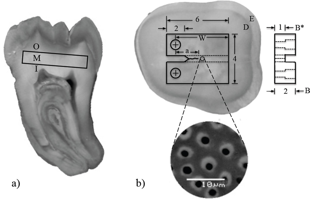
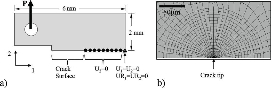
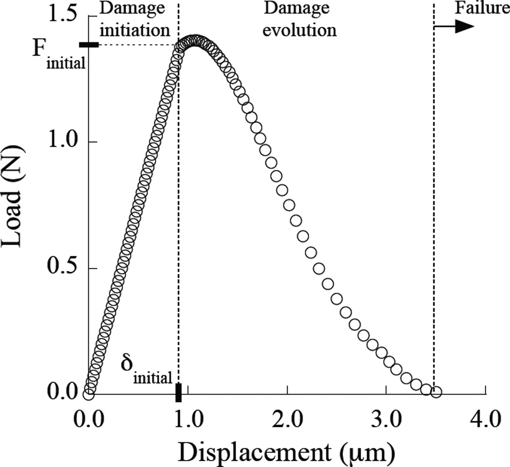
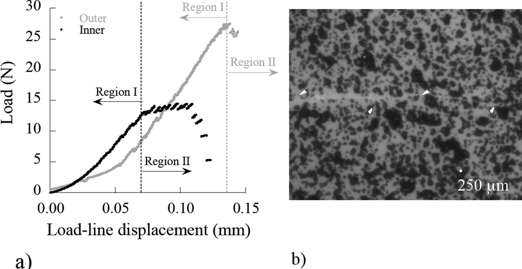
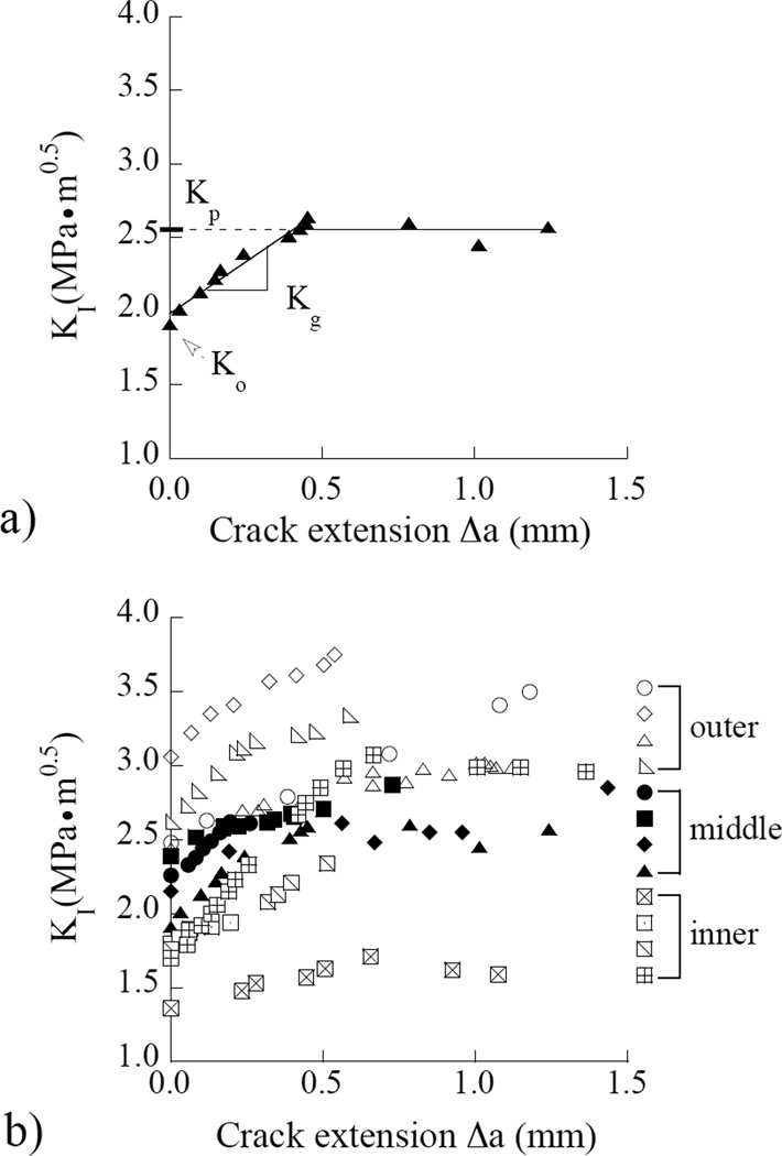
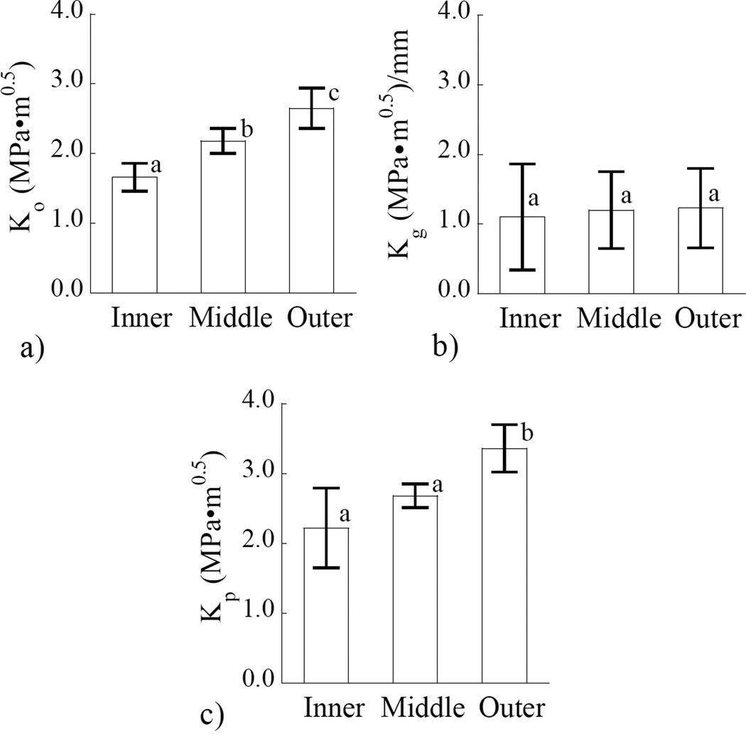
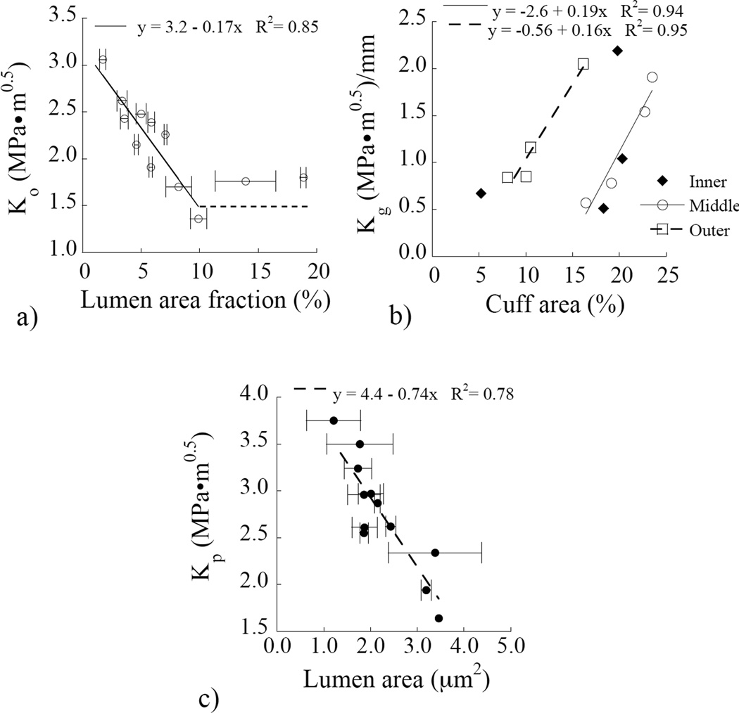
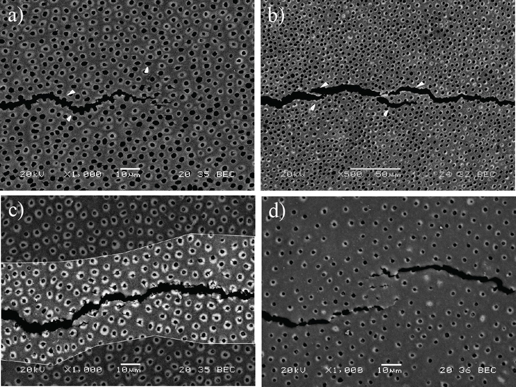
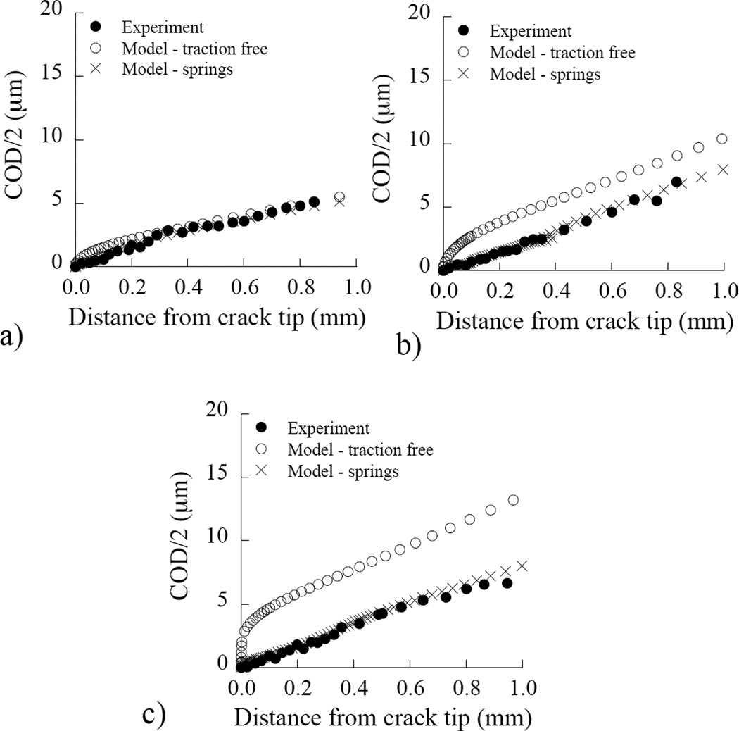
The crack growth resistance of human dentin was characterized as a function of relative distance from the DEJ and the corresponding microstructure. Compact tension specimens were prepared from the coronal dentin of caries-free 3rd molars. The specimens were sectioned from either the outer, middle or inner dentin. Stable crack extension was achieved under Mode I quasi-static loading, with the crack oriented in-plane with the tubules, and the crack growth resistance was characterized in terms of the initiation (K(o)), growth (K(g)) and plateau (K(p)) toughness. A hybrid approach was also used to quantify the contribution of dominant mechanisms to the overall toughness. Results showed that human dentin exhibits increasing crack growth resistance with crack extension in all regions, and that the fracture toughness of inner dentin (2.2 ± 0.5 MPa·m(0.5)) was significantly lower than that of middle (2.7 ± 0.2 MPa·m(0.5)) and outer regions (3.4 ± 0.3 MPa·m(0.5)). Extrinsic toughening, composed mostly of crack bridging, was estimated to cause an average increase in the fracture energy of 26% in all three regions. Based on these findings, dental restorations extended into deep dentin are much more likely to cause tooth fracture due to the greater potential for introduction of flaws and decrease in fracture toughness with depth.
Copyright © 2012 Elsevier Ltd. All rights reserved.
Figures
Similar articles
-
Aging and the reduction in fracture toughness of human dentin.J Mech Behav Biomed Mater. 2009 Oct;2(5):550-9. doi: 10.1016/j.jmbbm.2009.01.008. Epub 2009 Feb 5. J Mech Behav Biomed Mater. 2009. PMID: 19627862 Free PMC article.
-
Importance of tubule density to the fracture toughness of dentin.Arch Oral Biol. 2016 Jul;67:9-14. doi: 10.1016/j.archoralbio.2016.03.003. Epub 2016 Mar 15. Arch Oral Biol. 2016. PMID: 27010828
-
Fatigue crack propagation path across the dentinoenamel junction complex in human teeth.J Biomed Mater Res A. 2003 Jul 1;66(1):103-9. doi: 10.1002/jbm.a.10541. J Biomed Mater Res A. 2003. PMID: 12833436
-
The mechanical properties of human dentin: a critical review and re-evaluation of the dental literature.Crit Rev Oral Biol Med. 2003;14(1):13-29. doi: 10.1177/154411130301400103. Crit Rev Oral Biol Med. 2003. PMID: 12764017 Review.
-
Crystalline defects in bulk metallic glasses: consequences on fracture toughness determination and ductility.J Phys Condens Matter. 2020 Sep 4;32(48). doi: 10.1088/1361-648X/abaa7f. J Phys Condens Matter. 2020. PMID: 32726754 Review.
Cited by
-
Preparation of collagen fibrils from mineralized tissues and evaluation by atomic force microscopy.J Mech Behav Biomed Mater. 2023 Feb;138:105624. doi: 10.1016/j.jmbbm.2022.105624. Epub 2022 Dec 16. J Mech Behav Biomed Mater. 2023. PMID: 36543081 Free PMC article.
-
Importance of age on the dynamic mechanical behavior of intertubular and peritubular dentin.J Mech Behav Biomed Mater. 2015 Feb;42:229-42. doi: 10.1016/j.jmbbm.2014.11.021. Epub 2014 Nov 29. J Mech Behav Biomed Mater. 2015. PMID: 25498296 Free PMC article.
-
An inset CT specimen for evaluating fracture in small samples of material.J Mech Behav Biomed Mater. 2014 Feb;30:358-68. doi: 10.1016/j.jmbbm.2013.10.017. Epub 2013 Oct 31. J Mech Behav Biomed Mater. 2014. PMID: 24268892 Free PMC article.
-
On the Mechanics of Fatigue and Fracture in Teeth.Appl Mech Rev. 2014 May;66(3):0308031-3080319. doi: 10.1115/1.4027431. Epub 2014 Apr 30. Appl Mech Rev. 2014. PMID: 25516632 Free PMC article. Review.
-
Effect of Propolis on Root Dentine Microhardness When Used as an Intracanal Medicament: An In Vitro Study.J Funct Biomater. 2023 Mar 3;14(3):144. doi: 10.3390/jfb14030144. J Funct Biomater. 2023. PMID: 36976068 Free PMC article.
References
-
- Ten Cate AR. Oral Histology: Development, structure, and function. 5th Ed. St. Louis: Mosby- Year Book, Inc; 1998.
-
- Marshall GW, Jr, Marshall SJ, Kinney JH, Balooch M. The dentin substrate: structure and properties related to bonding. J Dent. 1997;25(6):441–458. - PubMed
-
- Kinney JH, Marshall SJ, Marshall GW. The mechanical properties of human dentin: a critical review and re-evaluation of the dental literature. Crit Rev Oral Biol Med. 2003;14(1):13–29. - PubMed
-
- Pashley DH. Dentin: A dynamic substrate - a review. Scan Microsc. 1989;3:161–176. - PubMed
-
- Mjör IA, Nordahl I. The density and branching of dentinal tubules in human teeth. Arch Oral Biol. 1996;41(5):401–412. - PubMed
Publication types
MeSH terms
Grants and funding
LinkOut - more resources
Full Text Sources
Other Literature Sources
Research Materials
