. 2013 May;6(3):626-38.
doi: 10.1038/mi.2012.104. Epub 2012 Nov 7.

Functional diversity of human vaginal APC subsets in directing T-cell responses

Affiliations

Functional diversity of human vaginal APC subsets in directing T-cell responses

D Duluc et al. Mucosal Immunol. 2013 May.

Abstract

Human vaginal mucosa is the major entry site of sexually transmitted pathogens and thus has long been attractive as a site for mounting mucosal immunity. It is also known as a tolerogenic microenvironment. Here, we demonstrate that immune responses in the vagina can be orchestrated by the functional diversity of four major antigen-presenting cell (APC) subsets. Langerhans cells (LCs) and CD14(-) lamina propria-dendritic cells (LP-DCs) polarize CD4(+) and CD8(+) T cells toward T-helper type 2 (Th2), whereas CD14(+) LP-DCs and macrophages polarize CD4(+) T cells toward Th1. Both LCs and CD14(-) LP-DCs are potent inducers of Th22. Owing to their functional specialties and the different expression levels of pattern-recognition receptors on the APC subsets, microbial products do not bias them to elicit common types of immune responses (Th1 or Th2). To evoke desired types of adaptive immune responses in the human vagina, antigens may need to be targeted to proper APC subsets with right adjuvants.


Figures
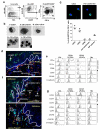
Figure 1
Figure 1
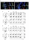
Human vaginal mucosa contains four major subsets of myeloid-originated APCs. (a) Flow cytometry analysis of cells in human vaginal mucosa after enzymatic digestion. Live HLA-DR+ cells were gated (left panel) and CD207+ cells (I) were gated (middle panel). HLA-DR+CD207 cells were further divided into four groups based on CD1c and CD14 expression (right panel, II: CD1c+CD14; III: CD1c+CD14+; IV: CD1c CD14+; V: CD1cCD14). Data are representative of 20 independent experiments. (b) Morphology of FACS-sorted subpopulations of vaginal cells (x100, bars are 20 μm). Data are representative of 4 independent experiments. (c) FACS-sorted HLA-DR+CD207 CD1cCD14 cells were stained for CD34, CD54, CD66, pan-cytokeratin or E/P selectin. One representative data from 3 independent experiments using cells from different donors (upper panel). Percentage of positive cells has been calculated (lower panel). Each dot represents data acquired from one donor. (d) Frozen tissue sections were stained for CD207 (green), CD14 (red), CD1c (light blue) and cell nuclei (dark blue) (x20, bar is 100 μm). Data are representative of 6 independent experiments. (e, g) Flow cytometry analysis of the vaginal APC subsets. Tissues were digested with enzymes and cell suspension was stained with indicated antibodies and gated as in a. Gray histograms represent isotype controls. Data are representative of 10 (e) or 6 (g) independent experiments. (f) Frozen tissue sections were stained for CD1a (green), CD1c (light blue) and CD14 (red) (upper panel) and CD163, (green), CD1c (light blue) and CD14 (red) (lower panel) (x20, bar is 100μm). Data are representative of 6 independent experiments. All of the independent experiments were performed with tissues or cells from different donors.
Figure 2
Figure 2
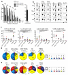
Functional specialties of the vaginal APC subsets in directing CD4+ T cell responses. CFSE-labeled allogeneic naïve total T cells were co-cultured for 7 days with different numbers of the vaginal APCs or IFNDCs. (a) Live CD4+ T cells were gated and CD4+ T cell proliferation was assessed by measuring CFSE dilution. Data are mean ± SD of 3 independent experiments with duplicates. (b) After 7 days, T cells were stimulated with PMA/ionomycin in the presence of brefeldin A. Cells were then stained for intracellular IFNγ, IL-13, IL-5, and TNFα. 6 independent experiments using cells from different donors showed similar data. (c) Summary of the data from independent experiments using APCs from 6 donors marked with different colors. (d-e) Boolean gate analysis. (d) IFNγ+, IL-13+, and IL-5+ CD4+ T cells (N=6) and (e) IFNγ+, IL-13+, and TNFα+ CD4+ T cells induced with different subsets of APCs (N=3). * indicates p<0.05 by ANOVA test.
Figure 3
Figure 3
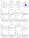
Functional specialties of the vaginal APC subsets in directing CD8+ T cell responses. CFSE-labeled allogeneic naïve total T cells were co-cultured for 7 days with the vaginal APC subsets or IFNDCs. (a) CD8+ T cell proliferation was assessed by measuring CFSE dilution. Data are mean ± SD of 3 independent experiments with duplicates. (b-e) After 7 days, T cells were stimulated with PMA/ionomycin in the presence of brefeldin A, and then stained for intracellular IFNγ, TNFα, and IL-5 expression. (b) Representative data from 6 independent experiments. (c) Summary of the data from 6 (IFNγ+ and IL-5+) and 4 (TNFα+) independent experiments using cells from different donors. (d) IFNγ+ and IL-5+ CD8+ T cells (N=6) or (e) IFNγ+ and TNFα+ CD8+ T cells induced with different APC subsets (N=3). * indicates p<0.05 by ANOVA test.
Figure 4
Figure 4
Vaginal APCs can induce naïve T cells to express CD103 and CCR4 that are found to be expressed in T cells in the vaginal mucosa. (a) Frozen tissue sections were stained for CD3, CD4 and CD103 and (b) CD3, CD8, CD103 expression (x20, bar is 100 μm). (c, e) CD103 (c) and CCR4 (e) expressions on CD4+ (top panel) and CD8+ (bottom panel) T cells from the vaginal mucosa. (d, f) CD103 (d) and CCR4 (f) expressions on naïve CFSE-labeled CD4+ (top panels) and CD8+ T cells (bottom panels) co-cultured for 7 days with the vaginal APC subsets or IFNDCs. (a-c and e) T cells in the vagina from 5 donors showed similar results. (d and f) 4 independent experiments using APCs from different donors showed similar results.
Figure 5
Figure 5
Expression levels of pattern-recognition receptors and their effects on the APC subset-driven T cell responses (a) TLRs, MDA-5 and RIG-I average gene expression in FACS-sorted vaginal LCs (N=2), CD14 LP-DCs (N=3), CD14+ LP-DCs (N=2), and Mφ (N=6). (b, c) APC subsets were stimulated with flagellin, R848 or poly IC for 16h and then CFSE-labeled naïve total T cells were co-cultured for 7 days. T cells were stimulated with PMA/ionomycin in the presence of brefeldin A and then stained for intracellular IFNγ and IL-5 expression. Each color represents the data generated with one tissue donor (* indicates p<0.05 and ** indicates p<0.01; paired t-test). CD4+ (b) and CD8+ T cells (c).
Figure 6
Figure 6
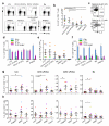
Vaginal LCs and CD14 LP-DCs can efficiently induce IL-22-producing T cell responses. CFSE-labeled allogeneic naïve total T cells were co-cultured for 7 days with the vaginal APCs or IFNDCs. T cells were restimulated with PMA/ionomycin in the presence of brefeldin A and then stained for intracellular IL-22. (a-d, g) Data for CD4+ T cells (e, f, h) Data for CD8+ T cells. (a) Representative data from one experiment are presented. (b, e) Summarized data from 6 independent experiments (* indicates p<0.05; One-way ANOVA test). (c, d, f) Frequency of IFNγ+, IL-5+ and IL-17+ cells among the IL-22-producing T cells. 5 independent experiments using APCs from different donors showed similar results. (c) Representative data from one experiment after co-culture with CD14 LP-DCs or (d, f) Combined data from 5 independent experiments using cells from different tissue donors. (g, h) Allogeneic naïve T cells were co-cultured for 7 days with the vaginal APCs non-activated or activated for 16 h with flagellin, R848 or poly IC. T cells were then stained for intracellular IL-22 expression. Data from 7 (CD14+ LP-DCs) to 10 (Mφ) independent experiments using APCs from different donors are presented (* indicates p<0.05 and ** indicates p<0.01;paired t-test). In (b, e, g, h), each color represents the data generated with one tissue donor.

References

    1. Mestecky J, Moldoveanu Z, Smith PD, Hel Z, Alexander RC. Mucosal immunology of the genital and gastrointestinal tracts and HIV-1 infection. J Reprod Immunol. 2009;83(1-2):196–200. - PMC - PubMed
    1. Iwasaki A. Antiviral immune responses in the genital tract: clues for vaccines. Nat Rev Immunol. 2010;10(10):699–711. - PMC - PubMed
    1. Mestecky J, Moldoveanu Z, Russell MW. Immunologic uniqueness of the genital tract: challenge for vaccine development. Am J Reprod Immunol. 2005;53(5):208–214. - PubMed
    1. Russell MW, Mestecky J. Tolerance and protection against infection in the genital tract. Immunol Invest. 2010;39(4-5):500–525. - PubMed
    1. Russell MW, Mestecky J. Humoral immune responses to microbial infections in the genital tract. Microbes Infect. 2002;4(6):667–677. - PubMed

Publication types

MeSH terms

Substances