. 2014 Feb;24(2):532-40.
doi: 10.1093/cercor/bhs333. Epub 2012 Nov 6.

Task difficulty manipulation reveals multiple demand activity but no frontal lobe hierarchy

Affiliations

Task difficulty manipulation reveals multiple demand activity but no frontal lobe hierarchy

Ben M Crittenden et al. Cereb Cortex. 2014 Feb.

Abstract

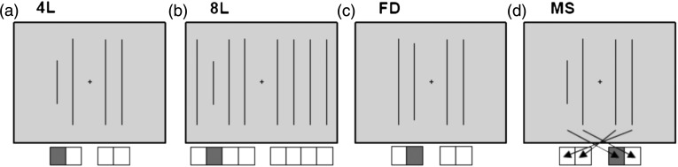
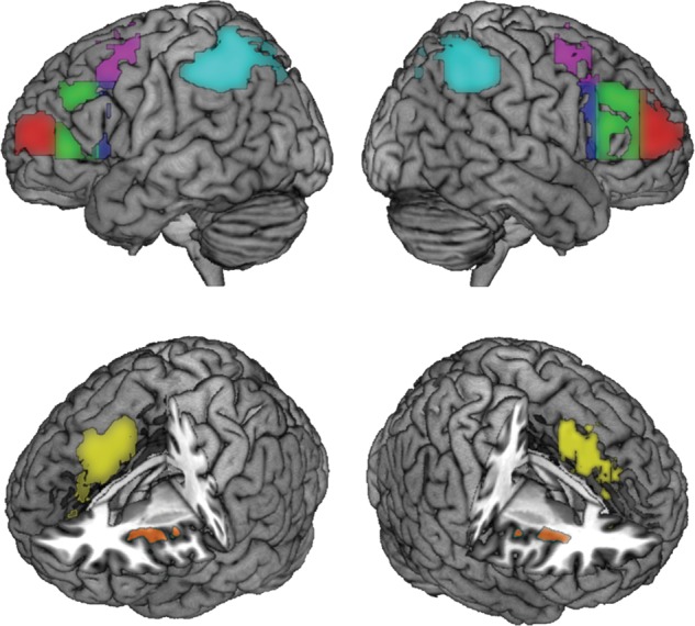
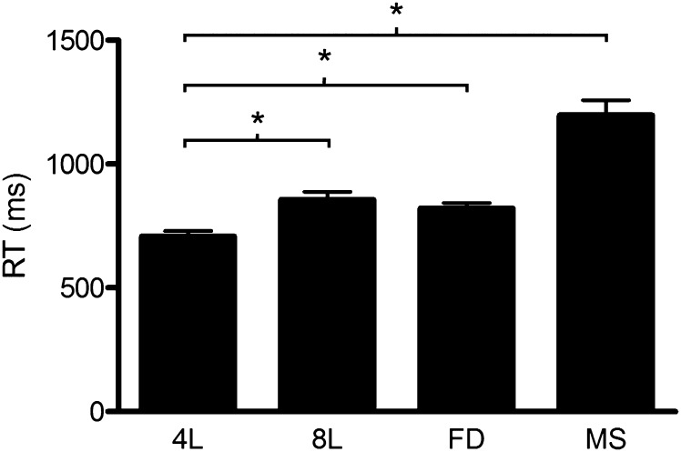
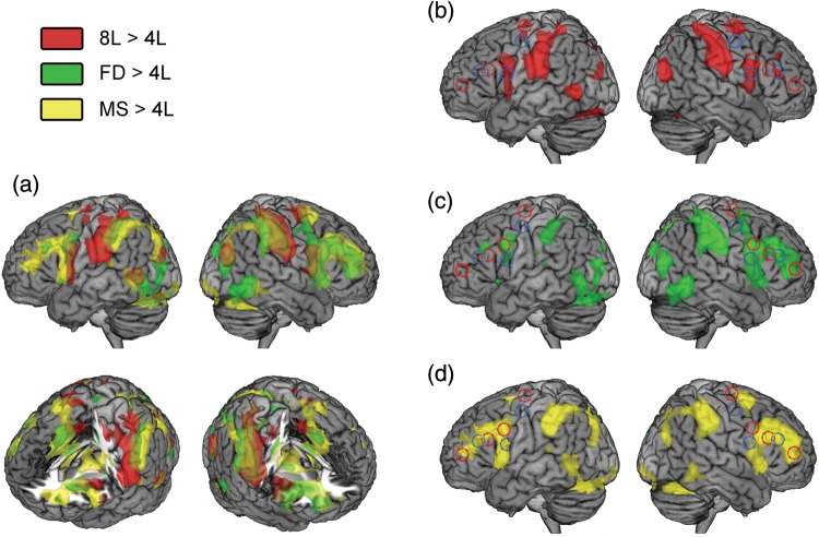
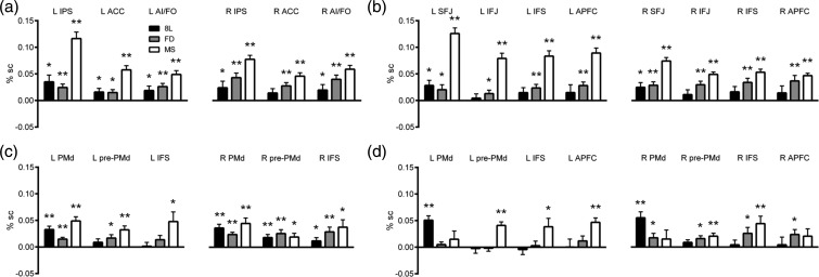
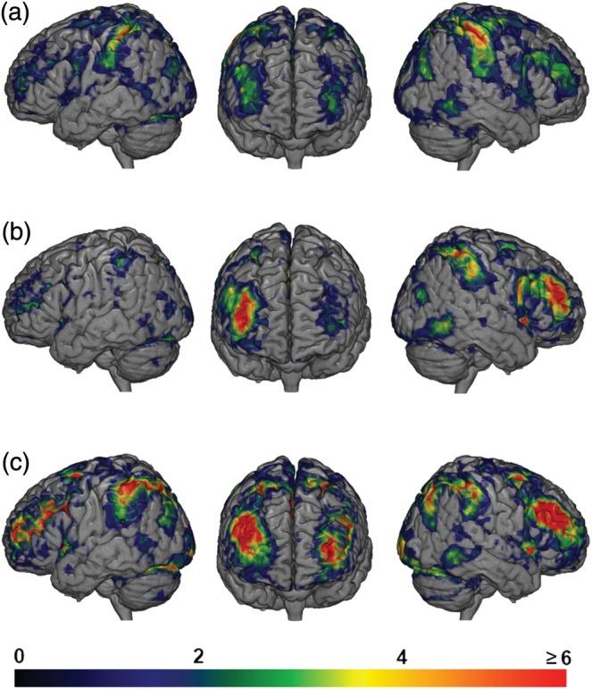
It has been proposed that task control is functionally implemented by a distributed frontoparietal system. It has been argued that one component of this system, the lateral frontal cortex, is functionally organized through a caudal to rostral gradient. Here, we tested 2 competing models, the Information Cascade and Rule Abstraction hypotheses, which suggest different principles underlying the rostrocaudal gradient. We presented participants with 4 vertical lines on a screen and asked them to indicate the position of the shortest line. We manipulated the difficulty of the task in 3 simple ways: By increasing the number of lines, by decreasing the difference between short and normal line length, and by changing the stimulus-response mapping. As expected, these manipulations evoked widespread frontoparietal activation, with activity much more anterior than predicted by Information Cascade and Rule Abstraction models. There were also striking individual differences in the rostrocaudal extent of activity. The results suggest an integrated frontoparietal system, which can be recruited as a whole even by very simple task demands.

Keywords: Information Cascade; Rule Abstraction; functional magnetic resonance imaging; multiple demand; prefrontal cortex.


Figures

Figure 1.
Figure 1.
Frontal regions from the Rule Abstraction and Information Cascade theories. Peak voxels taken from the Rule Abstraction theory (Badre and D'Esposito 2007), in red, and the Information Cascade theory (Koechlin et al. 2003), in blue. In MNI coordinates: A = (−30, −10, 68), PMd; B = (−37, 10, 34), pre-PMd; C = (−51, 26, 24), IFS; D = (−36, 51, 9), APFC; X = (−32, −8, 53) PMd; Y = (−43, 7, 20), pre-PMd; Z = (−50, 35, 23), IFS. Note that coordinates B and Y fall within the precentral sulcus, but for illustration are shown here on the surface.
Figure 2.
Figure 2.
Task conditions. In each case, the task was to respond to the position of the shorter line, using either 4 (conditions 4L, FD, and MS) or 8 alternative response buttons. Icons below each example display show the correct response for that display. (a) 4L condition, (b) 8L condition, (c) FD condition, and (d) MS condition; arrows show correct key presses for each target stimulus (shorter line) position.
Figure 3.
Figure 3.
Example MD ROIs from the localizer task. Pink—around the SFJ; blue—around the IFJ; green—around the mid-IFS; red—APFC; cyan—around the IPS; yellow—around pre-SMA and DACC; orange—around the AI.
Figure 4.
Figure 4.
Mean reaction times for each condition. Error bars represent the standard error of the mean.*P < 0.05.
Figure 5.
Figure 5.
Whole-brain analysis. Overlaid activation maps from 3 whole-brain analyses: 8L > 4L in red, FD > 4L in green, MS > 4L in yellow. (a) All 3 activation patterns overlaid on a single brain. Clockwise from top left: Lateral surface of the left hemisphere, lateral surface of the right hemisphere, right PFC cut away revealing medial PFC and right insula activations, and left PFC cut away revealing medial PFC and left insula activations. (b) Bilateral view of lateral surface showing activations from the 8L > 4L contrast only. ROI outlines from Figure 1 also shown: Rule Abstraction ROIs in red outline, Information Cascade ROIs in blue outline. (c) Bilateral view of the lateral surface showing activations from the FD > 4L contrast only. (d) Bilateral view of the lateral surface showing activations from the MS > 4L contrast only.
Figure 6.
Figure 6.
ROI results. Graphs plot the percentage signal change between each comparison condition (8L, FD, and MS) and the 4L baseline. Black bars: 8L; gray bars: FD; white bars: MS. Error bars represent the standard error of the mean signal change between that condition and the 4L baseline. *P < 0.05 for each condition > baseline comparison, **P < 0.005. (a) Localizer ROIs outside the lateral frontal lobe. (b) Localizer ROIs in the left and right frontal lobes. SFJ, superior fontal junction; IFJ, inferior frontal junction; IFS, inferior frontal sulcus; APFC, anterior prefrontal cortex, (c) Information Cascade ROIs. PMd, dorsal pre-motor cortex; pre-PMd, pre-dorsal pre-motor cortex. (d) Rule Abstraction ROIs.
Figure 7.
Figure 7.
Overlap of significant activations found in single subjects. Warmer colors indicate that more subjects showed activations in that region. (a) Contrast of 8L > 4L. Some overlap found in anterior regions shown in green (3 subjects) and yellow (4 subjects). (b) Contrast of FD > 4L. Most individuals show activity along the rostrocaudal extent of the right IFS, while a few show accompanying activity on the left. (c) Contrast of MS > 4L. Robust activations across most individuals over much of the IFS, APFC, and pre-motor regions.

Similar articles

Cited by

References

    1. Badre D. Cognitive control, hierarchy, and the rostro-caudal organization of the frontal lobes. Trends Cogn Sci. 2008;12:193–200. doi:10.1016/j.tics.2008.02.004. - DOI - PubMed
    1. Badre D, D'Esposito M. Functional magnetic resonance imaging evidence for a hierarchical organization of the prefrontal cortex. J Cogn Neurosci. 2007;19:2082–2099. doi:10.1162/jocn.2007.19.12.2082. - DOI - PubMed
    1. Badre D, D'Esposito M. Is the rostro-caudal axis of the frontal lobe hierarchical? Nat Rev Neurosci. 2009;10:659–669. doi:10.1038/nrn2667. - DOI - PMC - PubMed
    1. Bishop SJ, Fossella J, Croucher CJ, Duncan J. COMT val158met genotype affects recruitment of neural mechanisms supporting fluid intelligence. Cereb Cortex. 2008;18:2132–2140. doi:10.1093/cercor/bhm240. - DOI - PMC - PubMed
    1. Botvinick MM, Braver TS, Barch DM, Carter CS, Cohen JD. Conflict monitoring and cognitive control. Psychol Rev. 2001;108:624–652. doi:10.1037/0033-295X.108.3.624. - DOI - PubMed

Publication types