. 2013:955:527-37.
doi: 10.1007/978-1-62703-176-9_28.

Lipid monolayer and sparse matrix screening for growing two-dimensional crystals for electron crystallography: methods and examples

Affiliations

Lipid monolayer and sparse matrix screening for growing two-dimensional crystals for electron crystallography: methods and examples

Mark Yeager et al. Methods Mol Biol. 2013.

Abstract

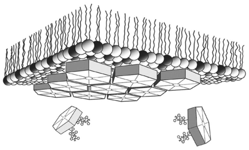
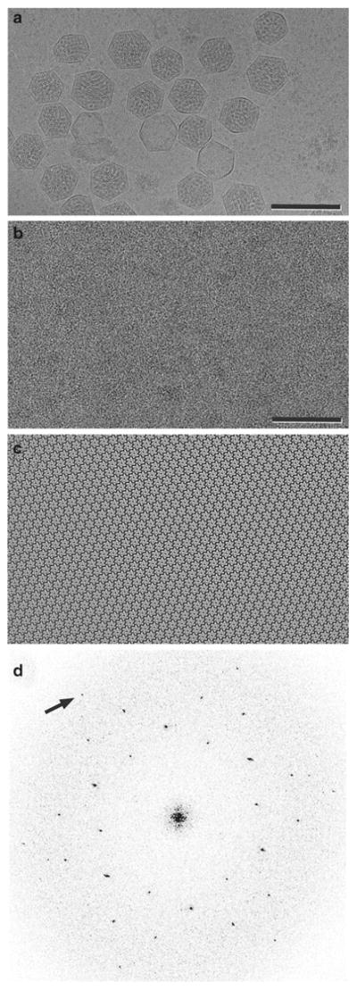
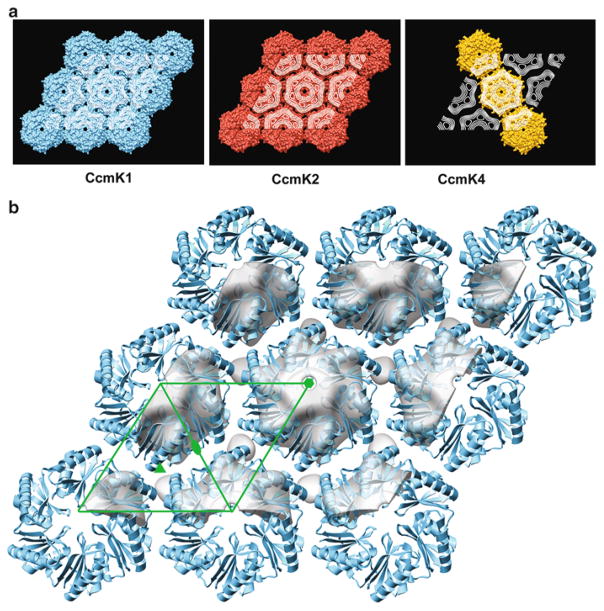
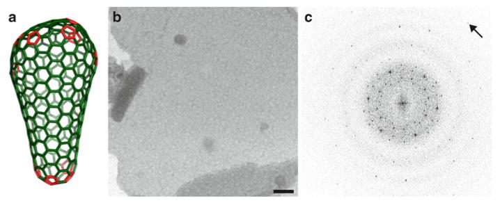
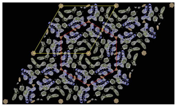
Electron microscopy provides an efficient method for rapidly assessing whether a solution of macromolecules is homogeneous and monodisperse. If the macromolecules can be induced to form two-dimensional crystals that are a single layer in thickness, then electron crystallography of frozen-hydrated crystals has the potential of achieving three-dimensional density maps at sub-nanometer or even atomic resolution. Here we describe the lipid monolayer and sparse matrix screening methods for growing two-dimensional crystals and present successful applications to soluble macromolecular complexes: carboxysome shell proteins and HIV CA, respectively. Since it is common to express recombinant proteins with poly-His tags for purification by metal affinity chromatography, the monolayer technique using bulk lipids doped with Ni(2+) lipids has the potential for broad application. Likewise, the sparse matrix method uses screening conditions for three-dimensional crystallization and is therefore of broad applicability.


Figures

Fig. 1
Fig. 1
Schematic depiction of 2D lipid monolayer crystallization. Carboxysome BMC shell protein hexamers were localized at the lipid-aqueous interface of a mixed lipid layer with 4:1 (wt:wt) l-α-phosphatidylcholine (light spheres) plus DOGS-NTA-Ni2+ (dark spheres) by means of polyhistidine tag chelation via Ni2+ bound within the polar lipid layer. Resulting 2D crystals were transferred to an EM grid and stained for image diffraction analysis (From ref. (13)).
Fig. 2
Fig. 2
(a) Electron cryomicrograph of carboxysomes, which are bacterial microcompartments formed by CcmK proteins that assemble as polyhedral shells containing key metabolic enzymes. Scale bar = 2,000 Å. (b) Image of negatively stained CcmK1 2D crystals. Scale bar = 500 Å. (c) The underlying order is observed when the image is Fourier filtered, and corrections are made for lattice distortions. (d) The Fourier transform of the negatively stained image displays hexagonal symmetry. The arrow identifies the (2,2) reflection at 17-Å resolution. Images were contrast stretched to emphasize salient features (From ref. (13)).
Fig. 3
Fig. 3
(a ) Projection maps from 2D crystals of three CcmK proteins (white contours) shown projected onto the molecular packings visualized in 3D crystals. In the case of CcmK4, hexamers in 3D crystals packed into uniformly ordered strips (illustrated) but not uniformly oriented layers. (b ) 3D density map (tilted by 15 ° to enhance depth) showing the hexamer packing of CcmK1 in 2D lipid monolayer crystals (gray surface), which recapitulates the packing in the ab lattice plane in 3D crystals (blue ribbon) (pdb id. 3dn9). Center-to-center hexamer spacing is ~68 Å for the EM map and ~70 Å for the crystal structure. For comparison, the X-ray structure was manually docked into the EM density obtained from 3D reconstruction. The green line outlines the 2D unit cell with standard symbols at the axes of rotational symmetry (From ref. (13)).
Fig. 4
Fig. 4
(a) Fullerene cone model of the HIV capsid in which a continuously variable hexagonal lattice of CA hexamers (green) is closed by insertion of exactly 12 CA pentamers (red), seven at the wide end, and five at the narrow end of the cone (16). (b ) 2D crystals of HIV CA grown by sparse matrix screening. The hexagonal lattice is an in vitro symmetric mimic of the variable hexagonal lattice in pleomorphic capsids of HIV. (c ) Fourier transform of an image of a flattened sphere (b) formed by a single layer of HIV CA hexamers preserved in vitreous ice, collected at 0 ° tilt (arrow points to a reflection at 9.8 Å). The transform displays two superimposed hexameric lattices that arise from the top and bottom layers of the flattened sphere (From ref. (14) and reproduced by permission from Cell press).
Fig. 5
Fig. 5
The 3D cryoEM density map of HIV-1 CA (gray-scale, mesh) at 9-Å in-plane resolution serves as a template for docking the high-resolution structures of the N-terminal (green ) and C-terminal (blue ) domains. A hexamer of N-terminal domains is outlined in red, and the p6 unit cell is outlined in yellow. The hexamers are linked together by dimers of the C-terminal domains (blue ). The six, three, and twofold symmetry axes are indicated by hexagons, triangles, and ellipses, respectively (From ref. (14) and reproduced by permission from Cell press).
Fig. 6
Fig. 6
Teflon block used for lipid monolayer crystallization. The wells are 3 mm in diameter to accommodate the EM grid and have a depth of 0.5 mm.
Fig. 7
Fig. 7
600 μl Eppendorf, 8-tube pcr strip, for sparse matrix screening. An aliquot of the precipitant solution is mixed with an aliquot of the solution containing the protein to be crystallized.

Similar articles

Cited by

References

    1. McPherson A. Preparation and analysis of protein crystals. Wiley; New York: 1982.
    1. Yeager M, Unger VM, Mitra AK. Three-dimensional structure of membrane proteins determined by two-dimensional crystallization, electron cryomicroscopy, and image analysis. Methods Enzymol. 1999;294:135–180. - PubMed
    1. Adair BD, Yeager M. Electron microscopy of integrins. Methods Enzymol. 2007;426:337–373. - PubMed
    1. Hemming SA, Bochkarev A, Darst SA, Kornberg RD, Ala P, Yang DSC, Edwards AM. The mechanism of protein crystal growth from lipid layers. J Mol Biol. 1995;246:308–316. - PubMed
    1. Brisson A, Olofsson A, Ringler P, Schmutz M, Stoylova S. Two-dimensional crystallization of proteins on planar lipid films and structure determination by electron crystallography. Biol Cell. 1994;80:221–228. - PubMed

Publication types

LinkOut - more resources