. 2012 Dec 1;139(23):4365-73.
doi: 10.1242/dev.083840.

Different assemblies of Notch receptors coordinate the distribution of the major bronchial Clara, ciliated and neuroendocrine cells

Affiliations

Different assemblies of Notch receptors coordinate the distribution of the major bronchial Clara, ciliated and neuroendocrine cells

Mitsuru Morimoto et al. Development. .

Abstract

In the developing lung, it is thought that the terminal buds of elongating airways contain a population of multipotent epithelial progenitors. As the bronchial tree extends, descendants of these cells give rise to lineage-restricted progenitors in the conducting airways via Notch signaling, which is involved in the establishment of epithelial Clara, ciliated and pulmonary neuroendocrine (NE) cell populations. However, the precise molecular details of this selection process are still emerging. Our stepwise removal of the three Notch receptors from the developing lung epithelium reveals that, whereas Notch2 mediates the Clara/ciliated cell fate decision with negligible contributions from Notch1 and Notch3, all three Notch receptors contribute in an additive manner to regulate the abundance of NE cells and the size of the presumptive pulmonary neuroepithelial body (pNEB) as a result of mutual interactions between NE cells and the Notch-dependent, SSEA-1(+), CC10(-) cell population surrounding the pNEB (SPNC cells). Ectopic expression of the Notch1 or Notch2 intracellular domain was sufficient to induce SSEA-1(+) cells and to suppress pNEB formation without expending Clara cells. We provide evidence that the additive functions of Notch receptors, together with other signaling pathways, maintains the expression of Hes1, a key regulator of NE cell fate, and that maintenance of Hes1 expression in epithelial cells is key to the regulation of pNEB size. These results suggest that two different assemblies of Notch receptors coordinate the numbers and distribution of the major epithelial cell types in the conducting airway during lung organogenesis.


Figures
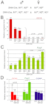
Fig. 1.
Fig. 1.
Endodermal epithelium-specific stepwise removal of Notch paralogs revealed the dominant contribution of Notch2 in Clara/ciliated cell fate decision. (A) Two mating strategies were employed to achieve endodermal epithelium-specific knockout of Notch prologs. Male genotypes are noted on the left of the cross. (B-D) For each genotype, total RNA was extracted from one or two whole lungs collected at E18.5. The relative gene expression levels of CC10 (B) and Foxj1 (C) in the lungs were measured by quantitative RT-PCR. (B) CC10 expression is progressively diminished by reduction in the Notch signal dose. Brackets highlight a comparison between SHH-Cre; N1flox/flox, N2flox/+ and SHH-Cre; N1flox/+, N2flox/flox mice that unveiled a unique contribution of Notch2 in Clara cell fate determination. (C) Accordingly, Foxj1 expression is upregulated in all mutants, reflecting the expansion of ciliated cells (see Fig. 2A). Although elevation of Foxj1 expression correlates with a reduction of Clara cells, SHH-Cre; N1flox/flox; N2flox/flox, SHH-Cre; N1flox/flox; N2flox/flox; N3−/+ and SHH-Cre; N1flox/flox, N2flox/flox; N3−/− lungs show lower levels of Foxj1 expression relative to Shh-RKO lungs (brackets). (D) Analysis of SHH-Cre; N2flox/flox lung also demonstrated the reduction of CC10 and the elevation of Foxj1 expression in Notch2-null lung epithelium, while the Cgrp level was unaltered. Error bars from technical triplicates indicate s.d. P values are shown above the bars.
Fig. 2.
Fig. 2.
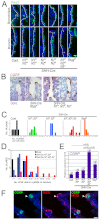
Loss of Notch receptors affects NE cell numbers. For each genotype, two or three whole lungs were collected at E18.5 and at least six paraffin wax-embedded sections were stained for each individual sample. (A) Immunofluorescence of E18.5 bronchioles (upper panels) and bronchus (lower panels) stained with anti-Foxj1 (green) revealed that the ciliated cell expansion progressed as the Notch dose decreased. In addition, foci of Foxj1 cells are observed in the Notch double knockout and triple knockout epithelia (asterisks). Dotted lines demarcate the mesenchyme (left) and the epithelium (right). (B) Immunohistochemistry for CGRP in E18.5 lungs shows a significant increase and enlargement of pNEBs (brown) in the Notch triple knockout but a moderate increase in Rbpj knockout epithelium. (C) Frequency distribution of pNEB abundance. Eight or nine photographs of proximal airways in 12 sections from two or three mice were taken at ×20 magnification for individual genotypes: control (black), SHH-Cre; N1flox/+; N2flox/flox (green), Notch double knockout (blue), triple knockout (red) and Rbpj knockout (orange) embryos. CGRP-positive clusters were determined as pNEBs on the monitor. Two to 11 pNEBs were observed in each photograph. (D) Frequency distribution of the numbers of NE cells per pNEB. The numbers of NE cells in a pNEB were counted in 30 pNEBs randomly selected from anti-CGRP-stained sections, for which at least two biological lung samples were collected from E18.5 control (black), Notch double knockout (blue) and triple knockout (red) embryos. (E) Quantification of Cgrp expression in E18.5 lungs estimates the magnitude of pNEB enlargement in each genotype. Total RNA was extracted from one to three whole lungs collected at E18.5 for each genotype. In comparison with the control, the Notch triple knockout displayed the greatest expansion, and Rbpj loss elevated the expression more than the loss of any two Notch receptors. (F) Double immunofluorescence of E18.5 bronchus with anti-CGRP (green) and anti-N1ICD (red) determined Notch activation in peri-pNEB cells (arrowheads). Scale bars: 10 μm in A,F; 20 μm in B. Error bars from technical triplicates and multiple samples indicate s.d. P values are shown above the bars.
Fig. 3.
Fig. 3.
Peri-pNEB, Notch signal-receiving cells in the bronchus visualized by SSEA-1 immunoreactivity. (A-C) Immunofluorescence staining of E18.5 lung with anti-CC10 (green), anti-CGRP (red) and anti-SSEA-1 (blue) shows localizations of Clara, NE and SSEA-1+ cells. At the bronchus, SSEA-1+ cells are restricted to the peri-pNEB area (white arrowheads in A and B). These are SSEA-1+ CGRP CC10 cells. The SSEA-1+/pNEB clusters are interspaced between Clara cells (gray arrowheads in B). At the bronchioles, SSEA-1+ cells that are often CC10+ are broadly distributed (white arrowheads in C). (D,E) Immunofluorescence staining with anti-SSEA-1 (green) and anti-CGRP (red) (D), or anti-SSEA-1 (green) and anti-N1ICD (red) (E) illustrates the SSEA-1 immunoreactivity of peri-pNEB, Notch signal-receiving cells at the E18.5 bronchus (white arrowheads). A gray arrowhead identifies N1ICD in a Clara cell. Some N1ICD-positive vascular smooth muscle and endothelial cells are observed in mesenchyme (asterisk). These distribution patterns were confirmed in at least eight sections from three biological samples. Scale bars: 40 μm in A; 20 μm in B,C, D (left), E (left); 10 μm in D (right), E (right).
Fig. 4.
Fig. 4.
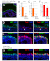
Notch signals coordinate the NE/SPNC cell populations. (A-C) Immunofluorescence staining of SSEA-1 (green) and CGRP (red) in E18.5 Ascl1 heterozygous (A) or null mutant (B,C) lungs. Wild-type and Ascl1+/− lungs contain pNEBs and SPNC cells (white arrowhead), whereas NE cells are not detected in Ascl1-null mutants. We also see a dramatic decrease in SSEA-1+ cells at the bronchus in Ascl1-null mutants, suggesting that the existence of this marker depends on a signal from Ascl1-dependent NE cells. SSEA-1-positive cells remain in Ascl1-deficient bronchioles, suggesting that the presence of SSEA1 antigen in this cell population is independent of Ascl1 and pNEBs. (D) Quantitative analysis of SSEA-1+ epithelial cells in the bronchi of E18.5 wild-type, Ascl1 heterozygous and null mutant mice. At least 10 photos of the proximal airways at ×20 magnification from three biological samples for individual genotype were examined for this analysis. In the mutant, a significant decrease in the number of SSEA-1+ cells is observed. Error bars indicate s.d. P values are shown above the bars. (E-H) Immunofluorescence staining for SSEA-1 (green) and CGRP (red) in wild-type (E), SHH-Cre;N2flox/flox (F), SHH-Cre;N1flox/+;N2flox/flox (G) and SHH-Cre;N1flox/flox;;N2flox/flox;N3−/− (H) lungs. The relative expansion of the SPNC population (green) or pNEBs (red) is represented below. Scale bars: 10 μm in A-C,E; 20 μm in F-H.
Fig. 5.
Fig. 5.
Artificial Notch1 or Notch2 activation promotes the SPNC cell-like population but not Clara cells. (A) Immunofluorescence staining for SSEA-1 (green) and CGRP (red) in SPC-rtTA;TetO-Cre;Rosa-GFP-N1ICD lungs. (B,C,F) The numbers of pNEBs (B), NE (C) and Clara cells (F) were quantitatively evaluated in wild-type or triple-transgenic lung. Ten or more photographs of proximal airways in nine sections from three mice were taken at ×20 magnification. The numbers of pNEBs and NE cells were determined by counting cells on the PC display. P values are shown above the bars. (D) Anti-Foxj1 (red) and anti-GFP (green) identified a few ciliated cells in E18.5 SPC-rtTA;TetO-Cre;Rosa-GFP-N1ICD mice that were most likely due to incomplete recombination induced by Dox feeding. (E) Immunofluorescence staining for anti-CC10 (red) and anti-SSEA1 (green) determined that forced Notch1 activation does not result in Clara cell hyper-expansion. (G) Immunofluorescence staining for SSEA-1 (green) and CC10 (red) in SHH-Cre;Rosa-N2ICD mice. Notch2 activation also failed to promote Clara cells. Scale bars: 20 μm in A; 10 μm in D,E,G.
Fig. 6.
Fig. 6.
Hes1 expression correlates with the severity of the pNEB defect in the Notch allelic series. (A-D) Hes1 protein levels (white arrowheads) in E18.5 wild-type (A), SHH-Cre;N1flox/+;N2flox/flox (B), SHH-Cre;Rbpjflox/flox (C) and SHH-Cre;N1flox/flox;N2flox/flox;N3−/− (D) mice were assessed by immunofluorescence. Scale bar: 10 μm. Asterisks indicate NE cells. (E) Mean Hes1 intensity within the epithelium was measured using ImageJ. Normalization was performed by dividing the mean by the intensity from Hes1-negative NE cells within the same picture. At least four pictures from two to three biological samples were measured for each genotype. Error bars indicate s.d. P values are shown above the bars. (F-H) The proposed regulatoy mechanisms involved in Notch-dependent cell fate determination during bronchial epithelial development, which integrate our findings with reports from other groups. In the pseudoglandular stage, epithelial progenitors (purple) located at the distal tip produce descendants that differentiate into Clara (pink), ciliated (green), SPNC (red) and NE (blue) cells. See the text for details.

References

    1. Borges M., Linnoila R. I., van de Velde H. J., Chen H., Nelkin B. D., Mabry M., Baylin S. B., Ball D. W. (1997). An achaete-scute homologue essential for neuroendocrine differentiation in the lung. Nature 386, 852-855 - PubMed
    1. Cardoso W. V., Lü J. (2006). Regulation of early lung morphogenesis: questions, facts and controversies. Development 133, 1611-1624 - PubMed
    1. Fujimura S., Jiang Q., Kobayashi C., Nishinakamura R. (2010). Notch2 activation in the embryonic kidney depletes nephron progenitors. J. Am. Soc. Nephrol. 21, 803-810 - PMC - PubMed
    1. Guha A., Vasconcelos M., Cai Y., Yoneda M., Hinds A., Qian J., Li G., Dickel L., Johnson J. E., Kimura S., et al. (2012). Neuroepithelial body microenvironment is a niche for a distinct subset of Clara-like precursors in the developing airways. Proc. Natl. Acad. Sci. USA 109, 12592-12597 - PMC - PubMed
    1. Guillemot F., Lo L. C., Johnson J. E., Auerbach A., Anderson D. J., Joyner A. L. (1993). Mammalian achaete-scute homolog 1 is required for the early development of olfactory and autonomic neurons. Cell 75, 463-476 - PubMed

Publication types

MeSH terms