Review
. 2013 Jan;10(1):111-23.
doi: 10.1007/s13311-012-0155-4.

Neuroinflammatory imaging biomarkers: relevance to multiple sclerosis and its therapy

Affiliations
Review

Neuroinflammatory imaging biomarkers: relevance to multiple sclerosis and its therapy

Thomas Tourdias et al. Neurotherapeutics. 2013 Jan.

Abstract

Magnetic resonance imaging is an established tool in the management of multiple sclerosis (MS). Loss of blood brain barrier integrity assessed by gadolinium (Gd) enhancement is the current standard marker of MS activity. To explore the complex cascade of the inflammatory events, other magnetic resonance imaging, but also positron emission tomographic markers reviewed in this article are being developed to address active neuroinflammation with increased sensitivity and specificity. Alternative magnetic resonance contrast agents, positron emission tomographic tracers and imaging techniques could be more sensitive than Gd to early blood brain barrier alteration, and they could assess the inflammatory cell recruitment and/or the associated edema accumulation. These markers of active neuroinflammation, although some of them are limited to experimental studies, could find great relevance to complete Gd information and thereby increase our understanding of acute lesion pathophysiology and its noninvasive follow-up, especially to monitor treatment efficacy. Furthermore, such accurate markers of inflammation combined with those of neurodegeneration hold promise to provide a more complete picture of MS, which will be of great benefit for future therapeutic strategies.


Figures
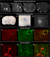
Fig. 1
Fig. 1
Active lesions in multiple sclerosis (MS). Features of MS active lesions in the human brain (A1, A2) and the rat brain (A3, A4, B, C, and D). Active MS lesions in the human are surrounded by blurry Fluid Attenuated Inversion Recovery (FLAIR) hypersignal related to edema (A1) and show gadolinium (Gd) enhancement (A2, arrowheads). A focal MS-like lesion induced in the rat brain shows the same MR features on T2-weighted image (A3, arrow) and T1-weighted image after Gd injection (A4) 3 days after its induction. Histological study of such focal prototypical MS lesion in the rat shows blood brain barrier (BBB) alteration as assessed by extravasation of Evans blue injected 2 h prior sacrifice (B1 and B2, enlarged view of the square dotted area) and serum protein extravasation depicted with IgG staining (B3, staining at the same level as the white rectangular dotted area delineated on T2). Cellular infiltration appears as perivascular infiltration (*indicates a capillary lumen) of recruited macrophages stained with Iba1 (C1, red) and ED1 (C2, green), whereas some of these cells could also represent amoeboid microglia with a fully activated profile. Some resident activated microglia Iba+, but ED1- are also seen (C3, merged image). Interstitial edema is difficult to depict histologically, but it is revealed by a strong expression of molecules involved in water regulation, such as aquaporin 4 (D1, red), which is expressed at the BBB level on the astrocyte end feet (D2, green; D3, merged image). Scale bars, 25 μm
Fig. 2
Fig. 2
Discrepant imaging profiles of active lesions. Images from 2 relapsing-remitting multiple sclerosis (MS) patients included in the European ultra-small superparamagnetic iron oxide (USPIO) study [53]. Axial T2-wi, T1-wi after a few minutes after gadolinium (Gd; Dotarem, Guerbet, France) injection and ADC maps obtained 7 (top row) and 8 (bottom row) days following a clinical relapse. Immediately after these first MR scans, ultra-small superparamagnetic iron oxide particles (Sinerem, Guerbet, France) were injected over 30 minutes and second MR scans were repeated 24 h later to assess T1-wi post-USPIO and T2-wi post-USPIO. The first patient (top row) showed a large MS lesion (arrowheads) with active features, assessed by Gd enhancement, reflecting blood brain barrier (BBB) breakdown, which was associated with USPIO enhancement (seen both on T1-wi and T2-wi), reflecting concomitant macrophage invasion, and high apparent diffusion coefficient (ADC) values probably related to ongoing vasogenic edema. The second patient (bottom row) also showed an MS lesion (arrows) that could be classified as active, according to USPIO enhancement (seen both on T1-wi and T2-wi) reflecting macrophage invasion while the BBB was not macroscopically altered (no Gd enhancement). The significance of the associated low ADC values is still debated, but could reflect hypoxic damage and the close association with USPIO enhancement could suggest toxic products from activated macrophages as an inducer of hypoxic-like conditions

Similar articles

Cited by

References

    1. Filippi M, Rocca MA. MR imaging of multiple sclerosis. Radiology. 2011;259:659–681. doi: 10.1148/radiol.11101362. - DOI - PubMed
    1. Bakshi R, Thompson AJ, Rocca MA, et al. MRI in multiple sclerosis: current status and future prospects. Lancet Neurol. 2008;7:615–625. doi: 10.1016/S1474-4422(08)70137-6. - DOI - PMC - PubMed
    1. Polman CH, Reingold SC, Banwell B, et al. Diagnostic criteria for multiple sclerosis: 2010 revisions to the McDonald criteria. Ann Neurol. 2011;69:292–302. doi: 10.1002/ana.22366. - DOI - PMC - PubMed
    1. Miller DH, Altmann DR, Chard DT. Advances in imaging to support the development of novel therapies for multiple sclerosis. Clin Pharmacol Ther. 2012;91:621–634. doi: 10.1038/clpt.2011.349. - DOI - PubMed
    1. Barkhof F, Calabresi PA, Miller DH, Reingold SC. Imaging outcomes for neuroprotection and repair in multiple sclerosis trials. Nat Rev Neurol. 2009;5:256–266. doi: 10.1038/nrneurol.2009.41. - DOI - PubMed

Publication types