. 2013 Jan;10(1):124-42.
doi: 10.1007/s13311-012-0157-2.

Scriptaid, a novel histone deacetylase inhibitor, protects against traumatic brain injury via modulation of PTEN and AKT pathway : scriptaid protects against TBI via AKT

Affiliations

Scriptaid, a novel histone deacetylase inhibitor, protects against traumatic brain injury via modulation of PTEN and AKT pathway : scriptaid protects against TBI via AKT

Guohua Wang et al. Neurotherapeutics. 2013 Jan.

Abstract

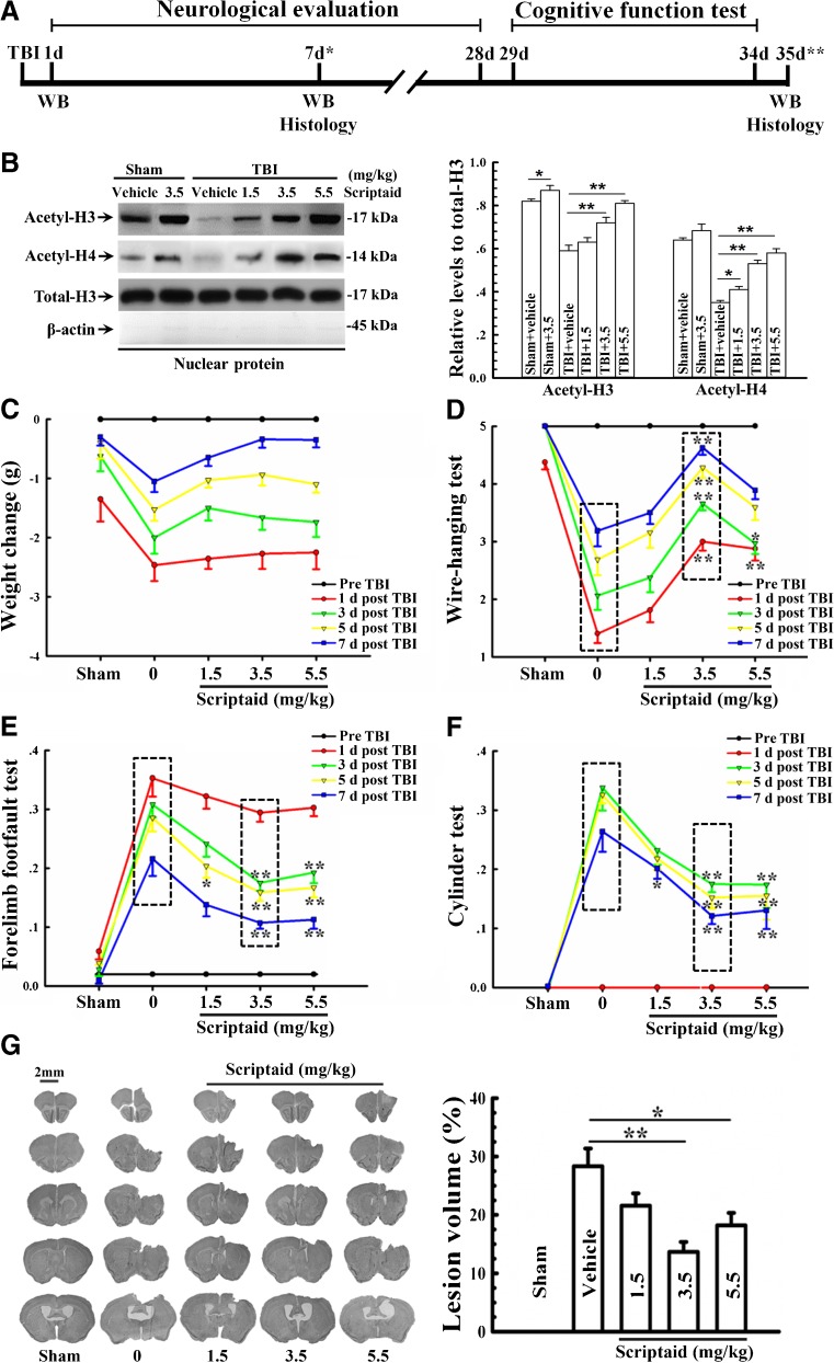
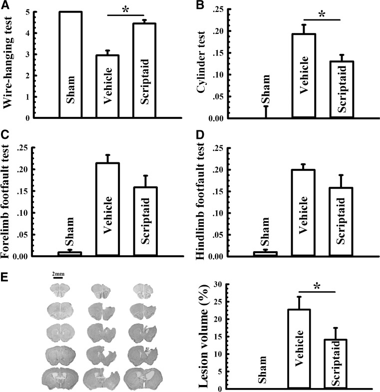
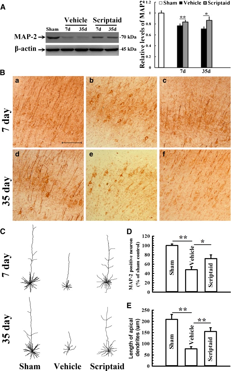
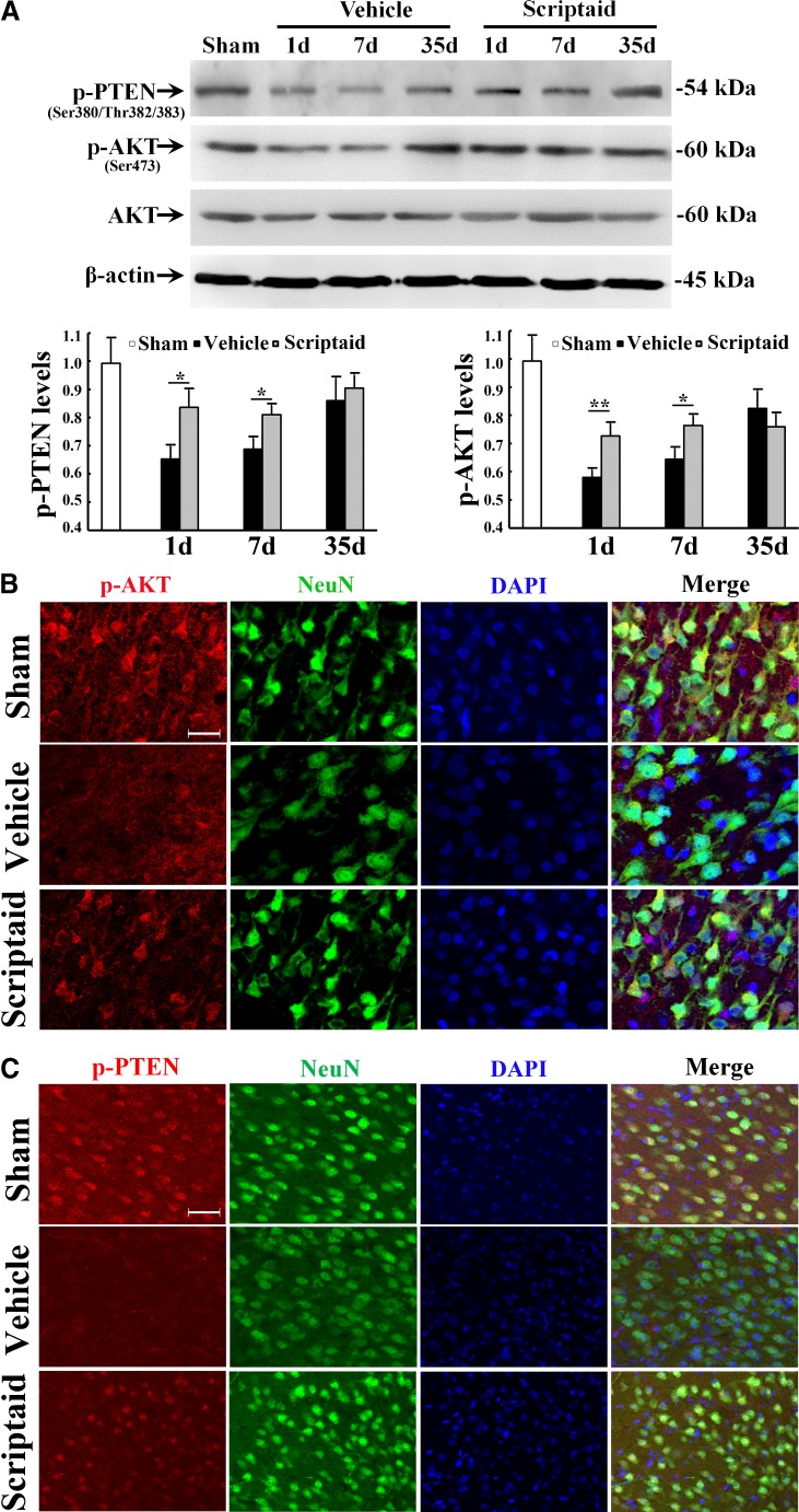
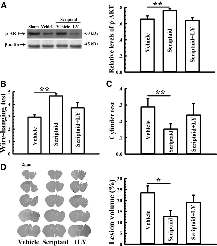
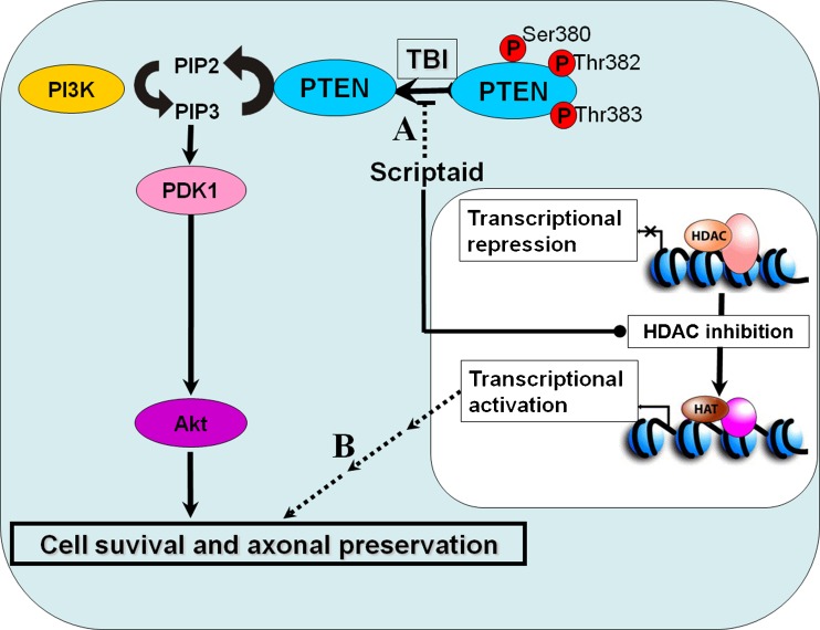
Traumatic brain injury (TBI) is a leading cause of motor and cognitive deficits in young adults for which there is no effective therapy. The present study characterizes the protective effect of a new histone deacetylase inhibitor, Scriptaid (Sigma-Aldrich Corporation, St. Louis, MO), against injury from controlled cortical impact (CCI). Scriptaid elicited a dose-dependent decrease in lesion size at 1.5 to 5.5 mg/kg and a concomitant attenuation in motor and cognitive deficits when delivered 30 minutes postinjury in a model of moderate TBI. Comparable protection was achieved even when treatment was delayed to 12 h postinjury. Furthermore, the protection of motor and cognitive functions was long lasting, as similar improvements were detected 35 days postinjury. The efficacy of Scriptaid (Sigma-Aldrich Corporation) was manifested as an increase in surviving neurons, as well as the number/length of their processes within the CA3 region of the hippocampus and the pericontusional cortex. Consistent with other histone deacetylase inhibitors, Scriptaid treatment prevented the decrease in phospho-AKT (p-AKT) and phosphorylated phosphatase and tensin homolog deleted on chromosome 10 (p-PTEN) induced by TBI in cortical and CA3 hippocampal neurons. Notably, the p-AKT inhibitor LY294002 attenuated the impact of Scriptaid, providing mechanistic evidence that Scriptaid functions partly by modulating the prosurvival AKT signaling pathway. As Scriptaid offers long-lasting neuronal and behavioral protection, even when delivered 12 h after controlled cortical impact, it is an excellent new candidate for the effective clinical treatment of TBI.


Figures

Fig. 1
Fig. 1
The dose-dependent protection conferred by Scriptaid against traumatic brain injury (TBI). (A) Temporal schematic of the experimental protocol and the time points of sample collection and analysis. (B) Changes in the levels of acetylated-H3, acetylated-H4, and total-H3 24 h postinjury, as visualized by Western blot analysis. Nuclear protein extracts were obtained from ipsilateral cortices. β-actin immunoblotting was used as the control to ensure nuclear extracts were not contaminated by cytoplasm. Quantification of the changes in acetylated-H3 and -H4 levels, normalized to total-H3 histone. Data are presented as mean ± standard error (n = 6; *p < 0.05, **p < 0.01 vs sham or vehicle). (C) No statistically significant change in weight by Scriptaid. (D) Wire-hanging test: a lower score represents more severe neurological deficits, ranging from a minimum score of 0 for severe impairments, to a maximum score of 5, indicating near-normal function. The scores for the wire-hanging test significantly decreased in the vehicle group after TBI, but performance of Scriptaid-treated (3.5 mg/kg) mice was significantly less affected from 1 to 7 days (dotted boxes; all p < 0.01). The high-dose group (5.5 mg/kg) exhibited greatly improved neurological function only at 1 and 3 days after TBI compared with the vehicle group (p < 0.05 or 0.01). For the low-dose (1.5 mg/kg) group, the lesion was similar to controls. Data are presented as mean ± standard error (n = 6; *p < 0.05, **p < 0.01 vs vehicle). (E) Forelimb foot-fault: was calculated as F/T, where F is the total number of foot faults for left forelimb and T is the total movement number of left forelimb. Controlled cortical impact increased foot-faults compared to sham. Treatment with Scriptaid at 3.5 mg/kg elicited the greatest reduction in forelimb foot-faults on days 3-7 (dotted boxes) postinjury. Data are presented as mean ± standard error (n = 6; *p < 0.05, **p < 0.01 vs vehicle). (F) Cylinder test: final score = (nonimpaired forelimb movement - impaired forelimb movement)/(nonimpaired forelimb movement + impaired forelimb movement + both movements). Contralateral forelimb placing for the vehicle group was significantly decreased at 3, 5, and 7 days after TBI, leading to an increase in the cylinder test score. In comparison, mice from the Scriptaid-treated group exhibited a higher placing frequency, resulting in lower scores than the vehicle group. Data are presented as mean ± standard error (n = 6; *p < 0.05, **p < 0.01 vs vehicle). (G) Visualization of the lesion volume with Nissl staining of brain sections from mice euthanized at day 7 after TBI and the comparison of various doses of Scriptaid and vehicle. Data were presented as mean ± standard error (n = 10; *p < 0.05, **p < 0.01 vs vehicle). WB = Western Blot
Fig. 2
Fig. 2
Scriptaid conferred protection against long-term motor and cognitive deficits and tissue loss from controlled cortical impact (CCI) injury. (A) Effect of Scriptaid on long-term motor deficits; a) wire-hanging test: Scriptaid treatment attenuated motor deficits leading to an increase in performance scores compared with the vehicle group for as many as 21 days postinjury. Data are presented as mean ± standard error (n = 6; *p < 0.05, **p < 0.01 vs vehicle); b) forelimb foot-fault test; c) hindlimb foot-fault test: Scriptaid treatment reduced forelimb and hindlimb foot-faults for as many as 4-weeks post-CCI when compared to vehicle. Data are presented as mean ± standard error (n = 6; *p < 0.05, **p < 0.01 vs vehicle; d) cylinder test: the administration of Scriptaid also diminished biased forelimb use for as many as 4-weeks postinjury. (B) Scriptaid treatment significantly reduced tissue loss (cavity size) produced by CCI trauma at 35 days after traumatic brain injury (TBI) compared to the vehicle group. TBI induced a macroscopic area of cortical tissue loss extending rostrocaudally from bregma + 2.8 mm to bregma -1.06 mm in the vehicle group. The region of interest lies between the internal and external circles (1.5-mm gap; pericontusional cortex). (C) Quantitation of the difference of lesion volume at 5-weeks postinjury between Scriptaid-treated and -nontreated mice. Data are presented as mean ± standard error (n = 6; *p < 0.05, **p < 0.01 vs vehicle. (D) Effect of Scriptaid on cognitive deficits postinjury: a) cued learning response: a typical swim pattern of mice from sham, vehicle, and Scriptaid-treated groups while locating the platform within the Morris water maze; b) probed trial response: a typical swim pattern of mice from sham, vehicle, and Scriptaid-treated groups while attempting to locate the platform within the Morris water maze based on memory; c) time required for mice from these 3 groups to locate the platform in the cued learning response at 29- to 33-days postinjury. Data are presented as mean ± standard error (n = 6; *p < 0.05, **p < 0.01 vs vehicle and ##p < 0.01 vs sham; d) the percentage of time spent in the same quadrant as the platform after training for mice from sham, vehicle, and Scriptaid-treated groups. Data are presented as mean ± standard error (n = 10; *p < 0.05 vs vehicle and #p < 0.05, ##p < 0.01 vs sham)
Fig. 3
Fig. 3
Scriptaid conferred comparable protection when administrated 12 h post-controlled cortical impact at day 7 after injury. (A) Wire-hanging test: Scriptaid treatment attenuated motor deficits, leading to an increase in performance scores compared with the vehicle group. Data are presented as mean ± standard error (n = 6; *p < 0.05 vs vehicle). (B) Cylinder test: the administration of Scriptaid also diminished biased forelimb use. Data are presented as mean ± standard error (n = 6; *p < 0.05 vs vehicle). (C) Forelimb foot-fault test: an overall decrease in foot-faults with Scriptaid treatment was not statistically significant. (D) Hindlimb foot-fault test: an overall decrease in foot-faults with Scriptaid treatment was not statistically significant. (E) Quantification of lesion volume by Nissl staining of brain sections. Scriptaid treatment (3.5 mg/kg) at 12-h postinjury significantly reduced lesion volume. Data are presented as mean ± standard error (n = 8; *p < 0.05 vs vehicle)
Fig. 4
Fig. 4
Scriptaid prevented neurodegeneration in cortex and hippocampus. (A) Viable neurons from the ipsilateral CA3 region of the hippocampus from mice in sham, vehicle, and Scriptaid-treated groups, as visualized by Nissl staining; scale bar, 100 μm. (B) Stereological quantification of viable CA3 neurons in the hippocampus ipsilateral to traumatic brain injury. Data are presented as mean ± standard error (n = 6; **p < 0.01 vs vehicle). (C) Correlation between spatial memory and number of viable neurons in the ipsilateral CA3 region from mice of sham, vehicle, and Scriptaid-treated groups. (D) Visualization of degenerating neurons in the ipsilateral cortex and hippocampus from mice of sham, vehicle, and Scriptaid-treated groups by Fluoro-Jade C (FJC) staining at day 7 postinjury. These sections are co-stained with 4'-6-Diamidino-2-phenylindole (DAPI) to reveal nuclear morphology. Scale bar, 25 μm
Fig. 5
Fig. 5
Reductions in neuronal microtubule-associated protein-2 (MAP-2) expression and neurites at the ipsilateral cortex were attenuated by Scriptaid treatment. (A) The decrease in MAP-2 levels after traumatic brain injury (TBI) was prevented by Scriptaid treatment at 7 and 35 days postinjury, as shown by Western blot analysis. β-actin was included as the loading control. Quantification of the changes in MAP-2 levels in vehicle and Scriptaid-treated groups normalized to levels in the sham group. Data are presented as mean ± standard error (n = 6; *p < 0.05, **p < 0.01 vs vehicle). (B) Morphological changes in neuronal neurites induced by TBI at days 7 and 35 postinjury with and without Scriptaid treatments. Images were taken from the pericontusional regions in the cortex, as indicated in Fig. 2B. a, d) sham groups; b, e) vehicle treated groups; c, f) Scriptaid treated groups. TBI significantly decreased the number of neurites and average neurites length when compared to sham controls. Scriptaid treatment prevented neurite loss and shortening of dendrites length compared to the vehicle group at both days 7 and 35 postinjury. Scale bar, 100 μm. (C) Drawings of neuron morphology in sham, vehicle, and Scriptaid-treated groups at 7 and 35 days postinjury. (D) Stereological analysis of MAP-2-positive neuron at 35 days postinjury. Data were normalized to the sham control and are presented as mean ± standard error (n = 6; *p < 0.05, **p < 0.01 vs vehicle). (E) Bar graphs illustrate the mean length of apical dendrites in layer III pyramidal neurons at 35 days postinjury. Degenerative changes were apparent in the length of the apical dendrites in the vehicle group, and were obviously prevented by Scriptaid treatment. Data are presented as mean ± standard error (n = 6; *p < 0.05, **p < 0.01 vs the vehicle group)
Fig. 6
Fig. 6
Scriptaid prevented the decrease in phosphorylated phosphatase and tensin homolog deleted on chromosome 10 (p-PTEN) and phospho-AKT (p-AKT) levels after injury. (A) p-AKT and p-PTEN levels were decreased by traumatic brain injury at days 1 and 7 postinjury compared to the vehicle group with no significant change in total AKT levels. However, these decreases were prevented with Scriptaid treatment. β-actin was used as the loading control. Quantification of p-PTEN and p-AKT in the vehicle and Scriptaid-treated groups normalized to sham levels at 1, 7, and 35 days postinjury. Data are presented as mean ± standard error (n = 6; *p < 0.05, **p < 0.01 vs vehicle). (B) The decrease in p-AKT in the ipsilateral cortex in vehicle-treated mice compared to the sham group was prevented by the Scriptaid group, as shown by immunofluorescent microscopy. Changes in p-AKT were localized to neuronal cell bodies in the sham, vehicle, and Scriptaid groups. Sections of cortex stained with p-AKT, neuronal marker (NeuN), and 4'-6-Diamidino-2-phenylindole (DAPI; Vector Laboratories Inc., Burlingame, CA). Scale bar, 25 μm, (C) The decrease in p-PTEN in the ipsilateral cortex in the vehicle group compared to the sham group was also prevented by Scriptaid, as shown by immunofluorescent microscopy. Changes in p-PTEN also occurred in neurons and not glia in the sham, vehicle, and Scriptaid groups. Sections of cortex stained with p-TEN, NeuN, and DAPI. Scale bar, 50 μm
Fig. 7
Fig. 7
Inhibiting AKT pathway with LY294002 significantly attenuated Scriptaid-induced protection against controlled cortical impact. (A) The increase in phospho-AKT (p-AKT) level due to Scriptaid was reversed by LY294002 treatment, as shown by Western blot analysis. β-actin was used as the loading control. Changes in p-AKT levels for vehicle, Scriptaid-treated, and LY294002 + Scriptaid-treated groups were quantified. The statistical increase in p-AKT due to Scriptaid was nullified by LY294002. Data are presented as mean ± standard error (n = 6; **p < 0.01 vs vehicle). (B) Wire-hanging test: Scriptaid treatment attenuated motor deficits leading to an increase in performance scores compared with the vehicle group. This improvement in motor function was inhibited by LY294002 treatment. Data are presented as mean ± standard error (n = 6; **p < 0.01 vs vehicle). (C) Cylinder test: LY294002 also inhibited the ability of Scriptaid to diminish biased forelimb use. Data are presented as mean ± standard error (n = 6; **p < 0.01 vs vehicle). (D) Visualization of the lesion volume with Nissl staining of brain sections from mice euthanized at day 7 after traumatic brain injury and quantification of lesion volumes from mice receiving vehicle, Scriptaid, or Scriptaid+LY294002. The decrease in lesion volume due to Scriptaid treatment was attenuated by pre-treatment with LY294002. Data are presented as mean ± standard error (n = 6; *p < 0.05 vs vehicle). LY = LY294002
Fig. 8
Fig. 8
Proposed mechanism of neuroprotection of histone deacetylase inhibitors (HDI) Scriptaid against traumatic brain injury (TBI). (A) Mechanistic relationships between phosphorylated phosphatase and tensin homolog deleted on chromosome 10 (p-PTEN), PTEN, and phospho-AKT (p-AKT), as well as the impact of Scriptaid on these pathways. A decrease in p-PTEN results in the concomitant increase in active PTEN, which converts PIP3 to PIP2, preventing the phosphorylation of PDK1 and the activation of AKT. Scriptaid treatment increases p-PTEN, leading to a concomitant decrease in active PTEN. This loss of PTEN function by phosphorylation results in the downstream activation of the AKT pathway. As a consequence of enhanced phospho-AKT (p-AKT) signaling, cell survival is promoted and neurite integrity is preserved. (B) The loss of histone acetyltransferase (HAT) function or increased HDAC activity leads to a decrease in histone/transcription factor acetylation and transcriptional repression. This may partly underlie neurological damage associated with TBI. HDAC inhibition by Scriptaid may be protective by shifting the HAT-HDAC balance in favor of HAT activity, so that the histones remain acetylated and gene expression is activated. We also propose that the accumulation of acetylated histones in nucleosomes leads to expression of specific genes, which, in turn, are responsible for the protection elicited by Scriptaid

Similar articles

Cited by

References

    1. Park HK, Fernandez II, Dujovny M, Diaz FG. Experimental animal models of traumatic brain injury: medical and biomechanical mechanism. Crit Rev Neurosurg. 1999;9:44–52. doi: 10.1007/s003290050108. - DOI - PubMed
    1. Stoica BA, Faden AI. Cell death mechanisms and modulation in traumatic brain injury. Neurotherapeutics. 2010;7:3–12. doi: 10.1016/j.nurt.2009.10.023. - DOI - PMC - PubMed
    1. Maas AIR, Stocchetti N, Bullock R. Moderate and severe traumatic brain injury in adults. Lancet Neurol. 2008;7:728–741. doi: 10.1016/S1474-4422(08)70164-9. - DOI - PubMed
    1. Wang GH, Zhang XG, Jiang ZL, et al. Neuroprotective effects of hyperbaric oxygen treatment on traumatic brain injury in the rat. J Neurotrauma. 2010;27:1733–1743. doi: 10.1089/neu.2009.1175. - DOI - PubMed
    1. Sande A, West C. Traumatic brain injury: a review of pathophysiology and management. J Vet Emerg Crit Care (San Antonio) 2010;20:177–190. doi: 10.1111/j.1476-4431.2010.00527.x. - DOI - PubMed

Publication types