

32-channel phased-array receive with asymmetric birdcage transmit coil for hyperpolarized xenon-129 lung imaging
- PMID: 23132336
- PMCID: PMC3568189
- DOI: 10.1002/mrm.24482
32-channel phased-array receive with asymmetric birdcage transmit coil for hyperpolarized xenon-129 lung imaging
Abstract
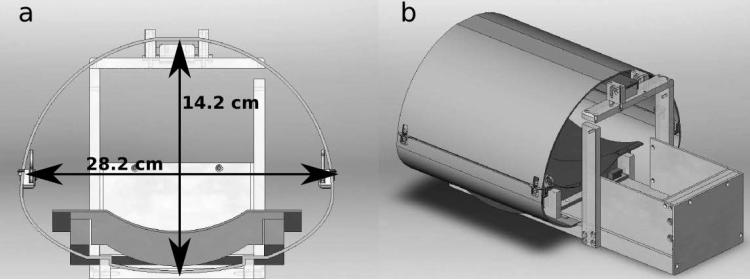
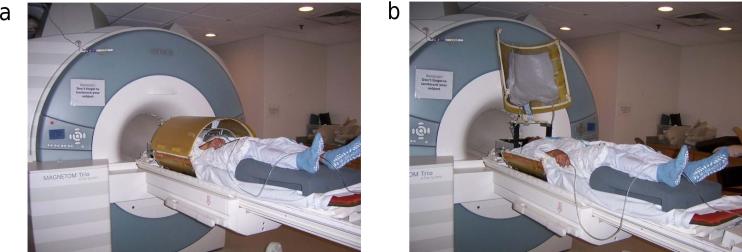
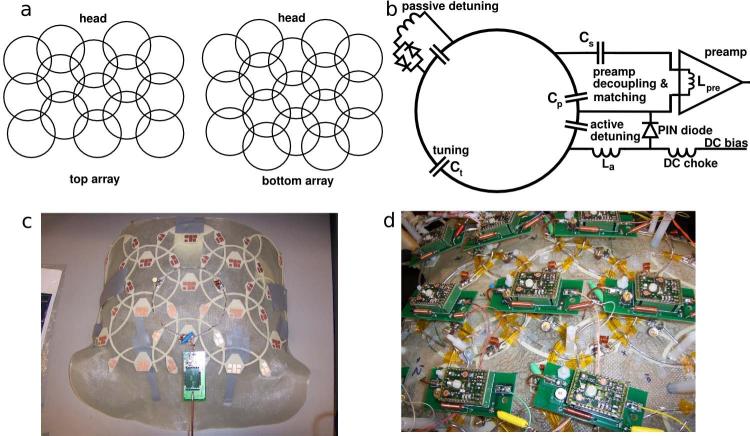
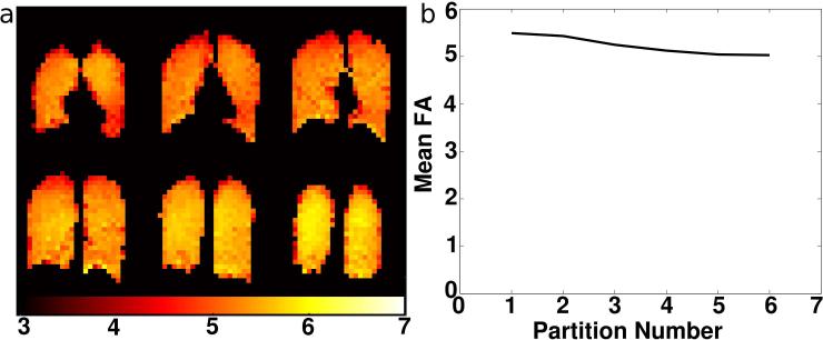
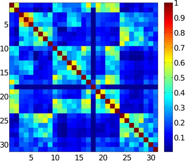
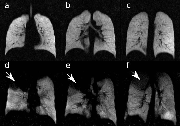
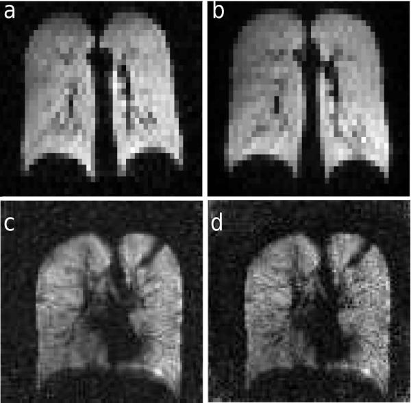
Hyperpolarized xenon-129 has the potential to become a noninvasive contrast agent for lung MRI. In addition to its utility for imaging of ventilated airspaces, the property of xenon to dissolve in lung tissue and blood upon inhalation provides the opportunity to study gas exchange. Implementations of imaging protocols for obtaining regional parameters that exploit the dissolved phase are limited by the available signal-to-noise ratio, excitation homogeneity, and length of acquisition times. To address these challenges, a 32-channel receive-array coil complemented by an asymmetric birdcage transmit coil tuned to the hyperpolarized xenon-129 resonance at 3 T was developed. First results of spin-density imaging in healthy subjects and subjects with obstructive lung disease demonstrated the improvements in image quality by high-resolution ventilation images with high signal-to-noise ratio. Parallel imaging performance of the phased-array coil was demonstrated by acceleration factors up to three in 2D acquisitions and up to six in 3D acquisitions. Transmit-field maps showed a regional variation of only 8% across the whole lung. The newly developed phased-array receive coil with the birdcage transmit coil will lead to an improvement in existing imaging protocols, but moreover enable the development of new, functional lung imaging protocols based on the improvements in excitation homogeneity, signal-to-noise ratio, and acquisition speed.
Keywords: asymmetric-birdcage; hyperpolarized; lung MRI; phased-array; xenon-129.
© 2012 Wiley Periodicals, Inc.
Figures
Similar articles
-
A flexible 32-channel receive array combined with a homogeneous transmit coil for human lung imaging with hyperpolarized 3He at 1.5 T.Magn Reson Med. 2011 Dec;66(6):1788-97. doi: 10.1002/mrm.22962. Epub 2011 May 13. Magn Reson Med. 2011. PMID: 21574180
-
Asymmetric quadrature split birdcage coil for hyperpolarized 3He lung MRI at 1.5T.Magn Reson Med. 2008 Aug;60(2):431-8. doi: 10.1002/mrm.21664. Magn Reson Med. 2008. PMID: 18666111
-
Design and evaluation of a 32-channel phased-array coil for lung imaging with hyperpolarized 3-helium.Magn Reson Med. 2010 Feb;63(2):456-64. doi: 10.1002/mrm.22265. Magn Reson Med. 2010. PMID: 20099333
-
Diffusion lung imaging with hyperpolarized gas MRI.NMR Biomed. 2017 Mar;30(3):10.1002/nbm.3448. doi: 10.1002/nbm.3448. Epub 2015 Dec 16. NMR Biomed. 2017. PMID: 26676342 Free PMC article. Review.
-
Hyperpolarized 129Xe MRI of the human lung.J Magn Reson Imaging. 2013 Feb;37(2):313-31. doi: 10.1002/jmri.23844. J Magn Reson Imaging. 2013. PMID: 23355432 Free PMC article. Review.
Cited by
-
In vivo methods and applications of xenon-129 magnetic resonance.Prog Nucl Magn Reson Spectrosc. 2021 Feb;122:42-62. doi: 10.1016/j.pnmrs.2020.11.002. Epub 2020 Dec 9. Prog Nucl Magn Reson Spectrosc. 2021. PMID: 33632417 Free PMC article. Review.
-
Dose and pulse sequence considerations for hyperpolarized (129)Xe ventilation MRI.Magn Reson Imaging. 2015 Sep;33(7):877-85. doi: 10.1016/j.mri.2015.04.005. Epub 2015 Apr 30. Magn Reson Imaging. 2015. PMID: 25936684 Free PMC article. Clinical Trial.
-
Acquisition strategies for spatially resolved magnetic resonance detection of hyperpolarized nuclei.MAGMA. 2020 Apr;33(2):221-256. doi: 10.1007/s10334-019-00807-6. Epub 2019 Dec 6. MAGMA. 2020. PMID: 31811491 Free PMC article. Review.
-
Application of a stretched-exponential model for morphometric analysis of accelerated diffusion-weighted 129Xe MRI of the rat lung.MAGMA. 2021 Feb;34(1):73-84. doi: 10.1007/s10334-020-00860-6. Epub 2020 Jul 6. MAGMA. 2021. PMID: 32632748
-
Massively parallel MRI detector arrays.J Magn Reson. 2013 Apr;229:75-89. doi: 10.1016/j.jmr.2013.02.001. Epub 2013 Feb 7. J Magn Reson. 2013. PMID: 23453758 Free PMC article. Review.
References
-
- Albert MS, Cates GD, Driehuys B, Happer W, Saam B, Springer CS, Wishnia A. Biological magnetic resonance imaging using laser-polarized 129Xe. Nature. 1994;370(6486):199–201. - PubMed
-
- Mugler JP, 3rd, Driehuys B, Brookeman JR, Cates GD, Berr SS, Bryant RG, Daniel TM, de Lange EE, Downs JH, Erickson CJ, Happer W, Hinton DP, Kassel NF, Maier T, Phillips CD, Saam BT, Sauer KL, Wagshul ME. MR imaging and spectroscopy using hyperpolarized 129Xe gas: preliminary human results. Magn Reson Med. 1997;37:809–815. - PubMed
-
- Fain SB, Korosec FR, Holmes JH, O'Halloran R, Sorkness RL, Grist TM. Functional lung imaging using hyperpolarized gas MRI. J Magn Reson Imaging. 2007;25:910–923. - PubMed
-
- Wagshul ME, Button TM, Li HF, Liang Z, Springer CS, Zhong K, Wishnia A. In vivo MR imaging and spectroscopy using hyperpolarized 129Xe. Magn Reson Med. 1996;36:183–191. - PubMed
Publication types
MeSH terms
Substances
Grants and funding
LinkOut - more resources
Full Text Sources
Other Literature Sources
Medical
