. 2013 Jan;81(1):311-6.
doi: 10.1128/IAI.00871-12. Epub 2012 Nov 6.

Major T cell response to a mycolyl glycolipid is mediated by CD1c molecules in rhesus macaques

Affiliations

Major T cell response to a mycolyl glycolipid is mediated by CD1c molecules in rhesus macaques

Daisuke Morita et al. Infect Immun. 2013 Jan.

Abstract

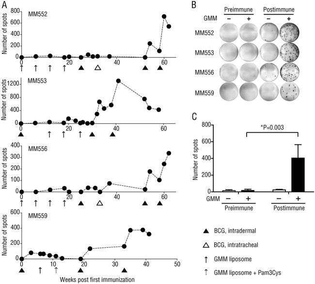
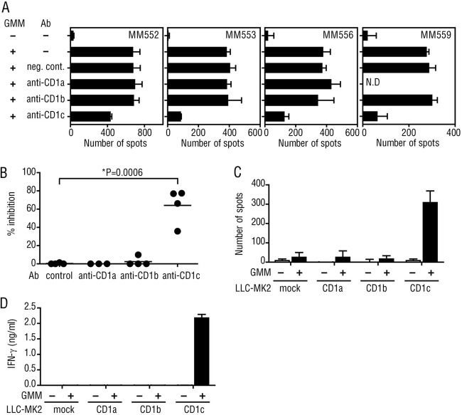
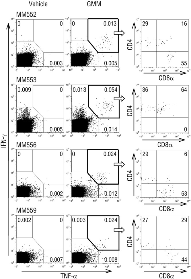
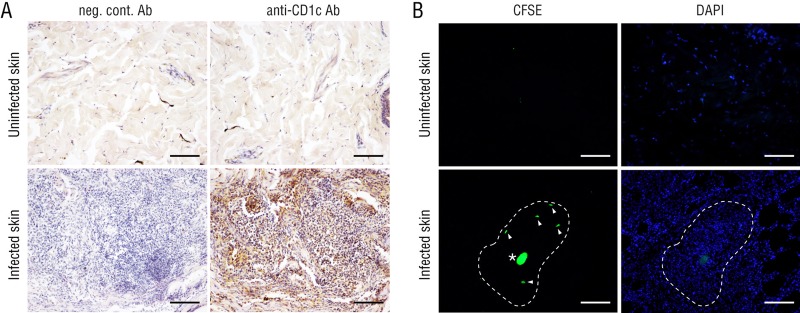
Human CD1b molecules contain a maze of hydrophobic pockets and a tunnel capable of accommodating the unusually long, branched acyl chain of mycolic acids, an essential fatty acid component of the cell wall of mycobacteria. It has been accepted that CD1b-bound mycolic acids constitute a scaffold for mycolate-containing (glyco)lipids stimulating CD1b-restricted T cells. Remarkable homology in amino acid sequence is observed between human and monkey CD1b molecules, and indeed, monkey CD1b molecules are able to bind glucose monomycolate (GMM), a glucosylated species of mycolic acids, and present it to specific human T cells in vitro. Nevertheless, we found, unexpectedly, that Mycobacterium bovis bacillus Calmette-Guerin (BCG)-vaccinated monkeys exhibited GMM-specific T cell responses that were restricted by CD1c rather than CD1b molecules. GMM-specific, CD1c-restricted T cells were detected in the circulation of all 4 rhesus macaque monkeys tested after but not before vaccination with BCG. The circulating GMM-specific T cells were detected broadly in both CD4(+) and CD8(+) cell populations, and upon antigenic stimulation, a majority of the GMM-specific T cells produced both gamma interferon (IFN-γ) and tumor necrosis factor alpha (TNF-α), two major host protective cytokines functioning against infection with mycobacteria. Furthermore, the GMM-specific T cells were able to extravasate and approach the site of infection where CD1c(+) cells accumulated. These observations indicate a previously inconceivable role for primate CD1c molecules in eliciting T cell responses to mycolate-containing antigens.


Figures

Fig 1
Fig 1
GMM-specific T cell responses induced in BCG-immunized monkeys. (A) Four monkeys were vaccinated intradermally with BCG and GMM liposome. In some cases, BCG was administered via an intratracheal route and the GMM liposome was injected along with the Pam3Cys-SKKKK adjuvant. At the indicated time points, PBMCs were obtained, and the number of GMM-specific cells was determined by IFN-γ ELISPOT assays. The numbers of the GMM-specific spots per 1 × 10⁶ PBMCs are plotted. (B and C) Preimmune PBMCs and those obtained after the third BCG immunization were incubated with either the GMM liposome (GMM +) or empty liposome (GMM −), followed by detection of IFN-γ-producing cells in an ELISPOT assay. Statistical analysis was performed using a one-way analysis of variance (ANOVA). Error bars show standard deviations.
Fig 2
Fig 2
The GMM-specific T cell response was dependent on CD1c molecules. (A and B) Postimmune PBMCs from the monkeys were subjected to IFN-γ ELISPOT assays in the presence or absence of anti-CD1 MAbs, and the numbers of GMM-specific spots per 1 × 10⁶ PBMCs are shown (A). The percentage of inhibition by anti-CD1 Abs in each monkey was plotted, and statistical analysis was performed using a one-way ANOVA (B). (C) Postimmune PBMCs were isolated from MM553, and negative selection with Abs to CD1c, CD14, CD16, CD20, and CD56 was performed. Subsequently, the cells were subjected to a IFN-γ ELISPOT assay using LLC-MK2 cell transfectants expressing monkey CD1a, CD1b, or CD1c molecules as APCs. (D) Cells of the GMM-specific monkey T cell line established from MM553 were incubated with the LLC-MK2 cell transfectants in the presence or absence of the GMM liposome, and the amount of IFN-γ released into the medium was measured by a IFN-γ ELISA. neg. cont., negative control. Error bars show standard deviations.
Fig 3
Fig 3
Profiles for surface marker expression on monkey GMM-specific T cells. PBMCs obtained from the BCG-vaccinated monkeys were incubated for 6 h with either empty liposome or the GMM liposome, and then brefeldin A was added to the culture. After an additional 6-h incubation, the cells were fixed, permeabilized, and labeled with Abs to CD4, CD8, IFN-γ, and TNF-α. This was followed by a multicolor flow cytometric analysis. The IFN-γ+ TNF-α+ cell population (boxed in bold lines) was further separated based on the expression of CD4 and CD8α molecules. The percentage of cells present in indicated regions is shown for each panel.
Fig 4
Fig 4
Recruitment of GMM-specific T cells to the site of infection. The CFSE-labeled, GMM-specific T cells were injected into the circulation of the donor. At the same time, BCG (1 × 10⁸ CFU) was inoculated into the skin. After 4 days, samples of the infected (bottom, BCG-positive) or uninfected (top, BCG-negative) skin were obtained and examined for infiltration by CD1c+ cells (A) and CFSE-labeled cells (B). The tissue sections were also counterstained with DAPI (B). Arrowheads indicate CFSE-positive cells, and dashed lines indicate the area of granulomatous macrophage aggregation. The amorphous fluorescence-positive structure (indicated with an asterisk) appeared to represent the necrotic center of the granuloma. neg. cont., negative control. Scale bars, 100 μm.

Similar articles

Cited by

References

    1. Beckman EM, Porcelli SA, Morita CT, Behar SM, Furlong ST, Brenner MB. 1994. Recognition of a lipid antigen by CD1-restricted alpha beta+ T cells. Nature 372:691–694 - PubMed
    1. Moody DB, Reinhold BB, Guy MR, Beckman EM, Frederique DE, Furlong ST, Ye S, Reinhold VN, Sieling PA, Modlin RL, Besra GS, Porcelli SA. 1997. Structural requirements for glycolipid antigen recognition by CD1b-restricted T cells. Science 278:283–286 - PubMed
    1. Batuwangala T, Shepherd D, Gadola SD, Gibson KJ, Zaccai NR, Fersht AR, Besra GS, Cerundolo V, Jones EY. 2004. The crystal structure of human CD1b with a bound bacterial glycolipid. J. Immunol. 172:2382–2388 - PubMed
    1. Layre E, Collmann A, Bastian M, Mariotti S, Czaplicki J, Prandi J, Mori L, Stenger S, De Libero G, Puzo G, Gilleron M. 2009. Mycolic acids constitute a scaffold for mycobacterial lipid antigens stimulating CD1-restricted T cells. Chem. Biol. 16:82–92 - PubMed
    1. Matsunaga I, Naka T, Talekar RS, McConnell MJ, Katoh K, Nakao H, Otsuka A, Behar SM, Yano I, Moody DB, Sugita M. 2008. Mycolyltransferase-mediated glycolipid exchange in Mycobacteria. J. Biol. Chem. 283:28835–28841 - PMC - PubMed

Publication types

MeSH terms