Free-breathing 3D cardiac MRI using iterative image-based respiratory motion correction
- PMID: 23132549
- PMCID: PMC3604140
- DOI: 10.1002/mrm.24538
Free-breathing 3D cardiac MRI using iterative image-based respiratory motion correction
Abstract
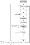
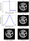
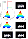
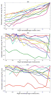
Respiratory motion compensation using diaphragmatic navigator gating with a 5 mm gating window is conventionally used for free-breathing cardiac MRI. Because of the narrow gating window, scan efficiency is low resulting in long scan times, especially for patients with irregular breathing patterns. In this work, a new retrospective motion compensation algorithm is presented to reduce the scan time for free-breathing cardiac MRI that increasing the gating window to 15 mm without compromising image quality. The proposed algorithm iteratively corrects for respiratory-induced cardiac motion by optimizing the sharpness of the heart. To evaluate this technique, two coronary MRI datasets with 1.3 mm(3) resolution were acquired from 11 healthy subjects (seven females, 25 ± 9 years); one using a navigator with a 5 mm gating window acquired in 12.0 ± 2.0 min and one with a 15 mm gating window acquired in 7.1 ± 1.0 min. The images acquired with a 15 mm gating window were corrected using the proposed algorithm and compared to the uncorrected images acquired with the 5 and 15 mm gating windows. The image quality score, sharpness, and length of the three major coronary arteries were equivalent between the corrected images and the images acquired with a 5 mm gating window (P-value > 0.05), while the scan time was reduced by a factor of 1.7.
Keywords: coronary MRI; diaphragmatic navigators; respiratory motion; retrospective motion correction.
Copyright © 2012 Wiley Periodicals, Inc.
Figures


Similar articles
-
Free-breathing cardiac MR with a fixed navigator efficiency using adaptive gating window size.Magn Reson Med. 2012 Dec;68(6):1866-75. doi: 10.1002/mrm.24210. Epub 2012 Feb 24. Magn Reson Med. 2012. PMID: 22367715 Free PMC article.
-
Comparison of image-based and reconstruction-based respiratory motion correction for golden radial phase encoding coronary MR angiography.J Magn Reson Imaging. 2015 Oct;42(4):964-71. doi: 10.1002/jmri.24858. Epub 2015 Feb 2. J Magn Reson Imaging. 2015. PMID: 25639861
-
Whole-heart coronary MRA with 3D affine motion correction using 3D image-based navigation.Magn Reson Med. 2014 Jan;71(1):173-81. doi: 10.1002/mrm.24652. Epub 2013 Feb 11. Magn Reson Med. 2014. PMID: 23400902
-
High efficiency free-breathing 3D thoracic aorta vessel wall imaging using self-gating image reconstruction.Magn Reson Imaging. 2024 Apr;107:80-87. doi: 10.1016/j.mri.2024.01.009. Epub 2024 Jan 17. Magn Reson Imaging. 2024. PMID: 38237694 Review.
-
T1-weighted Motion Mitigation in Abdominal MRI: Technical Principles, Clinical Applications, Current Limitations, and Future Prospects.Radiographics. 2024 Aug;44(8):e230173. doi: 10.1148/rg.230173. Radiographics. 2024. PMID: 38990776 Review.
Cited by
-
Faster 3D saturation-recovery based myocardial T1 mapping using a reduced number of saturation points and denoising.PLoS One. 2020 Apr 10;15(4):e0221071. doi: 10.1371/journal.pone.0221071. eCollection 2020. PLoS One. 2020. PMID: 32275668 Free PMC article.
-
Inversion recovery and saturation recovery pulmonary vein MR angiography using an image based navigator fluoro trigger and variable-density 3D cartesian sampling with spiral-like order.Int J Cardiovasc Imaging. 2024 Jun;40(6):1363-1376. doi: 10.1007/s10554-024-03111-0. Epub 2024 Apr 27. Int J Cardiovasc Imaging. 2024. PMID: 38676848
-
Nonrigid autofocus motion correction for coronary MR angiography with a 3D cones trajectory.Magn Reson Med. 2014 Aug;72(2):347-61. doi: 10.1002/mrm.24924. Epub 2013 Sep 4. Magn Reson Med. 2014. PMID: 24006292 Free PMC article.
-
Retrospective motion correction through multi-average k-space data elimination (REMAKE) for free-breathing cardiac cine imaging.Magn Reson Med. 2023 Jun;89(6):2242-2254. doi: 10.1002/mrm.29613. Epub 2023 Feb 10. Magn Reson Med. 2023. PMID: 36763898 Free PMC article.
-
Optimized respiratory-resolved motion-compensated 3D Cartesian coronary MR angiography.Magn Reson Med. 2018 Dec;80(6):2618-2629. doi: 10.1002/mrm.27208. Epub 2018 Apr 22. Magn Reson Med. 2018. PMID: 29682783 Free PMC article.
References
-
- Hauser TH, Manning WJ. Coronary MRI: more pretty pictures or present-day value? J Am Coll Cardiol. 2006;48(10):1951–1952. - PubMed
-
- Kim WY, Stuber M, Kissinger KV, Andersen NT, Manning WJ, Botnar RM. Impact of bulk cardiac motion on right coronary MR angiography and vessel wall imaging. J Magn Reson Imaging. 2001;14(4):383–390. - PubMed
-
- Felblinger J, Lehmann C, Boesch C. Electrocardiogram acquisition during MR examinations for patient monitoring and sequence triggering. Magn Reson Med. 1994;32(4):523–529. - PubMed
-
- Felblinger J, Debatin JF, Boesch C, Gruetter R, McKinnon GC. Synchronization device for electrocardiography-gated echo-planar imaging. Radiology. 1995;197(1):311–313. - PubMed
-
- Wang Y, Rossman PJ, Grimm RC, Riederer SJ, Ehman RL. Navigator-echo-based real-time respiratory gating and triggering for reduction of respiration effects in three-dimensional coronary MR angiography. Radiology. 1996;198(1):55–60. - PubMed
Publication types
MeSH terms
Grants and funding
LinkOut - more resources
Full Text Sources
Other Literature Sources

