

The distribution of transient receptor potential melastatin-8 in the rat soft palate, epiglottis, and pharynx
- PMID: 23132652
- PMCID: PMC11497895
- DOI: 10.1007/s10571-012-9888-1
The distribution of transient receptor potential melastatin-8 in the rat soft palate, epiglottis, and pharynx
Abstract
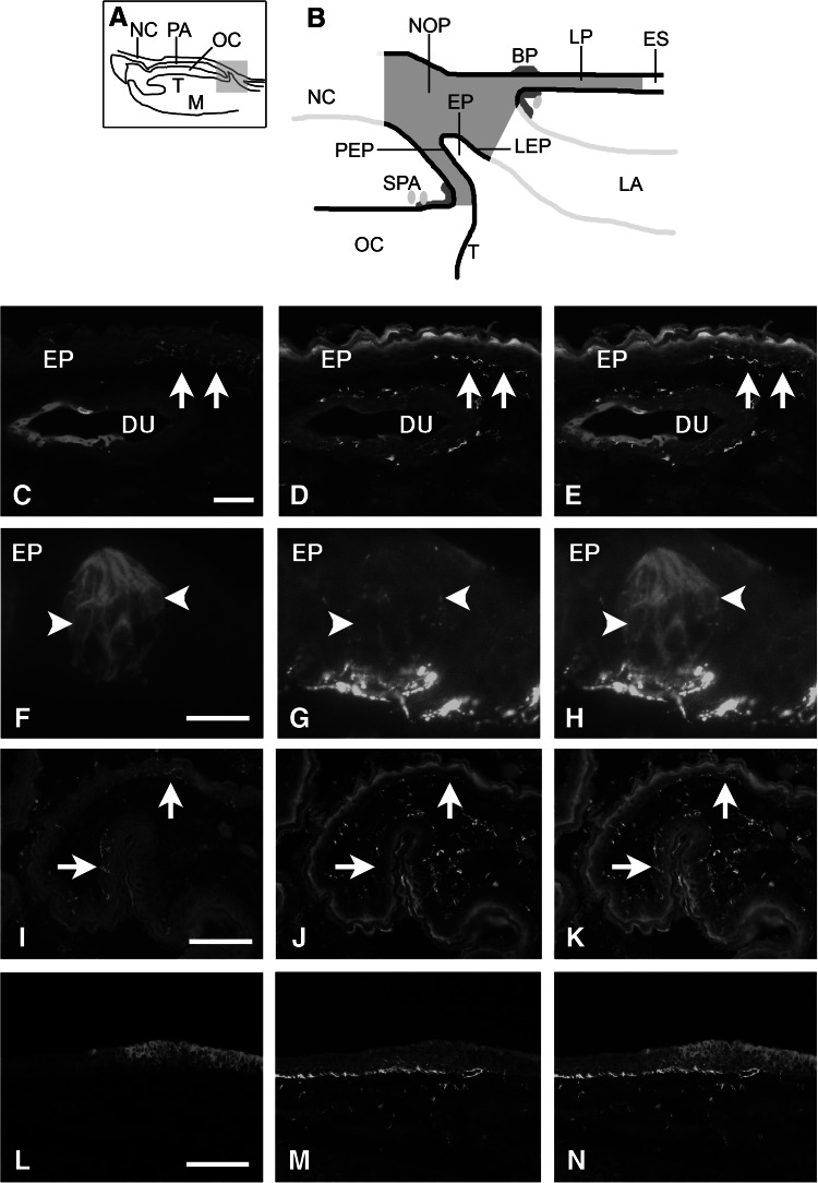
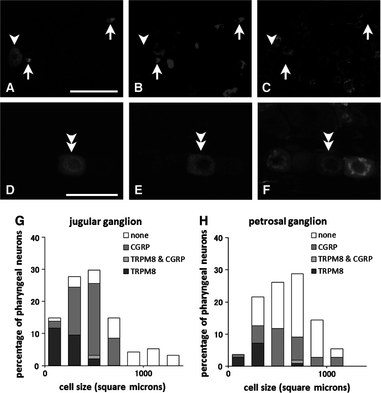
Immunohistochemistry for transient receptor potential melastatin-8 (TRPM8), the cold and menthol receptor, was performed on the rat soft palate, epiglottis and pharynx. TRPM8-immunoreactive (IR) nerve fibers were located beneath the mucous epithelium, and occasionally penetrated the epithelium. These nerve fibers were abundant in the posterior portion of the soft palate and at the border region of naso-oral and laryngeal parts of the pharynx. The epiglottis was free from such nerve fibers. The double immunofluorescence method demonstrated that TRPM8-IR nerve fibers in the pharynx and soft palate were mostly devoid of calcitonin gene-related peptide-immunoreactivity (CGRP-IR). The retrograde tracing method also demonstrated that 30.1 and 8.7 % of sensory neurons in the jugular and petrosal ganglia innervating the pharynx contained TRPM8-IR, respectively. Among these neurons, the co-expression of TRPM8 and CGRP-IR was very rare. In the nodose ganglion, however, pharyngeal neurons were devoid of TRPM8-IR. Taste bud-like structures in the soft palate and pharynx contained 4-9 TRPM8-IR cells. In the epiglottis, the mucous epithelium on the laryngeal side had numerous TRPM8-IR cells. The present study suggests that TRPM8 can respond to cold stimulation when food and drinks pass through oral and pharyngeal cavities.
Figures
Similar articles
-
The distribution of TRPV1 and TRPV2 in the rat pharynx.Cell Mol Neurobiol. 2013 Jul;33(5):707-14. doi: 10.1007/s10571-013-9938-3. Epub 2013 Apr 14. Cell Mol Neurobiol. 2013. PMID: 23584686 Free PMC article.
-
Pituitary adenylatecyclase-activating polypeptide-immunoreactive nerve fibers in the rat epiglottis and pharynx.Ann Anat. 2011 Dec 20;193(6):494-9. doi: 10.1016/j.aanat.2011.08.005. Epub 2011 Sep 14. Ann Anat. 2011. PMID: 21955674
-
Distribution of transient receptor potential melastatin-8-containing nerve fibers in rat oral and craniofacial structures.Ann Anat. 2015 Sep;201:1-5. doi: 10.1016/j.aanat.2015.04.003. Epub 2015 Apr 25. Ann Anat. 2015. PMID: 25978347
-
The distribution of galanin-immunoreactive nerve fibers in the rat pharynx.Neuropeptides. 2013 Aug;47(4):231-6. doi: 10.1016/j.npep.2013.05.001. Epub 2013 May 31. Neuropeptides. 2013. PMID: 23731834
-
Calcitonin gene-related peptide immunoreactive neurons innervating the soft palate, the root of tongue, and the pharynx in the superior glossopharyngeal ganglion of the rat.J Chem Neuroanat. 2010 Jul;39(4):221-7. doi: 10.1016/j.jchemneu.2009.12.003. Epub 2009 Dec 23. J Chem Neuroanat. 2010. PMID: 20034556
Cited by
-
TRPs in taste and chemesthesis.Handb Exp Pharmacol. 2014;223:827-71. doi: 10.1007/978-3-319-05161-1_5. Handb Exp Pharmacol. 2014. PMID: 24961971 Free PMC article. Review.
-
Transient receptor potential channels as an emerging therapeutic target for oropharyngeal dysphagia.Jpn Dent Sci Rev. 2023 Dec;59:421-430. doi: 10.1016/j.jdsr.2023.09.002. Epub 2023 Nov 10. Jpn Dent Sci Rev. 2023. PMID: 38022386 Free PMC article. Review.
-
Activation of TRPV1 and TRPM8 Channels in the Larynx and Associated Laryngopharyngeal Regions Facilitates the Swallowing Reflex.Int J Mol Sci. 2018 Dec 18;19(12):4113. doi: 10.3390/ijms19124113. Int J Mol Sci. 2018. PMID: 30567389 Free PMC article.
-
The interactions between different tastes on initiation of reflex swallow elicited by electrical stimulation in humans.Odontology. 2016 Sep;104(3):282-90. doi: 10.1007/s10266-015-0226-1. Epub 2015 Dec 24. Odontology. 2016. PMID: 26702624
-
The distribution of TRPV1 and TRPV2 in the rat pharynx.Cell Mol Neurobiol. 2013 Jul;33(5):707-14. doi: 10.1007/s10571-013-9938-3. Epub 2013 Apr 14. Cell Mol Neurobiol. 2013. PMID: 23584686 Free PMC article.
References
-
- Abe J, Hosokawa H, Okazawa M, Kandachi M, Sawada Y, Yamanaka K, Matsumura K, Kobayashi S (2005) TRPM8 protein localization in trigeminal ganglion and taste papillae. Brain Res Mol Brain Res 136:91–98 - PubMed
-
- Hayakawa T, Kuwahara S, Maeda S, Tanaka K, Seki M (2010) Calcitonin gene related peptide immunoreactive neurons innervating the soft palate, the root of tongue, and the pharynx in the superior glossopharyngeal ganglion of the rat. J Chem Neuroanat 39:221–227 - PubMed
-
- Hayashi T, Kondo T, Ishimatsu M, Yamada S, Nakamura K, Matsuoka K, Akasu T (2009) Expression of the TRPM8-immunoreactivity in dorsal root ganglion neurons innervating the rat urinary bladder. Neurosci Res 65:245–251 - PubMed
-
- Helke CJ, Hill KM (1988) Immunohistochemical study of neuropeptides in vagal and glossopharyngeal afferent neurons in the rat. Neuroscience 26:539–551 - PubMed
-
- Helke CJ, Niederer AJ (1990) Studies on the coexistence of substance P with other putative transmitters in the nodose and petrosal ganglia. Synapse 5:144–151 - PubMed
MeSH terms
Substances
LinkOut - more resources
Full Text Sources
Research Materials
