. 2013 Jan;154(1):16-24.
doi: 10.1210/en.2012-1579. Epub 2012 Nov 6.

Ligand-dependent actions of the vitamin D receptor are required for activation of TGF-β signaling during the inflammatory response to cutaneous injury

Affiliations

Ligand-dependent actions of the vitamin D receptor are required for activation of TGF-β signaling during the inflammatory response to cutaneous injury

Hilary F Luderer et al. Endocrinology. 2013 Jan.

Abstract

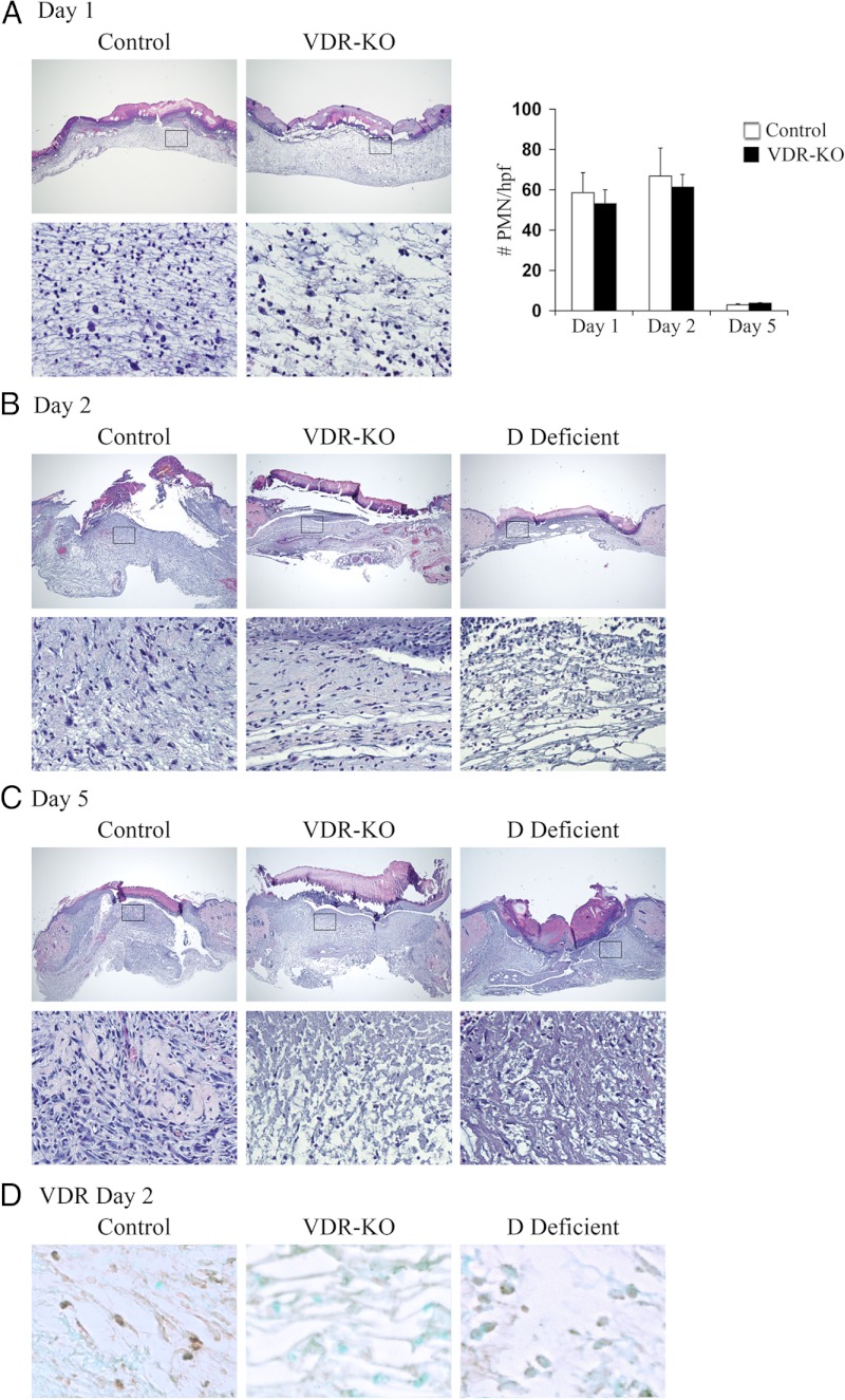
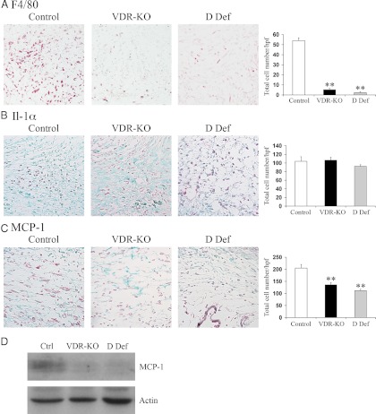
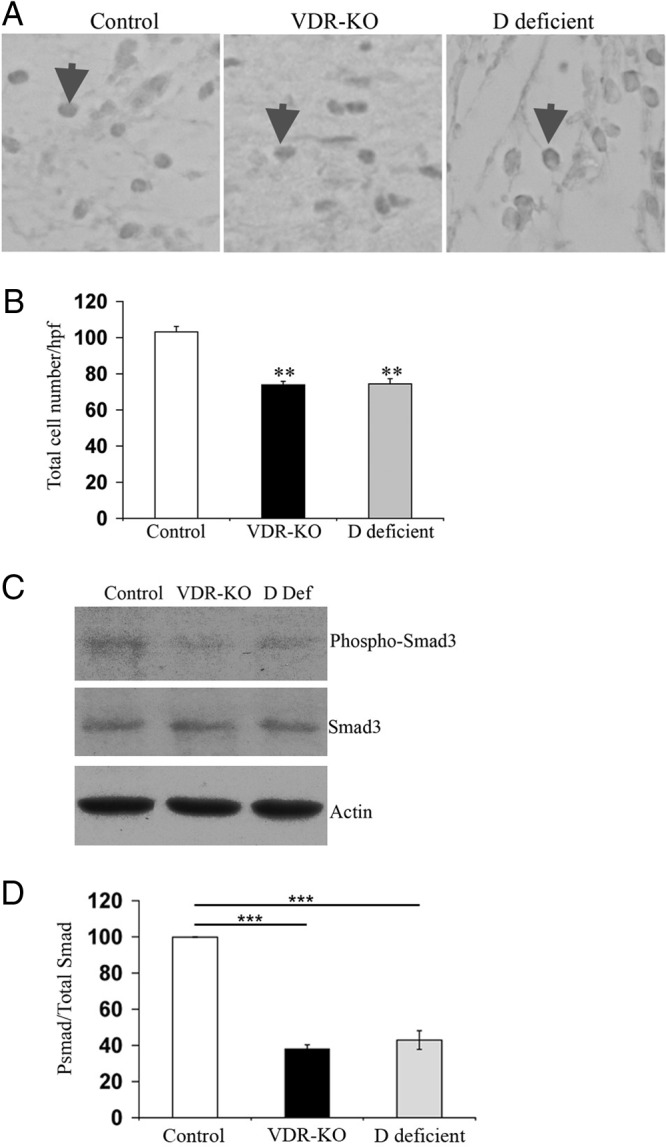
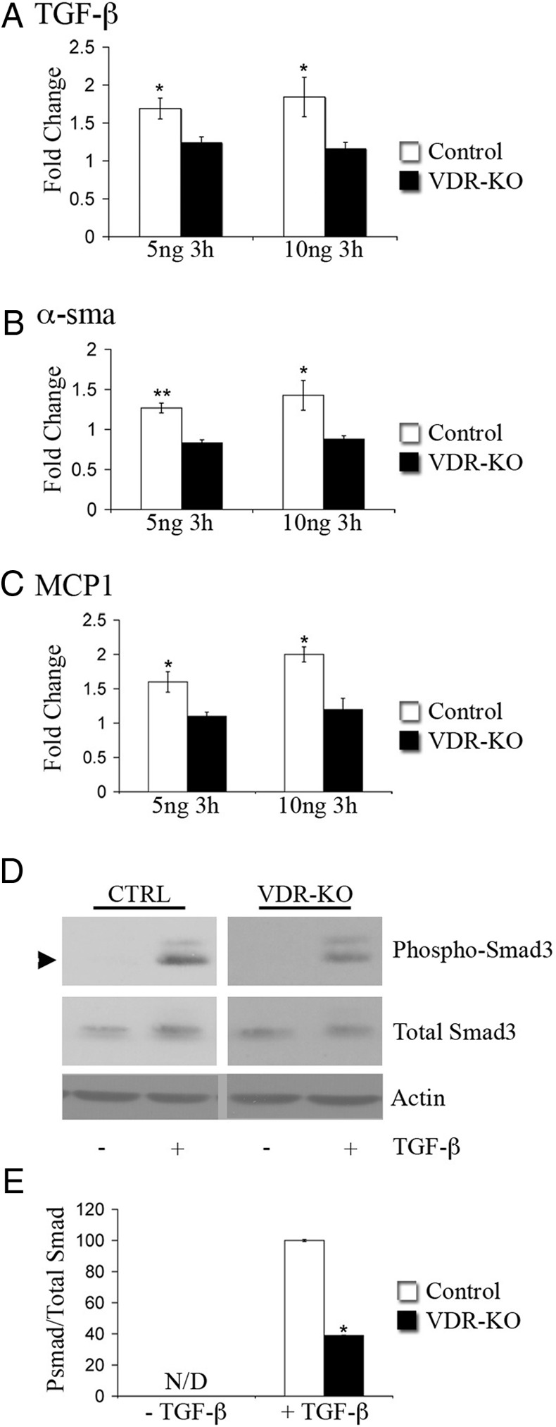
The vitamin D receptor (VDR) has both 1,25-dihydroxyvitamin D-dependent and -independent actions in the epidermis. Ligand-dependent actions of the VDR have been shown to promote keratinocyte differentiation and to regulate formation of the epidermal barrier. In contrast, the actions of the VDR that regulate postmorphogenic hair cycling do not require 1,25-dihydroxyvitamin D. The VDR also has immunomodulatory actions that are dependent on its ligand, 1,25-dihydroxyvitamin D. To determine whether the ligand-dependent or -independent actions of the VDR regulate the inflammatory response to cutaneous injury, studies were performed in control, VDR knockout, and vitamin D-deficient mice. These investigations demonstrate that absence of receptor or ligand impairs the dermal response to cutaneous injury. Although neutrophil recruitment is not affected, the absence of VDR signaling leads to defects in macrophage recruitment and granulation tissue formation. Studies performed to identify the molecular basis for this phenotype demonstrate that absence of the VDR, or its ligand, impairs TGF-β signaling in the dermis, characterized by decreased expression of monocyte chemotactic protein-1 and reduced phosphorylation of phosphorylated Smad-3 as well as attenuated phosphorylated Smad-3 phosphorylation in response to TGF-β in primary dermal fibroblasts lacking the VDR. Thus, these data demonstrate that the liganded VDR interacts with the TGF-β signaling pathway to promote the normal inflammatory response to cutaneous injury.


Figures

Fig. 1.
Fig. 1.
Abnormalities in the inflammatory response to cutaneous wounding are apparent in vdr−/− and vitamin D-deficient mice. Hematoxylin-and-eosin-stained sections from the center of wounds isolated 1 (A), 2 (B), or 5 (C) d after the injury from control, vdr−/− (VDR-KO), and vitamin D-deficient mice (D Deficient; housed in a UV free environment and fed a high calcium, high phosphorus, lactose supplemented diet lacking vitamin D metabolites). Boxes denote area magnified in bottom panel. Original magnification (top panel), ×4, (bottom panel), ×20. Data are based on at least two sections obtained per wound from wounds isolated from at least three mice per genotype or condition. Graph in A represents the number of polymorphonuclear cells/hpf. Data were manually quantified and averaged over 3 hpf from at least three mice per genotype/condition. Care was taken to exclude fibroblasts, macrophages, and endothelial cells and to avoid performing cell counts within the scale crust. D, Representative VDR IHC analyses 2 d after the injury for the VDR in the wound granulation tissue of control (left panel), vdr−/− (VDR-KO, middle panel), and vitamin D-deficient (D Def, right panel) mice. Original magnification, ×40. Data are based on at least two sections obtained per wound from wounds isolated from at least three mice per genotype or condition.
Fig. 2.
Fig. 2.
Impaired macrophage recruitment and decreased MCP-1 expression are observed in the wounds of vdr−/− and vitamin D-deficient mice. A, Representative IHC analyses 2 d after the injury for the macrophage marker F4/80 (red) in the wound granulation tissue of control (left panel), vdr−/− (VDR-KO; middle panel), and vitamin D-deficient (D Def; right panel) mice. The total number of F4/80-positive cells/hpf was quantified ±sem (n ≥ 3 mice). B, Representative IHC analyses 2 d after the injury for IL-1α (red) in the wound granulation tissue of control (left panel), vdr−/− (middle panel), and vitamin D-deficient (right panel) mice. The total number of IL-1α-positive cells/hpf was quantified ±sem (n ≥ 3 mice). C, Representative IHC analyses 2 d after the injury for MCP-1 (red) in the wound granulation tissue of control (left panel), vdr−/− (middle panel), and vitamin D-deficient (right panel) mice. The total number of MCP-1-positive cells/hpf was quantified ±sem (n ≥ 3 mice). Sections were counterstained with fast green. All images were taken at ×20 magnification. Statistical significance was determined using the Student's t test. **, P ≤ 0.001. Data were manually quantified and averaged over 3 hpf from at least three mice per genotype or condition. D, Total protein (20 μg) isolated from wounds of control (Ctrl), vdr−/− (VDR-KO), and vitamin D-deficient (D Def) mice 2 d after the injury was subjected to SDS-PAGE and immunoblotted for MCP-1 (top panel) and actin (bottom panel). Data are representative of protein lysates obtained from the wounds of four animals per genotype or condition.
Fig. 3.
Fig. 3.
Activation of the TGF-β signaling pathway in response to cutaneous injury requires ligand-dependent actions of the VDR. A, Representative IHC analyses 2 d after the injury for nuclear pSmad3 in the wound granulation tissue of control (left panel), vdr−/− (middle panel), and vitamin D-deficient (right panel) mice. Arrows indicate examples of pSmad3 nuclear immunoreactivity. B, The total number of pSmad3-positive nuclei/hpf was quantified ±sem (n ≥ 3 mice). Statistical significance was determined using the Student's t test. **, P ≤ 0.001. Data are based on at least two sections obtained per wound from wounds isolated from at least three mice per genotype or condition. C, Total protein (20 μg) isolated from wounds of control (Ctrl), vdr−/− (VDR-KO), and vitamin D-deficient (D Def) mice 2 d after the injury was subjected to SDS-PAGE and immunoblotted for pSmad3 (top panel), total Smad3 (middle panel), and actin (bottom panel). D, The ratio of pSmad3 to total Smad3 was determined by the quantitation of signal intensity of the relevant bands. The ratio of pSmad3 to total Smad3 in the wounds of vitamin D-deficient and vdr−/− mice was normalized to that obtained from wounds of control mice. Statistical significance was determined using the student's test. ***, P ≤ 0.0005. Data represent those obtained from the wound protein lysates of four animals per genotype or condition.
Fig. 4.
Fig. 4.
The VDR is required for the activation of TGF-β target gene expression in dermal fibroblasts. A–C, RNA isolated from primary wild-type (Control, white bars) or vdr−/− dermal fibroblasts(VDR-KO, black bars) treated for 3 h with 5 or 10 ng/ml TGF-β was subjected to qRT-PCR analysis for the genes indicated. Values are expressed as the relative fold change in expression of each transcript compared with unstimulated controls. RNA levels encoding each gene of interest were normalized for actin RNA in the same sample. Data shown are based on at least three independent RNA isolations ±sem. *, P ≤ 0.05, **, P ≤ 0.005. D, Total protein (7 μg) isolated from control (CTRL) or vdr−/− (VDR-KO) primary dermal fibroblasts treated with (+) or without (−) 10 ng TGF-β for 30 min was subjected to SDS-PAGE and immunoblotted for pSmad3, total Smad3, and actin. E, The ratio of pSmad3 to total Smad3 was determined by the quantitation of signal intensity of the relevant bands. The ratio of pSmad3 to total Smad3 of the TGF-β-treated vdr−/− dermal fibroblasts was normalized to that of the TGF-β-treated wild-type fibroblasts. Statistical significance was determined using the Student's t test, *, P ≤ 0.05. N/D, pSmad3 band was not detected in the absence of TGF-β. Data are representative of protein lysates obtained from four independent experiments.

Similar articles

Cited by

References

    1. Bikle DD, Oda Y, Xie Z. 2004. Calcium and 1,25(OH)2D: interacting drivers of epidermal differentiation. J Steroid Biochem Mol Biol 89–90:355–360 - PubMed
    1. Sakai Y, Demay MB. 2000. Evaluation of keratinocyte proliferation and differentiation in vitamin D receptor knockout mice. Endocrinology 141:2043–2049 - PubMed
    1. Xie Z, Komuves L, Yu QC, Elalieh H, Ng DC, Leary C, Chang S, Crumrine D, Yoshizawa T, Kato S, Bikle DD. 2002. Lack of the vitamin D receptor is associated with reduced epidermal differentiation and hair follicle growth. J Invest Dermatol 118:11–16 - PubMed
    1. Oda Y, Ishikawa MH, Hawker NP, Yun QC, Bikle DD. 2007. Differential role of two VDR coactivators, DRIP205 and SRC-3, in keratinocyte proliferation and differentiation. J Steroid Biochem Mol Biol 103:776–780 - PubMed
    1. Oda Y, Sihlbom C, Chalkley RJ, Huang L, Rachez C, Chang CP, Burlingame AL, Freedman LP, Bikle DD. 2003. Two distinct coactivators, DRIP/mediator and SRC/p160, are differentially involved in vitamin D receptor transactivation during keratinocyte differentiation. Mol Endocrinol (Baltimore, Md) 17:2329–2339 - PubMed

Publication types

MeSH terms