. 2013 Feb;31(2):397-407.
doi: 10.1002/stem.1270.

Mesenchymal stromal cells improve survival during sepsis in the absence of heme oxygenase-1: the importance of neutrophils

Affiliations

Mesenchymal stromal cells improve survival during sepsis in the absence of heme oxygenase-1: the importance of neutrophils

Sean R R Hall et al. Stem Cells. 2013 Feb.

Abstract

The use of mesenchymal stromal cells (MSCs) for treatment of bacterial infections, including systemic processes like sepsis, is an evolving field of investigation. This study was designed to investigate the potential use of MSCs, harvested from compact bone, and their interactions with the innate immune system, during polymicrobial sepsis induced by cecal ligation and puncture (CLP). We also wanted to elucidate the role of endogenous heme oxygenase (HO)-1 in MSCs during a systemic bacterial infection. MSCs harvested from the bones of HO-1 deficient (-/-) and wild-type (+/+) mice improved the survival of HO-1(-/-) and HO-1(+/+) recipient mice when administered after the onset of polymicrobial sepsis induced by CLP, compared with the administration of fibroblast control cells. The MSCs, originating from compact bone in mice, enhanced the ability of neutrophils to phagocytize bacteria in vitro and in vivo and to promote bacterial clearance in the peritoneum and blood after CLP. Moreover, after depleting neutrophils in recipient mice, the beneficial effects of MSCs were entirely lost, demonstrating the importance of neutrophils for this MSC response. MSCs also decreased multiple organ injury in susceptible HO-1(-/-) mice, when administered after the onset of sepsis. Taken together, these data demonstrate that the beneficial effects of treatment with MSCs after the onset of polymicrobial sepsis is not dependent on endogenous HO-1 expression, and that neutrophils are crucial for this therapeutic response.


Conflict of interest statement

Disclosure of Potential Conflicts of Interest

The authors indicate no potential conflicts of interest.

Figures
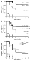
Figure 1
Figure 1
MSCs improve survival following CLP-induced polymicrobial sepsis and neutrophils are critical for this response. A) HO-1−/− mice were treated with HO-1+/+ MSCs (solid black line, n=10), HO-1−/− MSCs (dashed black line, n=10) or fibroblasts (solid gray line, n=9) at 2, 24 and 48 hours post-CLP. Mice were monitored for survival over 7 days, and data are presented as a Kaplan-Meier survival curve. Survival for HO-1+/+ MSCs versus fibroblasts, * P=0.031; HO-1−/− MSCs versus fibroblasts, * P=0.045. B) HO-1+/+ mice were treated with HO-1+/+ MSCs (solid black line, n=26), HO-1−/− MSCs (dashed black line, n=22), fibroblasts (solid gray line, n=17), or PBS (dashed gray line, n=18) at 2, 24 and 48 hours post-CLP. Mice were monitored for survival over 7 days, and data are presented as a Kaplan-Meier survival curve. Survival for HO-1+/+ versus fibroblasts (* P=0.049) and PBS († P=0.005); HO-1−/− MSCs versus PBS († P=0.032). C) HO-1+/+ mice receiving Ly6G antibody were treated with HO-1+/+ MSCs (solid black line, n=13), fibroblasts (solid gray line, n=13), or PBS (dashed gray line, n=6) administered 2, 24, and 48 hours post-CLP. Mice were monitored for survival over 7 days, and data are presented as a Kaplan-Meier survival curve.
Figure 2
Figure 2
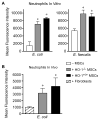
MSCs enhance the ability of peritoneal neutrophils to phagocytize bacteria. A) Activated peritoneal neutrophils were incubated with GFP-labeled E. coli or E. faecalis in the presence of MSCs (HO-1+/+, black bars or HO-1−/−, gray bars) or in the absence of MSCs (− MSCs, white bars) in vitro. Mean fluorescence intensity data are presented as mean±SEM from 3 independent experiments. *P<0.01 versus no MSCs, using Kruskal-Wallis one-way analysis of variance followed by the Dunn’s post test analysis. B) GFP-labeled E. coli were injected i.p. into HO-1−/− mice in the presence of MSCs (HO-1+/+, black bars or HO-1−/−, gray bars) or fibroblasts (striped bar) in vivo. Mean fluorescence intensity data are presented as mean±SEM of 3–4 mice per group. *P=0.043 versus fibroblasts, using Kruskal-Wallis one-way analysis of variance followed by the Dunn’s post test analysis.
Figure 3
Figure 3
MSCs promote bacterial clearance in mice after CLP, and this response is lost when neutrophils are depleted. HO-1−/− mice underwent sham (−) or CLP (+) surgery, and 2 hours after surgery were treated with MSCs or fibroblasts. At 24 hours after surgery, peritoneal fluid (A) and blood (B) were collected. CFUs were determined after incubating at 37°C overnight. Peritoneal fluid: sham, n=8; MSCs, n=8; fibroblasts, n=8. Blood: MSCs, n=6; fibroblasts, n=6. Horizontal bars represent mean values. Experiments were performed 2 independent times. *P<0.05 versus sham, using Kruskal-Wallis one-way analysis of variance followed by the Dunn’s post test analysis (A), and †P<0.05 MSC versus fibroblast, Mann-Whitney U testing (B). HO-1+/+ mice also underwent sham (−) or CLP (+) surgery, and 2 hours after surgery were treated with MSCs or fibroblasts. At 24 hours after surgery, peritoneal fluid (C) and blood (D) were collected. CFUs were determined after incubating at 37°C overnight. Peritoneal fluid: sham, n=8; MSCs, n=7; fibroblasts, n=6. Blood (no neutrophil depletion): MSCs, n=7; fibroblasts, n=6. Blood (neutrophil depletion): MSCs, n=6; fibroblasts, n=6. Horizontal bars represent mean values. Experiments were performed 2 independent times. *P<0.05 versus sham, and † P<0.05 versus fibroblasts, using Kruskal-Wallis one-way analysis of variance followed by the Dunn’s post test analysis (C), and †P<0.05 MSC versus fibroblast, Mann-Whitney U testing (D). NS, not significant.
Figure 4
Figure 4
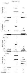
MSCs protect organs from injury after CLP-induced polymicrobial sepsis. HO-1−/− mice underwent sham (−) or CLP (+) surgery, and 2 hours after surgery were treated with MSCs or fibroblasts. At 24 hours after surgery, tissues were scored for injury (see Methods section). Horizontal bars represent mean values. Sham, n=8–10; MSCs, n=9–10; fibroblasts, n=9–10 for the kidney, liver and spleen. Sham, n=6; MSCs, n=7; fibroblasts, n=4 for the small bowel. *P<0.05 versus sham, †P<0.05 versus fibroblasts using Kruskal-Wallis one-way analysis of variance followed by the Dunn’s post test analysis.
Figure 5
Figure 5
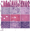
Representative images of organs from mice that received MSCs or fibroblasts after the onset of CLP-induced sepsis. H&E staining of sections from small bowel, liver, and spleen, and PAS staining of kidney, from mice 24 hours after sham (−) or CLP (+) surgery. In the CLP mice, 2 hours after the onset of sepsis, MSCs or fibroblasts were administered. The arrows point to subepithelial spaces in the villi (small bowel, black arrows), tubular epithelial cell swelling (kidney, black arrows), necrotic regions (liver, black arrows), and tingible body macrophages, predominantly in the white pulp, which have phagocytized apoptotic cells (spleen, yellow arrows). Light microscopy images, small bowel and kidney (20X objective), liver (10X objective), and spleen (40X objective).
Figure 6
Figure 6
MSC treatment decreases tissue injury 24 hours after CLP-induced polymicrobial sepsis. HO-1−/− mice underwent sham (−) or CLP (+) surgery, and 2 hours after surgery were treated with MSCs or fibroblasts. At 24 hours after surgery, tissues were harvested and injury was evaluated by TUNEL staining (brown), and quantitated (left panels). Light microscopy images were taken with a 40X objective, and the bar in the sham panels represents a length of 50 μm. Data are represented as mean±SEM. Sham, n=6; MSCs, n=7–8; fibroblasts, n=6–8. *P<0.05 versus sham, †P<0.05 versus fibroblasts using Kruskal-Wallis one-way analysis of variance followed by the Dunn’s post test analysis. Representative small bowel, kidney, liver and spleen sections of HO-1−/− mice are to the right of the quantitative graphs.
Figure 7
Figure 7
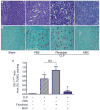
MSCs decrease splenic injury in wild-type mice after CLP-induced polymicrobial sepsis. A) PAS staining of spleen tissue from sham mice (− CLP) or mice 24 hours after CLP (+). In the CLP mice, 2 hours after the onset of sepsis, PBS, fibroblasts, or MSCs were administered. The white arrows point to representative tingible body macrophages, predominantly in the white pulp, which have phagocytized apoptotic cells. B) TUNEL staining was performed, in the same groups as described in (A), to identify apoptotic cells in splenic tissue after CLP. The brown staining identifies the apoptotic splenocytes. Light microscopy images, taken with a 20X objective. C) TUNEL staining was quantitated, and the data are represented as mean±SEM. For mice undergoing CLP, a minimum of 4–5 mice per group were analyzed. *P<0.05 versus sham, †P<0.05 versus fibroblasts and PBS using one-way analysis of variance followed by Newman-Keuls post test analysis. NS, not significant.

Similar articles

Cited by

References

    1. Friedenstein AJ, Chailakhyan RK, Latsinik NV, et al. Stromal cells responsible for transferring the microenvironment of the hemopoietic tissues. Cloning in vitro and retransplantation in vivo. Transplantation. 1974;17:331–340. - PubMed
    1. Prockop DJ. Marrow stromal cells as stem cells for nonhematopoietic tissues. Science. 1997;276:71–74. - PubMed
    1. Dennis JE, Merriam A, Awadallah A, et al. A quadripotential mesenchymal progenitor cell isolated from the marrow of an adult mouse. J Bone Miner Res. 1999;14:700–709. - PubMed
    1. Pittenger MF, Mackay AM, Beck SC, et al. Multilineage potential of adult human mesenchymal stem cells. Science. 1999;284:143–147. - PubMed
    1. da Silva Meirelles L, Chagastelles PC, Nardi NB. Mesenchymal stem cells reside in virtually all post-natal organs and tissues. J Cell Sci. 2006;119:2204–2213. - PubMed

Publication types

MeSH terms