. 2013 Jan;31(1):23-34.
doi: 10.1002/stem.1273.

Effective elimination of cancer stem cells by a novel drug combination strategy

Affiliations

Effective elimination of cancer stem cells by a novel drug combination strategy

Shuqiang Yuan et al. Stem Cells. 2013 Jan.

Abstract

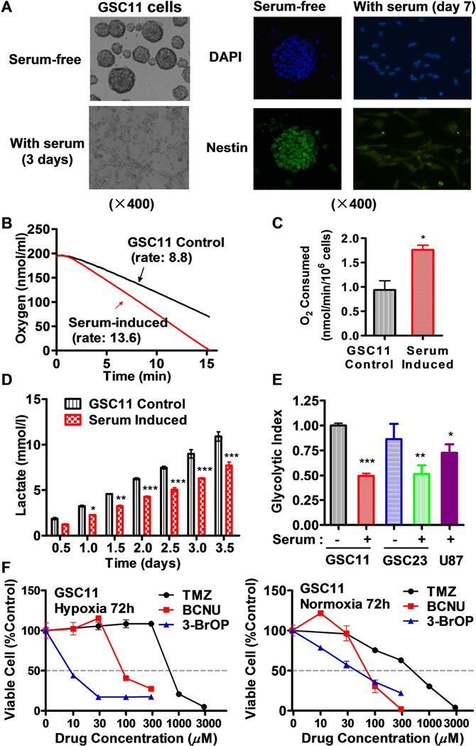
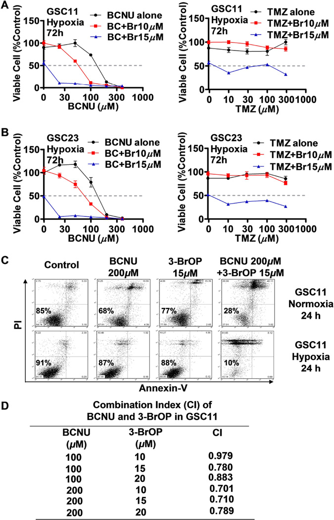
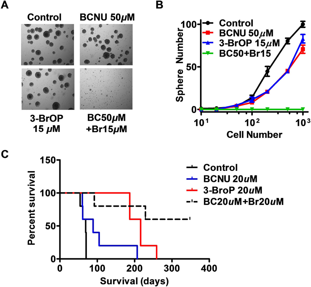
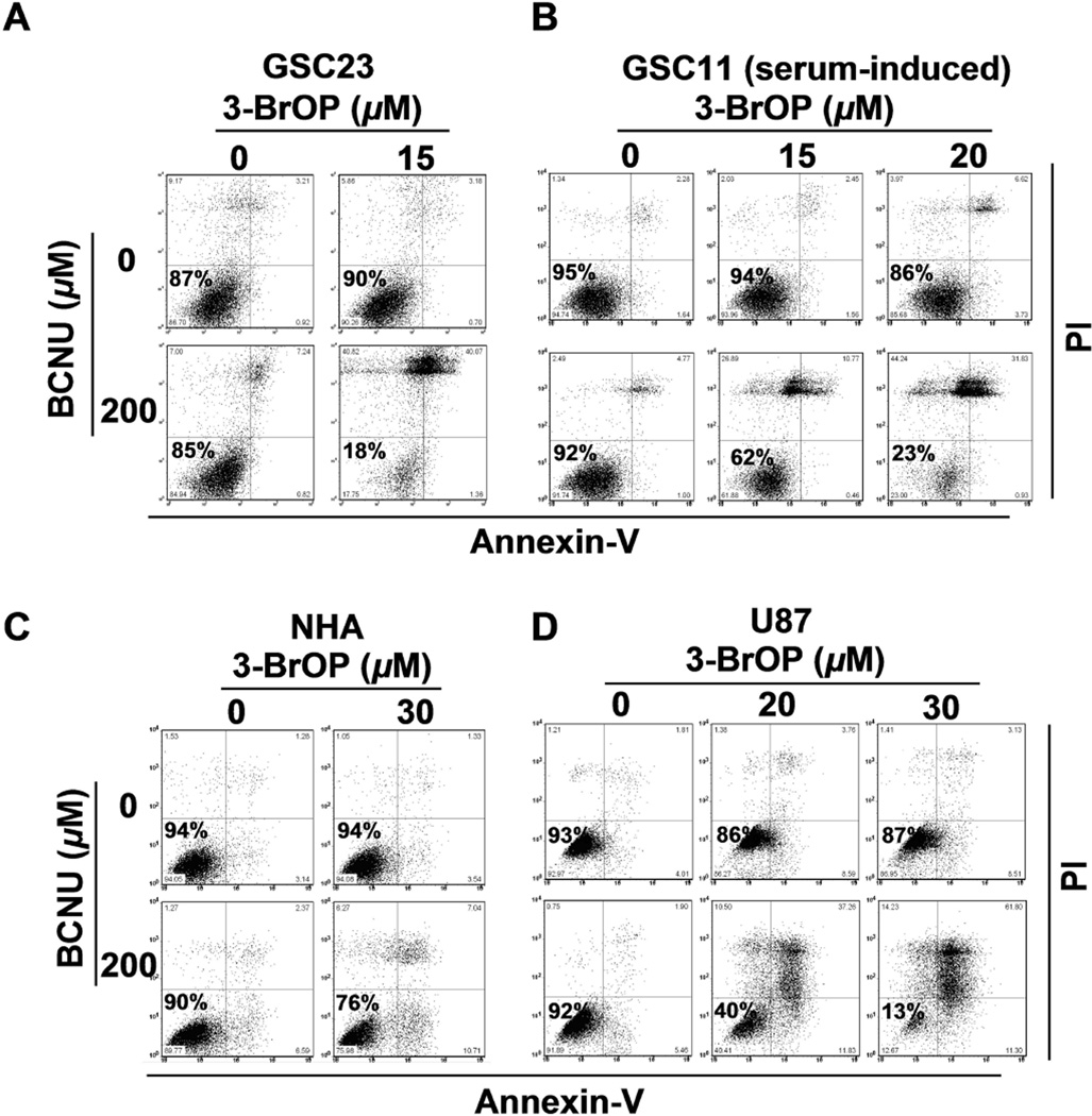
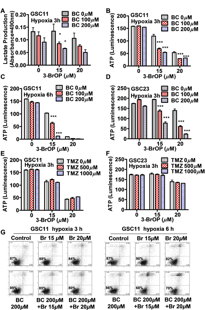
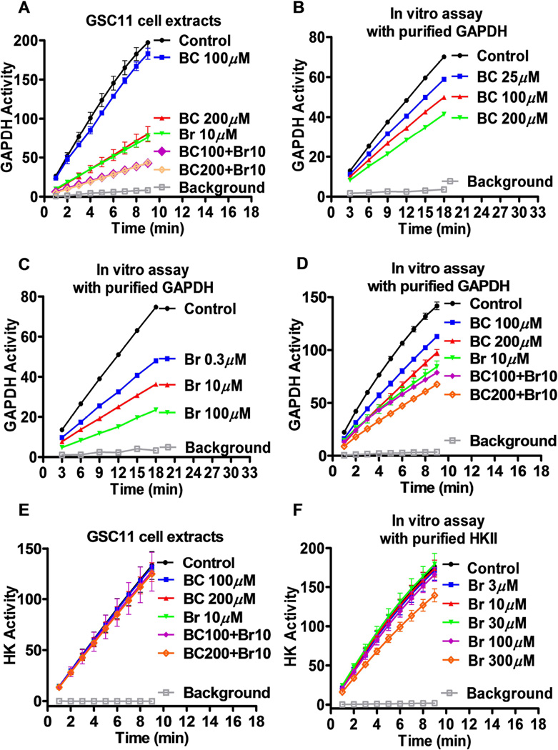
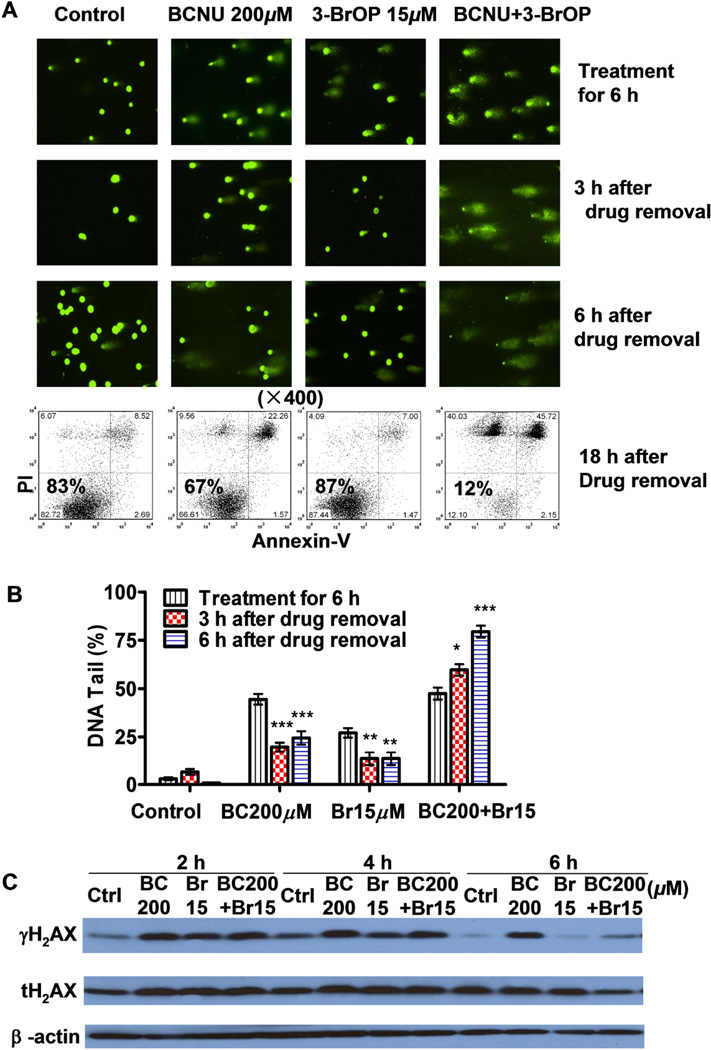
Development of effective therapeutic strategies to eliminate cancer stem cells, which play a major role in drug resistance and disease recurrence, is critical to improve cancer treatment outcomes. Our study showed that glioblastoma stem cells (GSCs) exhibited low mitochondrial respiration and high glycolytic activity. These GSCs were highly resistant to standard drugs such as carmustine and temozolomide (TMZ), but showed high sensitivity to a glycolytic inhibitor 3-bromo-2-oxopropionate-1-propyl ester (3-BrOP), especially under hypoxic conditions. We further showed that combination of 3-BrOP with carmustine but not with TMZ achieved a striking synergistic effect and effectively killed GSCs through a rapid depletion of cellular ATP and inhibition of carmustine-induced DNA repair. This drug combination significantly impaired the sphere-forming ability of GSCs in vitro and tumor formation in vivo, leading to increase in the overall survival of mice bearing orthotopic inoculation of GSCs. Further mechanistic study showed that 3-BrOP and carmustine inhibited glyceraldehyde-3-phosphate dehydrogenase and caused a severe energy crisis in GSCs. Our study suggests that GSCs are highly glycolytic and that certain drug combination strategies can be used to effectively overcome their drug resistance based on their metabolic properties.


Conflict of interest statement

Disclosure of Potential Conflicts of Interest

The authors indicate no potential conflicts of interest.

Figures

Figure 1
Figure 1
Low mitochondrial respiration and high glycolysis in glioblastoma stem cells (GSCs) and their resistance to standard chemotherapeutic agents TMZ and BCNU. (A) Loss of neurosphere formation and decrease in expression of neural stem cell marker nestin in GSC11 cells after exposure to serum (10% FBS) for the indicated time. (B) Representative of mitochondrial respiration rates in GSC11 cells cultured in stem cells medium and in serum-containing medium. Oxygen consumption was measured as an indicator of mitochondrial respiration. (C) Quantitative comparison of mitochondrial respiration rates in GSC11 cells cultured in stem cells medium and in serum-containing medium. (D) Lactate generation rates in GSC11 cells maintained in stem cell medium or in serum-containing medium. (E) Comparison of glycolytic index in GSC11, serum-induced GSC11, GSC23, serum-induced GSC23 and glioblastoma cell line (U87). Glycolytic index was calculated according to the formula (L × G)/O), in which L is the cellular lactate production, G is the glucose uptake, and O is the oxygen consumption rate. (F) Sensitivity of glioblastoma stem cells to TMZ, BCNU, and 3-BrOP under hypoxic and normoxic conditions. GSC11 cells were incubated with the indicated concentrations of drugs for 72 h in normoxia or hypoxia (2% O2) conditions, and cell viability was measured by MTS assay. *, p<0.05; **, p<0.01; ***, p<0.001.
Figure 2
Figure 2
Effective killing of glioblastoma stem cells by combination of 3-BrOP and BCNU. (A) Inhibition of GSC11 cells by BCNU (left panel), TMZ (right panel), or their combination with 3-BrOP under hypoxic conditions (2% O2 for 72 h, MTS assay). (B) Inhibition of GSC23 cells by BCNU (left panel), TMZ (right panel), or their combination with 3-BrOP under hypoxic conditions (2% O2 for 72 h, MTS assay). (C) Induction of cell death in GSC11 cells treated with BCNU, 3-BrOP, or their combination. Cells were incubated with the indicated concentrations of compounds for 24 h under normoxic and hypoxic conditions (2% O2). Cell viability was then measured using annexin-V/PI double staining followed by flow cytometry analysis. (D) Drug combination index (CI) calculated using the Calcusyn software in GSC11 cells treated with BCNU, 3-BrOP alone and in combination for 24 h under hypoxia (2% O2).
Figure 3
Figure 3
Impairment on GSC sphere formation in vitro and tumor formation in vivo by combination of 3-BrOP and BCNU. (A) Representative morphology of neurosphere formation of glioblastoma stem cells. GSC11 cells were incubated with the indicated concentrations of BCNU, 3-BrOP, or their combination for 3 h, and then cultured in drug-free medium for formation of neurospheres. (B) Quantitative data of neurosphere formation in the presence or absence of 3-BrOP and BCNU. (C) Effect of 3-BrOP and BCNU on tumor development in vivo. GSC11 cells were treated with PBS (control), 20 µM 3-BrOP, 20 µM BCNU, or their combination for 6 h. The cells were then inoculated orthotopically into the brains of mice (5 mice in each group), and mouse survival was monitored (without further drug treatment) as an indication of in vivo tumor formation and disease progression.
Figure 4
Figure 4
Preferential killing of glioblastoma cells by 3-BrOP and BCNU. The cytotoxic effects of 3-BrOP, BCNU, or their combination was compared in (A) GSC23 cells, (B) serum (10% FBS)-induced GSC11 cells, (C) non-malignant human astrocytes (NHA), and (D) U87 glioma cells. Cells were incubated with the indicated drugs for 24 h under hypoxia (2% O2), and cell viability was measured by annexin-V/PI double staining followed by flow cytometry analysis.
Figure 5
Figure 5
Effect of 3-BrOP and BCNU or TMZ on energy metabolism in glioblastoma stem cells. (A) Inhibition of lactate production in GSC11 cells by the indicated concentrations of BCNU and 3-BrOP under hypoxia (2% O2). (B) ATP depletion in GSC11 cells by the indicated concentrations of BCNU and 3-BrOP for 3 h under hypoxia (2% O2). (C) ATP depletion in GSC11 cells by the indicated concentrations of BCNU and 3-BrOP for 6 h under hypoxia (2% O2). (D) ATP depletion in GSC23 cells by the indicated concentrations of BCNU and 3-BrOP for 3 h under hypoxia. (E) ATP depletion in GSC11 cells by the indicated concentrations of TMZ and 3-BrOP for 3 h under hypoxia (2% O2). (F) ATP depletion in GSC23 cells by the indicated concentrations of TMZ and 3-BrOP for 3 h under hypoxia (2% O2). (G) Viability of GSC11 cells after incubation with 3-BrOP and BCNU for 3 h or 6 h under hypoxic conditions (2% O2). Cell viability was measured by annexin-V/PI double staining followed by flow cytometry analysis. *, p<0.05; ***, p<0.001.
Figure 6
Figure 6
Inhibition of glyceraldehyde-3-phosphate dehydrogenase (GAPDH) and hexokinase by BCNU and 3-BrOP. (A) Effect of BCNU (BC) and 3-BrOP (Br) on GAPDH enzyme activity in GSC11 cells. Cells were first incubated with the indicated concentrations of BCNU or 3-BrOP for 30 min, and protein extracts were prepared for analysis of GAPDH activity without further addition of the compounds in vitro. GAPDH activity was measured as described in Materials and Methods. (B) Inhibition of purified GAPDH enzyme by BCNU in vitro. (C) Inhibition of purified GAPDH enzyme by 3-BrOP in vitro. (D) Inhibition of purified GAPDH enzyme by the combination of BCNU and 3-BrOP in vitro. (E) Effect of BCNU, 3-BrOP, or their combination on hexokinase activity in GSC11 cells. Cells were first incubated with the indicated concentrations of compounds for 30 min, and protein extracts were prepared for analysis of hexokinase activity without further addition of the compounds in vitro. (F) Inhibition of purified hexokinase II enzyme by 3-BrOP in vitro.
Figure 7
Figure 7
Effect of 3-BrOP on repair of DNA damage induced by BCNU and cytotoxicity in glioblastome stem cells. (A) Comet assay of DNA damage in GSC11 cells treated with BCNU, 3-BrOP, or their combination. Cells were treated with the indicated concentrations of compounds for 6 h, and then either immediately processed for comet assay or cultured in drug-free medium for additional 3–6 h for potential DNA repair. The bright green dots represent the positions of cellular nuclei; the “tail” length and intensity and on the right side of each nucleus represent the degree of DNA strand breaks eluted out from the cell during electrophoresis. Flow cytometry analysis (annexin-V/PI staining) was also used to measure cell death at 24 h (18 h after drug removal, lower panel). (B) Quantification of DNA damage in GSC11 cells treated with or without BCNU and 3-BrOP as indicated. At least 30 cells in each sample were quantitatively analyzed for % of DNA tail that eluted from the cellular nuclei. (C) Western blot analysis of γH2AX and total H2AX (tH2AX) proteins in GSC11 cells treated with the indicated compounds for 2–6 h. *, p<0.05; **, p<0.01; ***, p<0.001.

References

    1. Gilbert CA, Ross AH. Cancer stem cells: cell culture, markers, and targets for new therapies. J Cell Biochem. 2009;108:1031–1038. - PMC - PubMed
    1. Bonnet D, Dick JE. Human acute myeloid leukemia is organized as a hierarchy that originates from a primitive hematopoietic cell. Nat Med. 1997;3:730–737. - PubMed
    1. Singh SK, Clarke ID, Terasaki M, et al. Identification of a cancer stem cell in human brain tumors. Cancer Res. 2003;63:5821–5828. - PubMed
    1. Al-Hajj M, Wicha MS, Benito-Hernandez A, et al. Prospective identification of tumorigenic breast cancer cells. Proc Natl Acad Sci U S A. 2003;100:3983–3988. - PMC - PubMed
    1. O'Brien CA, Pollett A, Gallinger S, et al. A human colon cancer cell capable of initiating tumour growth in immunodeficient mice. Nature. 2007;445:106–110. - PubMed

Publication types

MeSH terms