. 2012 Dec 14;287(51):42826-34.
doi: 10.1074/jbc.M112.405027. Epub 2012 Nov 6.

Transmembrane topology and oligomeric structure of the high-affinity choline transporter

Affiliations

Transmembrane topology and oligomeric structure of the high-affinity choline transporter

Takashi Okuda et al. J Biol Chem. .

Abstract

The high-affinity choline transporter CHT1 mediates choline uptake essential for acetylcholine synthesis in cholinergic nerve terminals. CHT1 belongs to the Na(+)/glucose cotransporter family (SLC5), which is postulated to have a common 13-transmembrane domain core; however, no direct experimental evidence for CHT1 transmembrane topology has yet been reported. We examined the transmembrane topology of human CHT1 using cysteine-scanning analysis. Single cysteine residues were introduced into the putative extra- and intracellular loops and probed for external accessibility for labeling with a membrane-impermeable, sulfhydryl-specific biotinylating reagent in intact cells expressing these mutants. The results provide experimental evidence for a topological model of a 13-transmembrane domain protein with an extracellular amino terminus and an intracellular carboxyl terminus. We also constructed a three-dimensional homology model of CHT1 based on the crystal structure of the bacterial Na(+)/galactose cotransporter, which supports our conclusion of CHT1 transmembrane topology. Furthermore, we examined whether CHT1 exists as a monomer or oligomer. Chemical cross-linking induces the formation of a higher molecular weight form of CHT1 on the cell surface in HEK293 cells. Two different epitope-tagged CHT1 proteins expressed in the same cells can be co-immunoprecipitated. Moreover, co-expression of an inactive mutant I89A with the wild type induces a dominant-negative effect on the overall choline uptake activity. These results indicate that CHT1 forms a homo-oligomer on the cell surface in cultured cells.


Figures

FIGURE 1.
FIGURE 1.
External accessibility of hCHT1 cysteine mutants. A, choline uptake and HC-3 binding activities of the mutants in HEK293 cells. The data are normalized to activity levels of the WT. HC-3 binding activity of T401C was not graphed because it was relatively high. B, biotinylation assays of hCHT1 cysteine mutants. MTSEA-biotin labeling of the introduced cysteine within the mutants was assessed by immunoblotting with anti-CHT1 antibody (shown as MTSEA-biotinylated). The cell surface expression of the mutants was monitored by biotinylation assays using membrane-impermeable sulfo-NHS-SS-biotin, which interacts with amino groups of proteins on the extracellular surface (shown below as cell surface).
FIGURE 2.
FIGURE 2.
hCHT1 homology model based on the vSGLT structure. Ribbon representation of the constructed model of hCHT1, in which α-helices are numbered. Horizontal bars represent the boundaries of the hydrophobic core of the lipid bilayer (M: membrane). Color changes gradually from the amino terminus (N, blue) to the carboxyl terminus (C, red). The unmodeled loops and carboxyl terminus are represented by dotted and a dashed line, respectively. The residues examined by MTSEA-biotin labeling in Fig. 1 are represented by space filling model. Carbon atoms of amino acid residues that are extracellularly accessible or inaccessible are shown in pink or cyan, respectively. Oxygen atoms are shown in red. The accessible or inaccessible residues located on unmodeled regions are represented by circles in pink or cyan, respectively.
FIGURE 3.
FIGURE 3.
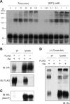
Characterization of hCHT1 oligomers. A, cross-linking of cell surface proteins in hCHT1-expressing HEK293 cells using a cross-linker, BS3. Cells were treated with 4 mm BS3 at 4 °C for the indicated time (left) or with the indicated concentration of BS3 for 120 min (right). Cells were lysed after cross-linking and cell lysates were immunoblotted (IB) with anti-CHT1 antibody. B, co-immunoprecipitation assay using two different versions of epitope-tagged CHT1. Lysates from cells expressing CHT1-FLAG and/or CHT1-HA were precipitated by anti-FLAG antibody and immunoblotted with anti-HA or anti-FLAG antibody. The input lanes represent 10% of cell lysates used in the co-immunoprecipitation assays (shown as lysate). C, co-immunoprecipitation assay using CHT1 and SMVT. Lysates from cells expressing CHT1-FLAG and/or SMVT-HA were precipitated by anti-FLAG antibody and immunoblotted with anti-HA antibody. D, cross-linking immunoprecipitation assay. Cells expressing CHT1-FLAG and/or CHT1-HA were first cross-linked with 4 mm BS3 at 4 °C for 30 min, after which cell lysates were precipitated by anti-FLAG antibody as in B. The input lanes represent 10% of cell lysates used in the co-immunoprecipitation (shown as lysate).
FIGURE 4.
FIGURE 4.
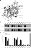
Functional characterization of hCHT1 mutants with altered negatively charged residues. A, the positions of 11 negatively charged residues mutated in this study are indicated in space filling model within the constructed model. B, cell surface biotinylation assay of the mutants. Cells expressing each mutant were labeled with membrane-impermeable, sulfo-NHS-SS-biotin, and biotinylated proteins were precipitated with streptavidin gel. Precipitated proteins (Biotinylated) or cell lysate (Total lysate) were immunoblotted with anti-CHT1 antibody. The mutants D48N, D242N, and D411N are not efficiently expressed in cells, possibly because these residues are required for proper trafficking to the cell membrane. C, choline uptake and HC-3 binding activities of the mutants in HEK293 cells. The data are normalized to activity levels of the WT.
FIGURE 5.
FIGURE 5.
Functional properties of hCHT1 E451D mutant. A, cell surface biotinylation assay using stable cell lines expressing the WT or E451D. Biotinylated proteins were prepared from 5-fold the amount of total lysate loaded on the gel. B, saturation analysis of [3H]choline uptake (shown left) and [3H]HC-3 binding activities (right) of the WT and E451D. C, the displacement of [3H]HC-3 binding by nonlabeled choline in intact cells expressing either the WT or E451D. The data are normalized to [3H]HC-3 binding in the absence of nonlabeled choline.
FIGURE 6.
FIGURE 6.
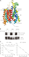
Dominant-negative effect by co-expression of an inactive mutant of CHT1 on the choline uptake activity of the WT. A, the positions of Ile-89 and Glu-451 are indicated in space filling model within the constructed model. Carbon and oxygen atoms are shown in yellow and red, respectively. The molecule is rotated by 90º from the orientation of the model in Figs. 2 and 4A. B, co-immunoprecipitation (IP) assay. For CHT1 WT, I89A, and E451Q, lysates from cells co-expressing CHT1-FLAG (WT) and CHT1-HA (WT or each mutant) were precipitated by anti-FLAG antibody, and the precipitates were immunoblotted (IB) with anti-HA or anti-FLAG antibody. The input lanes represent 10% of cell lysates used in the immunoprecipitation. C, dominant-negative effect by co-expression of the inactive hCHT1 mutant. Various ratios of plasmids encoding hCHT1 mutant (I89A or E451Q) relative to the WT were co-transfected into HEK293 cells, whereas the total DNA amount for each transfection was kept constant. The cells were assayed for [3H]choline uptake and [3H]HC-3 binding activities. The data are normalized to activity of cells transfected with the WT only. For the HC-3 binding activity of I89A, 5 nm [3H]HC-3 was used in the binding assay.

References

    1. Okuda T., Haga T. (2003) High-affinity choline transporter. Neurochem. Res. 28, 483–488 - PubMed
    1. Ferguson S. M., Blakely R. D. (2004) The choline transporter resurfaces. New roles for synaptic vesicles? Mol. Interv. 4, 22–37 - PubMed
    1. Sarter M., Parikh V. (2005) Choline transporters, cholinergic transmission and cognition. Nat. Rev. Neurosci. 6, 48–56 - PubMed
    1. Ribeiro F. M., Black S. A., Prado V. F., Rylett R. J., Ferguson S. S., Prado M. A. (2006) The “ins” and “outs” of the high-affinity choline transporter CHT1. J. Neurochem. 97, 1–12 - PubMed
    1. Ribeiro F. M., Alves-Silva J., Volknandt W., Martins-Silva C., Mahmud H., Wilhelm A., Gomez M. V., Rylett R. J., Ferguson S. S., Prado V. F., Prado M. A. (2003) The hemicholinium-3 sensitive high affinity choline transporter is internalized by clathrin-mediated endocytosis and is present in endosomes and synaptic vesicles. J. Neurochem. 87, 136–146 - PubMed

Publication types

LinkOut - more resources