. 2012 Nov 20;109(47):19232-7.
doi: 10.1073/pnas.1211665109. Epub 2012 Nov 6.

RNA polymerase III subunit architecture and implications for open promoter complex formation

Affiliations

RNA polymerase III subunit architecture and implications for open promoter complex formation

Chih-Chien Wu et al. Proc Natl Acad Sci U S A. .

Abstract

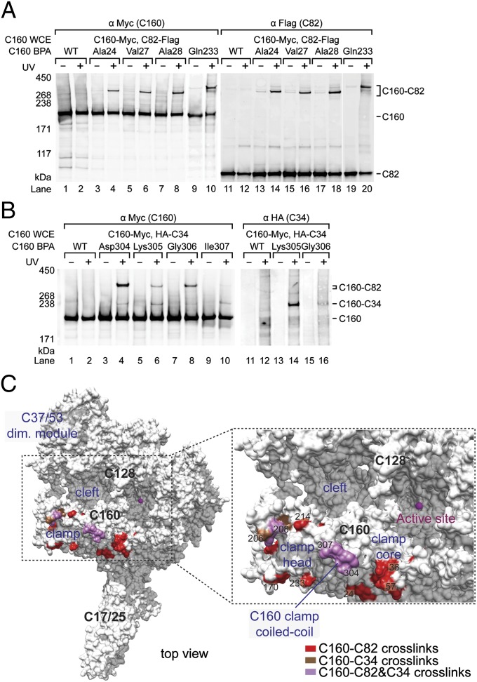
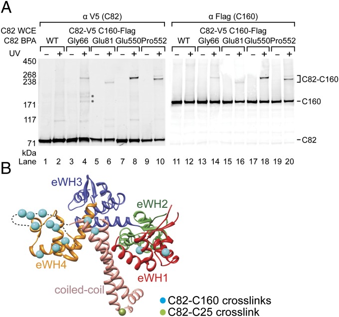
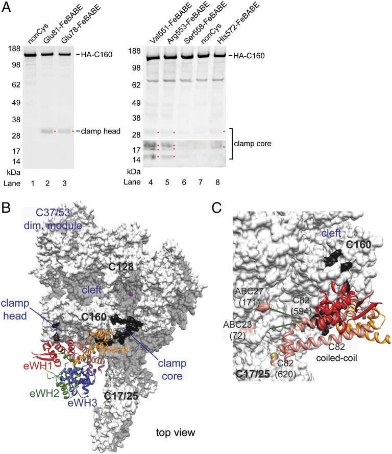
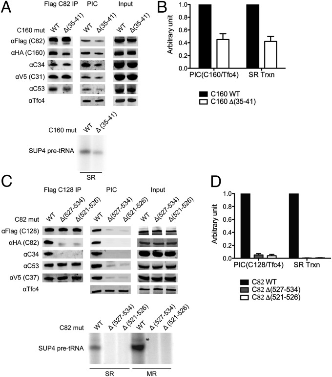
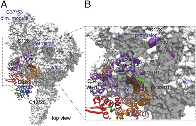
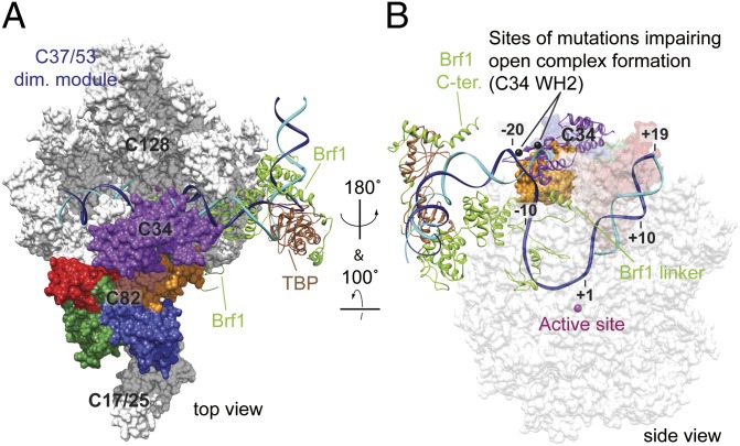
Transcription initiation by eukaryotic RNA polymerase (Pol) III relies on the TFIIE-related subcomplex C82/34/31. Here we combine cross-linking and hydroxyl radical probing to position the C82/34/31 subcomplex around the Pol III active center cleft. The extended winged helix (WH) domains 1 and 4 of C82 localize to the polymerase domains clamp head and clamp core, respectively, and the two WH domains of C34 span the polymerase cleft from the coiled-coil region of the clamp to the protrusion. The WH domains of C82 and C34 apparently cooperate with other mobile regions flanking the cleft during promoter DNA binding, opening, and loading. Together with published data, our results complete the subunit architecture of Pol III and indicate that all TFIIE-related components of eukaryotic and archaeal transcription systems adopt an evolutionarily conserved location in the upper part of the cleft that supports their functions in open promoter complex formation and stabilization.


Conflict of interest statement

The authors declare no conflict of interest.

Figures

Fig. 1.
Fig. 1.
C160 BPA photo-cross-linking indicates C82 and C34 reside on the C160 clamp domain. (A) Western blot of C82–C160 cross-links. Amino acid positions in C160 clamp replaced by BPA are indicated above the lanes. C160 and cross-linking bands were visualized by probing with anti-Myc antibody (C160-Myc, Left), and the cross-linking bands are confirmed to be C160–C82 fusion by probing Flag-tagged C82 (Right). C160 WCE, C160 whole-cell extract; UV + or −, with or without UV irradiation; WT, wild-type C160 with no BPA replacement. (B) C34-C160 cross-links. C160 and C34–C160 cross-linking bands were visualized by anti-Myc antibody (Left) and anti-HA antibody to reveal N-terminally HA-tagged C34 (Right). As indicated, these C160-BPA derivatives also generate simultaneous C82-C160 cross-links. (C) Positions in C82– and C34–C160 cross-links in polymerase clamp. The yeast Pol III core-C37/53 surface model is colored white. Magenta sphere: magnesium ion in the polymerase active site. As indicated, C160 residues yielding C82 and C34 cross-links are shown in red and brown, respectively. The residues cross-linking to both C82 and C34 are colored mauve.
Fig. 2.
Fig. 2.
C82 BPA photo-cross-linking. (A) Western blot of C82–C160 cross-linking. BPA-substituted residues in C82 are indicated above. C82 and cross-linking fusion bands are revealed by anti-V5 antibody (Left). The Flag-epitope tagged C160 in the cross-linking bands is revealed by anti-Flag antibody (Right). Asterisks indicate unidentified cross-linked polypeptides. (B) Amino acid positions cross-linked to C160 in the yeast C82 model. The yeast C82 homology model (displayed as the ribbon model of Cα trace) is generated by the Modeler program (42) using the human RPC62 structure (PDB 2XUB) (20) as the template. The dashed line indicates the missing residues (aa547–567 in C82) in the eWH4. C82 structural domains are colored in red (eWH1), green (eWH2), blue (eWH3), orange (eWH4), and salmon (coiled-coil). The color scheme is used for all following figures. Residues cross-linking to C160 and C25 are highlighted with cyan and olive spheres, respectively.
Fig. 3.
Fig. 3.
Localization of C82 on the C160 clamp. (A) Directed hydroxyl radical cleavage of C160. Western blots of C160 cleavage from tethered FeBABE in C82 eWH1 and eWH4 are respectively shown in the left and right panels. C-terminally HA-tagged C160 fragments are revealed by anti-HA antibody. FeBABE positions in C82 are indicated above the lanes. Cleavage fragments in the lower-molecular-weight range are displayed with signals amplified (Bottom Right). Red asterisks mark identified cleavage fragments. Approximate locations of cleavage sites in C160 are indicated. (B) C82-Pol III core model. C82 (ribbon model) is manually docked as a rigid body on the Pol III core model (white surface model) based on directed hydroxyl radical cleavage and cross-linking-MS analyses. Hydroxyl radical cleavage regions in C160 clamp are highlighted in black covering 11 residues centered at the deduced cut site. (C) Cross-linked lysine pairs between C82 coiled-coil and subunits of the Pol III core. Green lines connect cross-linked lysine pairs from C82 (Lys594) to ABC27 (Lys171) and ABC23 (Lys72). The BPA substitution at C82 Leu620, highlighted as an olive sphere, cross-links to C25.
Fig. 4.
Fig. 4.
Association of the C82/34/31 subcomplex with the Pol III core. (A) Western blot analyses of coimmunoprecipitation and Pol III PIC formation with yeast whole-cell extract containing a C160 mutation. Coimmunoprecipitation was conducted with anti-Flag agarose to immobilize Flag-epitope-tagged C82 (Flag C82 IP; Upper Left). Pol III PIC formation assay was conducted with the immobilized SUP4 tDNA (Upper Middle). The level of a TFIIIC subunit Tfc4 in the PIC is also shown. As indicated, whole-cell extracts containing wild-type (WT) or internal deletion Δ (–41) C160 were used. Single-round in vitro transcription of the isolated Pol III PIC was assayed (Lower). (B) Immunoblot and in vitro transcription signals were quantified from three independent experiments with WT signals set to 1. Error bars indicate SD. (C) Western blot analyses of coimmunoprecipitation and Pol III PIC formation with yeast whole-cell extracts containing C82 mutations. Similar to the analyses in A, whole-cell extracts containing WT or the indicated mutations in C82 eWH4 were used in coimmunoprecipitation, PIC formation, and in vitro transcription analyses. Different from the analysis in A, immune pull-down was conducted by immobilizing Flag-epitope-tagged C128, and both single-round (SR) and multiple-round (MR) transcription were assayed (Lower). An asterisk indicates the read-through transcripts. (D) The results for PIC formation and SR transcription from C are quantified and plotted with WT signals set to 1. Errors bars indicate SD from three independent experiments.
Fig. 5.
Fig. 5.
Model of C34 WH domains in Pol III based on lysine–lysine cross-links. Model of C34 and C82 on the Pol III core is shown on the left. C34 WH1 and WH2 ribbon models are colored purple. In the Pol III cleft view on the right, green lines connect intrasubunit and intersubunit cross-linked lysines (purple spheres with labels indicating subunit names and residue numbers). Brown patches on C160 clamp indicate the BPA-substituted residues in C160 that cross-linked to C34.
Fig. 6.
Fig. 6.
Pol III architecture and open promoter complex. (A) Model for the Pol III open promoter complex. The model of Pol III-Brf1-TBP-DNA open promoter complex was built based on the Pol II-TFIIB-TBP open complex (37) and the Brf1-TBP-DNA structure (43). Template and nontemplate DNA strands are in blue and cyan, respectively. The Pol III core-C37/53 model is displayed as the white surface model. Ribbon models for Brf1 and TBP are colored yellow-green and brown, respectively. The C82 surface model is colored according to the domain color scheme as above, and both C34 WH surface models are colored purple. (B) Protein–DNA organization in the Pol III active center. The Pol III open promoter complex in A is displayed with different orientation to view the Pol III active center. The Pol III core is semitransparent and C34 is changed to the ribbon model. Lys135 and Lys138 in C34 WH2 are highlighted as black spheres. These two amino acids are required for DNA binding and open complex formation (12, 20).

Similar articles

Cited by

References

    1. Cramer P, et al. Structure of eukaryotic RNA polymerases. Annu Rev Biophys. 2008;37:337–352. - PubMed
    1. Dieci G, Fiorino G, Castelnuovo M, Teichmann M, Pagano A. The expanding RNA polymerase III transcriptome. Trends Genet. 2007;23(12):614–622. - PubMed
    1. Schramm L, Hernandez N. Recruitment of RNA polymerase III to its target promoters. Genes Dev. 2002;16(20):2593–2620. - PubMed
    1. Bernard G, et al. Mutations of POLR3A encoding a catalytic subunit of RNA polymerase Pol III cause a recessive hypomyelinating leukodystrophy. Am J Hum Genet. 2011;89(3):415–423. - PMC - PubMed
    1. Tétreault M, et al. Recessive mutations in POLR3B, encoding the second largest subunit of Pol III, cause a rare hypomyelinating leukodystrophy. Am J Hum Genet. 2011;89(5):652–655. - PMC - PubMed

Publication types

MeSH terms

LinkOut - more resources