. 2012 Nov 20;109(47):19397-402.
doi: 10.1073/pnas.1217519109. Epub 2012 Nov 6.

MiR-495 is a tumor-suppressor microRNA down-regulated in MLL-rearranged leukemia

Affiliations

MiR-495 is a tumor-suppressor microRNA down-regulated in MLL-rearranged leukemia

Xi Jiang et al. Proc Natl Acad Sci U S A. .

Abstract

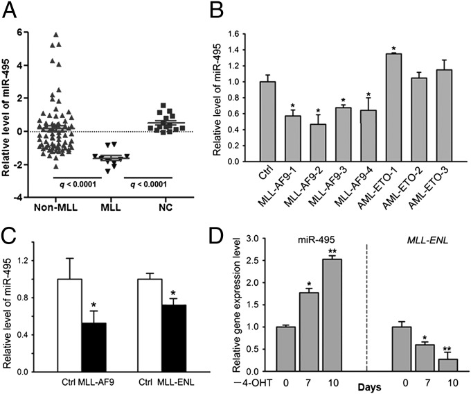
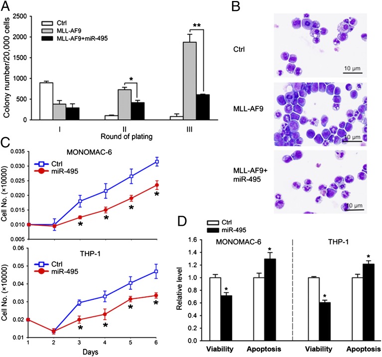
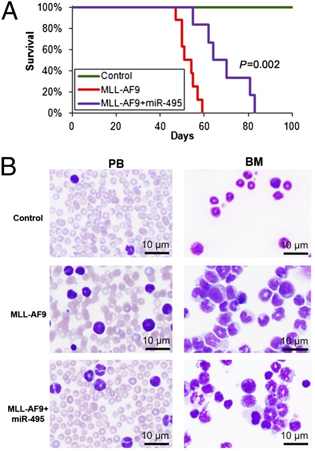
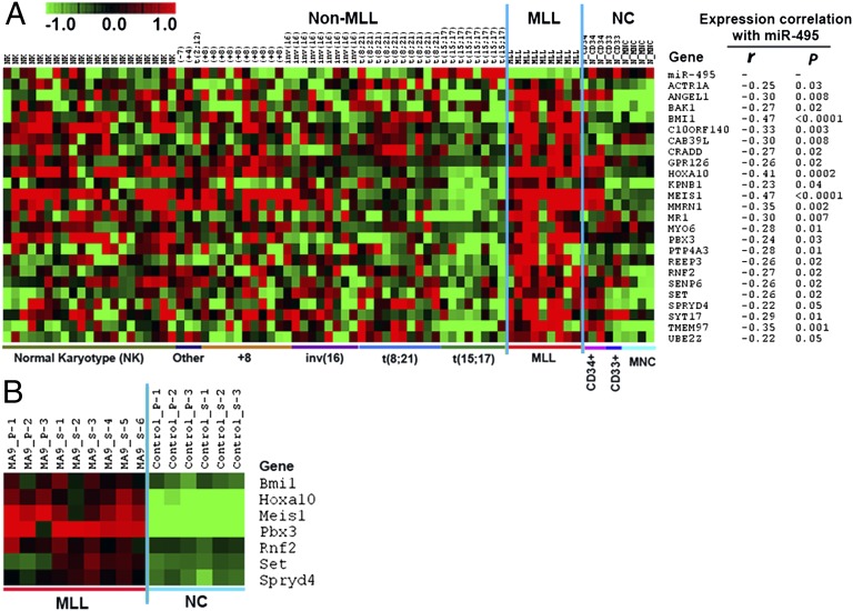
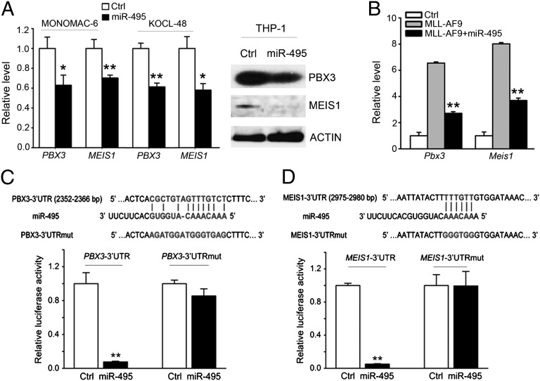
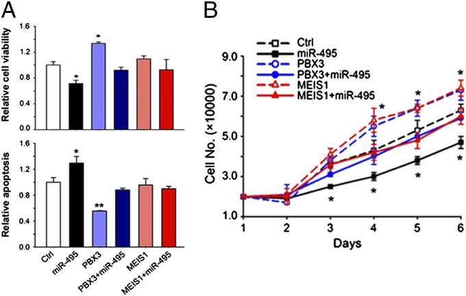
Acute myeloid leukemia (AML) is a heterogeneous group of hematopoietic malignancies with variable response to treatment. AMLs bearing MLL (mixed lineage leukemia) rearrangements are associated with intermediate or poor survival. MicroRNAs (miRNAs), a class of small noncoding RNAs, have been postulated to be important gene expression regulators virtually in all biological processes, including leukemogenesis. Through a large-scale, genome-wide miRNA expression profiling assay of 85 human AML and 15 normal control samples, we show that among 48 miRNAs that are significantly differentially expressed between MLL- and non-MLL-rearranged AML samples, only one (miR-495) is expressed at a lower level in MLL-rearranged AML than in non-MLL-rearranged AML; meanwhile, miR-495 is also significantly down-regulated in MLL-rearranged AML samples compared with normal control samples. Through in vitro colony-forming/replating assays and in vivo bone marrow transplantation studies, we show that forced expression of miR-495 significantly inhibits MLL-fusion-mediated cell transformation in vitro and leukemogenesis in vivo. In human leukemic cells carrying MLL rearrangements, ectopic expression of miR-495 greatly inhibits cell viability and increases cell apoptosis. Furthermore, our studies demonstrate that PBX3 and MEIS1 are two direct target genes of miR-495, and forced expression of either of them can reverse the effects of miR-495 overexpression on inhibiting cell viability and promoting apoptosis of human MLL-rearranged leukemic cells. Thus, our data indicate that miR-495 likely functions as a tumor suppressor in AML with MLL rearrangements by targeting essential leukemia-related genes.


Conflict of interest statement

The authors declare no conflict of interest.

Figures

Fig. 1.
Fig. 1.
miR-495 is significantly down-regulated in human MLL-rearranged AML compared with non–MLL-rearranged AML or normal controls. (A) Relative expression levels of miR-495 in 10 human MLL-rearranged AML (MLL), 75 non–MLL-rearranged AML (non-MLL), and 15 normal control (NC, including 6 CD34+ hematopoietic stem/progenitor cell, 5 CD33+ myeloid progenitor cell, and 4 MNC) samples, as detected by Exiqon miRNA microarray assays. (B) Quantitative PCR (qPCR) analysis of miR-495 expression level in human cord blood CD34+ cells that were retrovirally transduced with MSCV-MLL-AF9, MSCV-AML-ETO, or empty vector (Ctrl). (C) Down-regulation of miR-495 in MLL-AF9 or MLL-ENL–transduced mouse BM progenitor cells. (D) Withdrawal of 4-OHT (days 0, 7, and 10 are shown) results in a significant increase of miR-495 expression but a significant decrease of MLL-ENL expression in MLL-ENL-ERtm cells. *P < 0.05; **P < 0.01, two-tailed t test.
Fig. 2.
Fig. 2.
miR-495 functions as a tumor-suppressor miRNA in vitro. (A) miR-495 inhibits MLL-AF9–induced cell transformation of normal mouse BM progenitor cells. Normal mouse BM cells were retrovirally transduced with MSCVneo+MSCV-PIG (as control), MSCVneo-MLL-AF9+MSCV-PIG (MLL-AF9), or MSCVneo-MLL-AF9+MSCV-PIG-miR-495 (MLL-AF9+miR-495), and colony forming/replating assays were done thereafter. (B) miR-495 promotes the differentiation of MLL-AF9–transduced mouse BM progenitor cells. Colony-forming cells were collected for cytospin analysis from the secondary round of replating. (C) Ectopic expression of miR-495 inhibits cell growth/proliferation of MONOMAC-6 and THP-1 cells. Cells were transfected with MSCV-PIG (Ctrl) or MSCV-PIG-miR-495 (miR-495). Cell numbers were counted every day after transfection for 6 d. (D) Forced expression of miR-495 decreases cell viability and promotes apoptosis of MONOMAC-6 and THP-1 cells. Cells were transfected with MSCV-PIG (Ctrl) or MSCV-PIG-miR-495 (miR-495), and cell viability and apoptosis were assessed 48 h after transfection. *P < 0.05; **P < 0.01, two-tailed t test.
Fig. 3.
Fig. 3.
miR-495 inhibits MLL-fusion–mediated leukemogenesis in vivo. (A) Mouse BM transplantation assay of the control group (Control; MSCVneo+MSCV-PIG; n = 5), the MLL-AF9 group (MSCVneo-MLL-AF9+MSCV-PIG; n = 8), and the MLL-AF9+miR-495 group (MSCVneo-MLL-AF9+MSCV-PIG-miR495; n = 6). The MLL-AF9+miR-495 group developed leukemia significantly (P = 0.002, log–rank test) slower than the MLL-AF9 alone group. (B) miR-495 increased cell differentiation in both peripheral blood (PB) and BM of the recipient mice.
Fig. 4.
Fig. 4.
Expression profiles of candidate target genes of miR-495. (A) Expression profiles of the 24 candidate target genes of miR-495 and their expression correlation with miR-495 in the set of 79 human samples (including 9 MLL-rearranged AML, 61 nonMLL-rearranged AML, and 9 normal control). r, correlation coefficient; P, P value. Pearson correlation was applied to analyze the correlation. (B) Expression profiles of the seven potential target genes of miR-495 in the set of 15 mouse BM cell samples collected from mouse BM transplantation assays, which include 9 MLL-AF9 mouse leukemic BM cell samples (3 from primary BM transplantation and 6 from secondary BM transplantation recipient mice) and 6 normal control BM cell samples (3 each from primary and secondary BM transplantation recipient mice). MA9, MLL-AF9; _P, primary transplantation recipient mouse; _S, secondary transplantation recipient mouse. Expression data were mean centered, and the relative value for each sample is represented by a color, with red representing a high expression and green representing a low expression (scale shown at upper left).
Fig. 5.
Fig. 5.
PBX3 and MEIS1 are direct target genes of miR-495. (A) Ectopic expression of miR-495 significantly (P < 0.05) represses endogenous expression of PBX3 and MEIS1 in MLL-rearranged AML cells. The cells were transfected with MSCV-PIG (control) or MSCV-PIG-miR-495, and then the effect of miR-495 overexpression was analyzed 48 h after transfection at both mRNA (Left; detected by qPCR, and the GAPDH expression level was used for normalization) and the protein (Right; detected by Western blot) levels. (B) Inhibitory effect of miR-495 on the endogenous expression of Pbx3 and Meis1 in BM cells of the BM transplantation recipient mice shown in Fig. 3A. Gene levels were normalized to the level of endogenous Gapdh. (C and D) miR-495 directly targets PBX3 (C) and MEIS1 (D) as detected by luciferase reporter and mutagenesis assays. In HEK293T cells, plasmids encoding the wild-type 3′ UTR of PBX3 or MEIS1 (namely, PBX3/MEIS1-3′UTR) or the mutant 3′ UTR in which the predicted miR-495 binding site was mutated (namely, PBX3/MEIS1-3′UTRmut), together with MSCV-PIG or MSCV-PIG-miR-495, were cotransfected with β-gal reporter control vector. Luciferase reporter assays were done 48 h after transfection. Forced expression of miR-495 could significantly repress luciferase activity of the reporter gene bearing the 3′ UTR of PBX3 or MEIS1 in human 293T cells, whereas mutation at the predicted target site in the 3′ UTR abrogated the repression. The normalized luciferase activities represent the firefly: β-gal ratios normalized to the control sample. Error bars present SD obtained from three independent experiments. *P < 0.05; **P < 0.01, two-tailed t test.
Fig. 6.
Fig. 6.
Both PBX3 and MEIS1 are functionally important target genes of miR-495 in MLL-rearranged leukemic cells. (A) Analysis of the effects of forced expression of miR-495 (MSCV-PIG-miR-495+MSCVneo), PBX3 (MSCV-PIG+MSCVneo-PBX3), PBX3+miR-495 (MSCV-PIG-miR-495+MSCVneo-PBX3), MEIS1 (MSCV-PIG+MSCVneo-MEIS1), and MEIS1+miR-495 (MSCV-PIG-miR-495+MSCVneo-MEIS1), respectively, on cell viability (Upper) and apoptosis (Lower) of MONOMAC-6 cells. Cell viability and apoptosis were detected 48 h after transfection. (B) Analysis of their effects on cell growth/proliferation of MONOMAC-6 cells. Cell numbers were counted every day after transfection for 6 d. The coding regions (CDS) of PBX3 and MEIS1 were cloned into MSCVneo, and thus their ectopic expression would not be repressed by endogenous or cotransfected miR-495. The cells transfected with MSCV-PIG+MSCVneo (Ctrl) were used as controls. *P < 0.05; **P < 0.01, two-tailed t test.

References

    1. Löwenberg B, Downing JR, Burnett A. Acute myeloid leukemia. N Engl J Med. 1999;341(14):1051–1062. - PubMed
    1. Zhang Y, Rowley JD. Chromatin structural elements and chromosomal translocations in leukemia. DNA Repair (Amst) 2006;5(9-10):1282–1297. - PubMed
    1. Rowley JD. Chromosome translocations: Dangerous liaisons revisited. Nat Rev Cancer. 2001;1(3):245–250. - PubMed
    1. Peterson LF, et al. Acute myeloid leukemia with the 8q22;21q22 translocation: Secondary mutational events and alternative t(8;21) transcripts. Blood. 2007;110(3):799–805. - PMC - PubMed
    1. Ziemin-van der Poel S, et al. Identification of a gene, MLL, that spans the breakpoint in 11q23 translocations associated with human leukemias. Proc Natl Acad Sci USA. 1991;88(23):10735–10739. - PMC - PubMed

Publication types

MeSH terms

Associated data