. 2012;8(10):e1002978.
doi: 10.1371/journal.ppat.1002978. Epub 2012 Oct 25.

Deciphering the acylation pattern of Yersinia enterocolitica lipid A

Affiliations

Deciphering the acylation pattern of Yersinia enterocolitica lipid A

Mar Reinés et al. PLoS Pathog. 2012.

Abstract

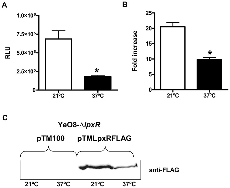
Pathogenic bacteria may modify their surface to evade the host innate immune response. Yersinia enterocolitica modulates its lipopolysaccharide (LPS) lipid A structure, and the key regulatory signal is temperature. At 21°C, lipid A is hexa-acylated and may be modified with aminoarabinose or palmitate. At 37°C, Y. enterocolitica expresses a tetra-acylated lipid A consistent with the 3'-O-deacylation of the molecule. In this work, by combining genetic and mass spectrometric analysis, we establish that Y. enterocolitica encodes a lipid A deacylase, LpxR, responsible for the lipid A structure observed at 37°C. Western blot analyses indicate that LpxR exhibits latency at 21°C, deacylation of lipid A is not observed despite the expression of LpxR in the membrane. Aminoarabinose-modified lipid A is involved in the latency. 3-D modelling, docking and site-directed mutagenesis experiments showed that LpxR D31 reduces the active site cavity volume so that aminoarabinose containing Kdo(2)-lipid A cannot be accommodated and, therefore, not deacylated. Our data revealed that the expression of lpxR is negatively controlled by RovA and PhoPQ which are necessary for the lipid A modification with aminoarabinose. Next, we investigated the role of lipid A structural plasticity conferred by LpxR on the expression/function of Y. enterocolitica virulence factors. We present evidence that motility and invasion of eukaryotic cells were reduced in the lpxR mutant grown at 21°C. Mechanistically, our data revealed that the expressions of flhDC and rovA, regulators controlling the flagellar regulon and invasin respectively, were down-regulated in the mutant. In contrast, the levels of the virulence plasmid (pYV)-encoded virulence factors Yops and YadA were not affected in the lpxR mutant. Finally, we establish that the low inflammatory response associated to Y. enterocolitica infections is the sum of the anti-inflammatory action exerted by pYV-encoded YopP and the reduced activation of the LPS receptor by a LpxR-dependent deacylated LPS.


Conflict of interest statement

The authors have declared that no competing interests exist.

Figures

Figure 1
Figure 1. Lipid A analysis from Y. enterocolitica lpxR mutant.
(A) Negative ion MALDI-TOF mass spectrometry spectra of lipid A isolated from YeO8 grown at 21°C and 37°C. (B) Negative ion MALDI-TOF mass spectrometry spectra of lipid A isolated from YeO8-ΔlpxRKm (ΔlpxR) grown at 21°C and 37°C. (C) Negative ion MALDI-TOF mass spectrometry spectra of lipid A isolated from YeO8-ΔlpxRKm carrying pTMLpxR grown at 21°C and 37°C. The results in all panels are representative of three independent lipid A extractions.
Figure 2
Figure 2. Temperature regulates the expression of Y. enterocolitica lpxR.
(A) Analysis of the expression of lpxR by measuring luciferase activity of YeO8 carrying lpxR::lucFF transcriptional fusion, which was grown at 21°C (white bars) or 37°C (black bars). Data are presented as mean ± SD (n = 3). *, results are significantly different (p<0.05; two-tailed t test) from the results for bacteria grown at 21°C. (B) Analysis of lpxR mRNA levels by RT-qPCR. Total RNA was extracted from bacteria grown at 21°C (white bar) or 37°C (black bar). Data are presented as mean ± SD (n = 3). *, results are significantly different (p<0.05; two-tailed t test) from the results for bacteria grown at 21°C. (C) Western blot analysis of LpxR FLAG tagged levels. Cell envelopes were purified from YeO8-ΔlpxRKm mutant carrying pTM100 or pTMLpxRFLAG plasmids. 80 µg of proteins were run in SDS-12% polyacrylamide gel, electrotransferred onto a nitrocellulose membrane, and developed by using anti-Flag antibodies.
Figure 3
Figure 3. Lipid A analysis from E. coli expressing Y. enterocolitica lpxR.
Negative ion MALDI-TOF mass spectrometry spectra of lipid A isolated from: (A) E. coli MG1655 (E. coli) grown at 21°C. (B) E. coli MG1655 (E. coli) grown at 37°C. (C) E. coli MG1655 (E. coli) carrying pTMLpxR grown at 21°C. (D) E. coli MG1655 (E. coli) carrying pTMLpxR grown at 37°C. The results in all panels are representative of three independent lipid A extractions.
Figure 4
Figure 4. Lipid A analysis from Y. enterocolitica lipid A mutants.
Negative ion MALDI-TOF mass spectrometry spectra of lipid A isolated from the indicated Y. enterocolitica strains grown at 21°C (A,C,E) and 37°C (B,D,F). The results in all panels are representative of three independent lipid A extractions. (G) Analysis of the expression of lpxR by measuring luciferase activity of YeO8 (white bars) and YeO8-ΔpmrF (gray bars) carrying lpxR::lucFF transcriptional fusion, which were grown at 21°C or 37°C. Data are presented as mean ± SD (n = 3).
Figure 5
Figure 5. Modelling of Y. enterocolitica O:8 LpxR.
(A) The model of YeLpxR based on the StLpxR crystal structure. The β-barrel is colored as rainbow, the helices are green and loops are wheat. (B) Close-up view of the active site. The amino acids mutated in this study are shown as sticks in yellow and pink.
Figure 6
Figure 6. Docking of Kdo2-lipid A to LpxR.
(A) The differences in surface cavities between YeLpxR (pink) and StLpxR (green) with K67 as sticks with pink carbon atoms for YeLpxR and green carbon atoms for StLpxR and YeLpxR D31 as sticks with pink carbon atoms. G31 in StLpxR is hidden behind D31 in YeLpxR. (B) The YeLpxR model with Kdo2-lipid A (sticks with yellow carbon atoms) in the active site. The surface cavity of YeLpxR is shown in pink and K67 and D31 as sticks with pink carbon atoms. (C) The StLpxR model with Kdo2-lipid A (yellow sticks) in the active site. The surface cavity of StLpxR is shown in green and K67 and G31 as sticks with green carbon atoms. (D) The StLpxR model with Kdo2-lipid A including aminoarabinose in the active site. The surface cavity of StLpxR is shown in green along with K67 and G31 (green sticks).
Figure 7
Figure 7. Presence of D31 in the active site pocket of Y. enterocolitica O:8 LpxR affects the deacylation activity of the enzyme.
Negative ion MALDI-TOF mass spectrometry spectra of lipid A isolated from: (A) E. coli MG1655 (E. coli) carrying pTMLpxR(D31G) grown at 37°C. (B) YeO8-ΔlpxRKm (ΔlpxR) carrying pTMLpxR(D31G) grown at 21°C. (C) YeO8-ΔlpxRKm (ΔlpxR) carrying pTMLpxR(D31G) grown at 37°C. The results in all panels are representative of three independent lipid A extractions.
Figure 8
Figure 8. Y. enterocolitica PhoPQ, PmrAB two-component systems and RovA control the expression of lpxR.
(A) Analysis of the expression of lpxR by YeO8 (white bar), and mutants (grays bars) YeO8-ΔphoPQphoPQ), YeO8-ΔpmrABpmrABand YeO8-ΔphoPQpmrABphoPQ-pmrAB), Yvm927 (ΔrovAYvm927-ΔphoPQpmrABrovAΔphoPQ-ΔpmrAB) carrying the transcriptional fusion lpxR::lucFF grown at 21°C or 37°C. Data are presented as mean ± SD (n = 3). *, results are significantly different (p<0.05; two-tailed t test) from the results for YeO8 grown at the same temperature. (B) This panel displays the same results shown in panel A for YeO8 and the double mutant Yvm927-ΔphoPQrovAphoPQ) and it is included for the sake of clarity.
Figure 9
Figure 9. Flagellar regulon is downregulated in the Y. enterocolitica O:8 lpxR mutant.
(A) Motility assays were performed with YeO8, and YeO8-ΔlpxRKm (ΔlpxR) in a semisolid agar plate (3% agar and 1% tryptone). Plates were incubated at 22°C for 24 h. (B) Analysis of flhDC expression by YeO8, YeO8-ΔlpxRKm (ΔlpxR), and YeO8-ΔlpxRKm with the plasmids pTMYeLpxR (ΔlpxR/pTMYeLpxR); pTMYeLpxR(N9A) (ΔlpxR/pTMYeLpxR(N9A), and pTMYeLpxR(S34A) (ΔlpxR/pTMYeLpxR(S34A) carrying the transcriptional fusion flhDC::lucFF grown at 21°C and 37°C. (C) β-galactosidase activity production by yplA'::lacZYA present in YeO8, YeO8-ΔlpxRKm (ΔlpxR), and YeO8-ΔlpxRKm with the plasmids pTMYeLpxR (ΔlpxR/pTMYeLpxR); pTMYeLpxR(N9A) (ΔlpxR/pTMYeLpxR(N9A), and pTMYeLpxR(S34A) (ΔlpxR/pTMYeLpxR(S34A) [β-galactosidase values given in Miller units, mean ± SD (n = 3)]. *, results are significantly different (p<0.05; two-tailed t test) from the results for YeO8.
Figure 10
Figure 10. Inv expression is altered in Y. enterocolitica O:8 lpxR mutant.
(A) Alkaline phosphatase (AP) activities exhibited by inv::phoA translational fusion present in YeO8, YeO8-ΔlpxRKm (ΔlpxR), and YeO8-ΔlpxR with the plasmids pTMYeLpxR (ΔlpxR/pTMYeLpxR); pTMYeLpxR(N9A) (ΔlpxR/pTMYeLpxR(N9A), and pTMYeLpxR(S34A) (ΔlpxR/pTMYeLpxR(S34A) [AP is expressed in enzyme units per OD600 unit; mean ± SD (n = 3)]. (B) Invasion of HeLa cells by YeO8, and YeO8-ΔlpxRKm (ΔlpxR). Invasion assays were done in triplicate without centrifugation (n = 3). (C) Analysis of rovA expression by YeO8, YeO8-ΔlpxRKm (ΔlpxR), and YeO8-ΔlpxR with the plasmids pTMYeLpxR (ΔlpxR/pTMYeLpxR); pTMYeLpxR(N9A) (ΔlpxR/pTMYeLpxR(N9A), and pTMYeLpxR(S34A) (ΔlpxR/pTMYeLpxR(S34A) carrying the transcriptional fusion rovA::lucFF. *, results are significantly different (p<0.05; one-tailed t test) from the results for YeO8.
Figure 11
Figure 11. The productions of Yops and YadA are not affected in Y. enterocolitica O:8 lpxR mutant.
(A) SDS-PAGE (the acrylamide concentration was 4% in the stacking gel and 12% in the separation one) and Coomasie brilliant blue staining of proteins from the supernatants of Ca2+- deprived cultures from YeO8 and YeO8-ΔlpxRKm. Result is representative of four independent experiments. (B) Actin disruption by Yersinia infection. A549 cells (monolayer of 70% confluence) were infected with YeO8, YeO8-ΔlpxRKm or YeO8-ΔyopE (MOI 25∶1) for 1 h. After fixing and permeabilization of cells actin was stained with OregonGreen 514-phalloidin (1∶100) and cells were analyzed by fluorescence microscopy. Result is representative of four independent experiments. (C) Translocation of YopE into A549 cells by YeO8, or YeO8-ΔlpxRKm (ΔlpxR) (MOI 25∶1 and 1 h of infection). After digitonin extraction, aliquots corresponding to approximately 6×10⁴ infected A549 cells were analysed by SDS-polyacrylamide gel electrophoresis and Western blotting using rabbit polyclonal antiserum raised against YopE (1∶2000 dilution). Result is representative of four independent experiments. (D) SDS-PAGE (the acrylamide concentration was 4% in the stacking gel and 10% in the separation one) followed by Coomasie brilliant blue staining of cell extracts from strains grown in RPMI 1640 at 37°C. White arrow marks YadA protein. Result is representative of four independent experiments. (E) Y. enterocolitica strains were allowed to adhere to collagen-coated coverslips. Weakly-bound bacteria were washed off and adherent bacteria stained with Hoechst 33342. YeO8c, pYV-cured derivative of YeO8 (Table 1). (F) Adhering bacteria to collagen-coated coverslips were counted. Wild-type bacteria (YeO8) adherence was set to 100%. Bars represent mean ± SD (n = 4). *, results are significantly different (p<0.05; two-tailed t test) from the results for YeO8.
Figure 12
Figure 12. Impact of lpxR on Y. enterocolitica O:8 interplay with the innate immune system.
(A) YeO8 (black circle) or YeO8-ΔlpxRKm (white circle) grown at 21°C or 37°C were exposed to different concentrations of polymyxin B. Each point represents the mean and standard deviation of eight samples from four independently grown batches of bacteria. (B) TNFα secretion by infected macrophages with YeO8 (WT), YeO8-ΔlpxRKm (ΔlpxR), YeO8-ΔyoPyopP), YeO8- ΔyopPlpxRKm (ΔyopPlpxR).“c” denotes bacteria without the virulence plasmid. Strains were grown at 21°C (denoted as 21) and 37°C (denoted as 37). The data are means and s.e.m. *, p<0.05 (for the indicated comparisons).

Similar articles

Cited by

References

    1. Raetz CR, Whitfield C (2002) Lipopolysaccharide endotoxins. Annu Rev Biochem 71: 635–700. - PMC - PubMed
    1. Raetz CR, Reynolds CM, Trent MS, Bishop RE (2007) Lipid A modification systems in gram-negative bacteria. Annu Rev Biochem 76: 295–329. - PMC - PubMed
    1. Poltorak A, He X, Smirnova I, Liu MY, Van HC, et al. (1998) Defective LPS signaling in C3H/HeJ and C57BL/10ScCr mice: mutations in Tlr4 gene. Science 282: 2085–2088. - PubMed
    1. Trent MS, Stead CM, Tran AX, Hankins JV (2006) Diversity of endotoxin and its impact on pathogenesis. J Endotoxin Res 12: 205–223. - PubMed
    1. Murata T, Tseng W, Guina T, Miller SI, Nikaido H (2007) PhoPQ-mediated regulation produces a more robust permeability barrier in the outer membrane of Salmonella enterica serovar typhimurium . J Bacteriol 189: 7213–7222. - PMC - PubMed

Publication types

MeSH terms