. 2012;8(10):e1002982.
doi: 10.1371/journal.ppat.1002982. Epub 2012 Oct 25.

Neuroimmunological blood brain barrier opening in experimental cerebral malaria

Affiliations

Neuroimmunological blood brain barrier opening in experimental cerebral malaria

Adela Nacer et al. PLoS Pathog. 2012.

Abstract

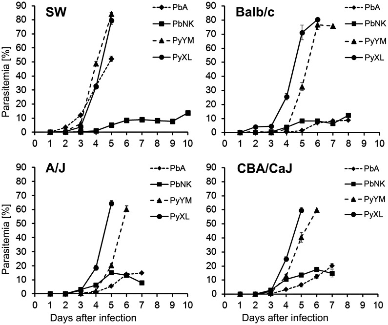
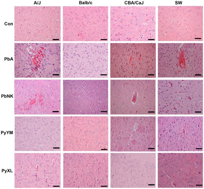
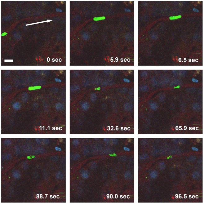
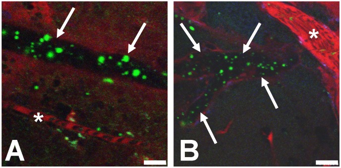
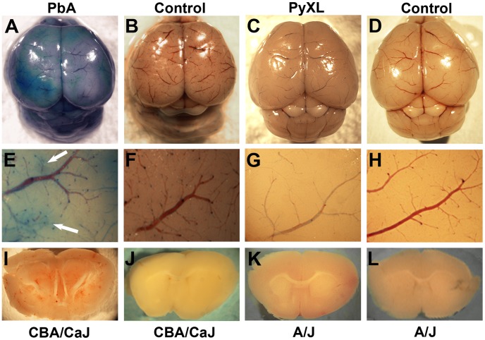
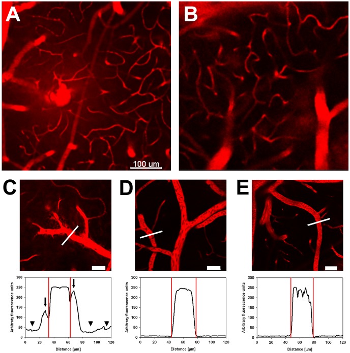
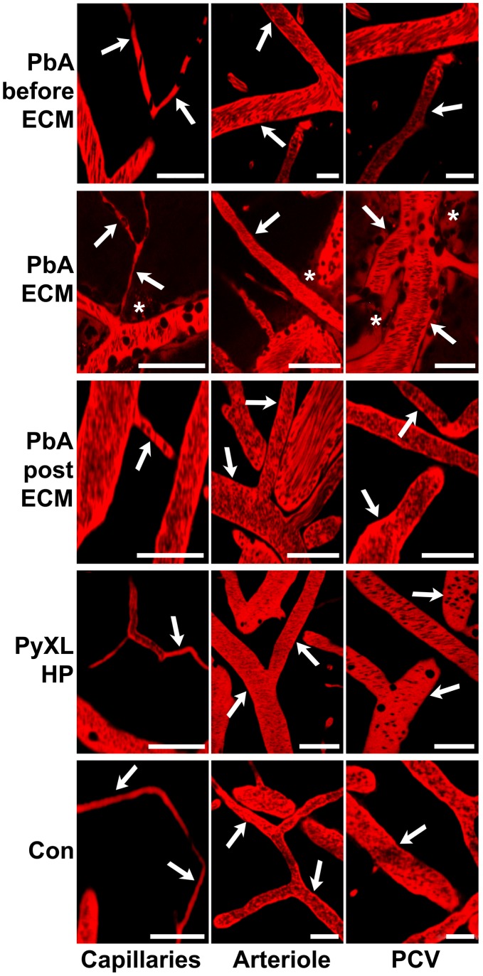
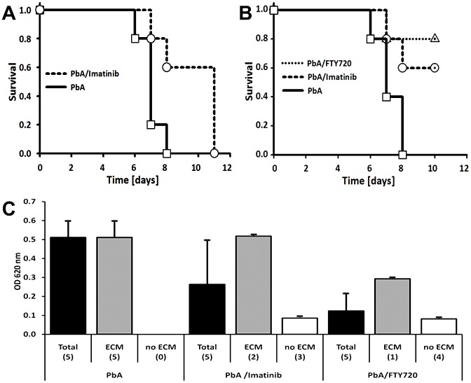
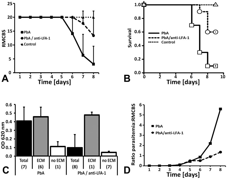
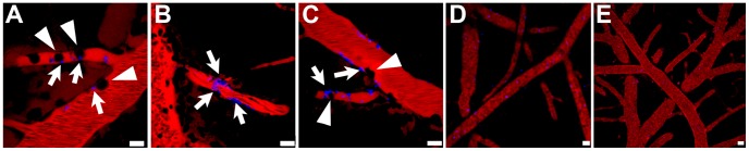
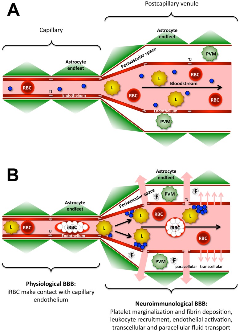
Plasmodium falciparum malaria is responsible for nearly one million annual deaths worldwide. Because of the difficulty in monitoring the pathogenesis of cerebral malaria in humans, we conducted a study in various mouse models to better understand disease progression in experimental cerebral malaria (ECM). We compared the effect on the integrity of the blood brain barrier (BBB) and the histopathology of the brain of P. berghei ANKA, a known ECM model, P. berghei NK65, generally thought not to induce ECM, P. yoelii 17XL, originally reported to induce human cerebral malaria-like histopathology, and P. yoelii YM. As expected, P. berghei ANKA infection caused neurological signs, cerebral hemorrhages, and BBB dysfunction in CBA/CaJ and Swiss Webster mice, while Balb/c and A/J mice were resistant. Surprisingly, PbNK induced ECM in CBA/CaJ mice, while all other mice were resistant. P. yoelii 17XL and P. yoelii YM caused lethal hyperparasitemia in all mouse strains; histopathological alterations, BBB dysfunction, or neurological signs were not observed. Intravital imaging revealed that infected erythrocytes containing mature parasites passed slowly through capillaries making intimate contact with the endothelium, but did not arrest. Except for relatively rare microhemorrhages, mice with ECM presented no obvious histopathological alterations that would explain the widespread disruption of the BBB. Intravital imaging did reveal, however, that postcapillary venules, but not capillaries or arterioles, from mice with ECM, but not hyperparasitemia, exhibit platelet marginalization, extravascular fibrin deposition, CD14 expression, and extensive vascular leakage. Blockage of LFA-1 mediated cellular interactions prevented leukocyte adhesion, vascular leakage, neurological signs, and death from ECM. The endothelial barrier-stabilizing mediators imatinib and FTY720 inhibited vascular leakage and neurological signs and prolonged survival to ECM. Thus, it appears that neurological signs and coma in ECM are due to regulated opening of paracellular-junctional and transcellular-vesicular fluid transport pathways at the neuroimmunological BBB.


Conflict of interest statement

The authors have declared that no competing interests exist.

Figures

Figure 1
Figure 1. Time course of parasitemia in young mice of different genetic background.
Groups of 6 three week-old SW, Balb/c, A/J, or CBA/CaJ mice were infected with 106 iRBC of the indicated parasite lines. As expected, infection with PbA resulted in a slow rise in parasitemia, which typically reached a plateau at below 20%. While PbA-infected SW mice developed ECM signs at around 50% parasitemia on day 5, CBA/CaJ mice displayed ECM signs on day 6 after infection. Balb/c and A/J were resistant. PbNK-infected CBA/CaJ mice also developed ECM. In contrast, infection with PyXL and PyYM resulted in a rapid rise in parasitemia between days 4 and 5 irrespective of the mouse strain. Mice were typically moribund by day 5 with >60% parasitemia. Data represented the mean parasitemia ± SD.
Figure 2
Figure 2. Histopathology of P. berghei and P. yoelii infection in different mouse strains.
Representative images of brains from groups of 3 A/J, Balb/c, CBA/CaJ, or SW mice, infected with PbA, PbNK, PyXL, or PyYM, as well as uninfected control brains were paraffin embedded and sections stained with H&E. PbA and PbNK infection caused focal vascular congestion and hemorrhages throughout the gray and white matter in the brains from A/J, CBA/CaJ, and SW, but not Balb/c mice. Brains from PbA-infected mice with symptomatic ECM exhibited congested microvessels containing leukocytes, which were restricted to the vascular lumen. Other than rare foci of large numbers of iRBC, the microvasculature of PyXL- or PyYM-infected brains exhibited no histological alterations in any of the mouse strains compared to uninfected controls. All images derive from coronal brain sections at Bregma and correspond to neocortical layer I tissue. Scale bars = 50 µm.
Figure 3
Figure 3. iRBC make intimate contact with capillary endothelia.
Selected frames from a two-minute IVM movie, which shows several late- and early-stage PbA-GFP iRBC traveling through a capillary. While iRBC harboring early-stage parasites (green, small) travel at blood velocity, iRBC containing late-stage parasites (green, large) squeeze slowly through the narrow capillary lumen, presumably due to the known increase in iRBC rigidity. Note the elongated shape of the two iRBC that contain large parasites, most likely trophozoites or schizonts. The vascular lumen is labeled with BSA-TX (red); nuclei are stained with Hoechst (blue). The arrow in the top left (0 sec) panel indicates the direction of blood flow. Scale bar = 10 µm. Video S3.
Figure 4
Figure 4. Microvascular occlusion can cause iRBC trapping.
A) A blocked larger vessel contains PbA-GFP iRBC (arrows), while a neighboring capillary (white star) displays normal blood flow. B) A large number of iRBC are visible in a blocked vessel (arrows) while a neighboring vessel (white star) exhibits normal flow. The vascular lumen was labeled with BSA-TX. Scale bars = 10 µm. Video S4 and S5.
Figure 5
Figure 5. Blood brain barrier integrity during ECM and HP.
A–D) Appearance of the cortical microvasculature of CBA/CaJ mice infected with PbA (A) no parasites (B) and A/J mice infected with PyXL (C) or no parasites (D). Mice were injected with Evans blue upon appearance of neurological signs (A) or reaching >50% parasitemia (B). Note that focal leakage of the vascular tracer into the parenchyma occurs during ECM (A), but not HP (C), and is absent from uninfected control mice. E–H) Higher magnification of the cortical microvasculature of mice infected with PbA (E), PyXL (G), or no parasites (F and H). Note the petechiae and diffuse Evans blue staining of the PbA-infected brain (E, arrows). I–L) Coronal slices of brains infected with PbA (I), PyXL (K), or no parasites (J and L). Numerous small hemorrhages are visible in both the gray and the white matter of the PbA-infected (I), but not the PyXL (K) or uninfected brains (J, L). The figure shows representative images of 3 mice per group.
Figure 6
Figure 6. Site of the ECM-associated BBB opening within the cerebral vascular tree.
CBA/CaJ mice were infected with PbA, PyXL, or no parasites. Microhemorrhages were occasionally found in the cortical vasculature of PbA-infected mice with ECM (A), but not PyXL-infected mice with HP (B). C–E, upper panels: C) PbA-infected mice exhibited extensive Evans blue leakage from PCV, but not capillaries, into the PVS and the parenchyma. In contrast, there was no Evans blue leakage into the cerebral parenchyma of PyXL (D) or age-matched uninfected control mice (E). C–E, lower panels: Graphic representation of Evans blue emission in relation to PCV from PbA, PyXL, and control mouse brains. The graphs show the Evans blue emission profile along the lines shown in (C–E). Note the emission peaks on either side of the PCV in the PbA-infected brain, which represent Evans blue leakage into the PVS (arrows). These peaks are absent in the PyXL-infected and control brain. Further, the overall Evans blue signal in the parenchyma surrounding the PCV is elevated in the PbA-infected brain (C, arrowheads) compared to the controls (D, E). The serrated shape of the control profile (E) is due to the presence of (dark) intravascular RBC. Scale bars = 100 µm (A, B), 50 µm (C–E).
Figure 7
Figure 7. Site of neuroimmunological BBB opening in symptomatic ECM mice.
Capillaries, arterioles, and PCV from PbA-infected CBA/CaJ mice before ECM (day 5), at the time of neurological signs (typically between days 6 and 7), and post ECM (day 8). The images show that leakage of Evans blue occurs only from PCV and only during, not before or after, symptomatic ECM. Neither PCV nor arterioles or capillaries from PyXL-infected mice with HP or uninfected control mice (Con) exhibit any leakage. Scale bars = 50 µm.
Figure 8
Figure 8. Vascular leakage is regulated and preventable.
Groups of 5 PbA-infected CBA/CaJ mice received two daily oral doses of 250 mg/kg imatinib on days 5, 6, and 7 post infection, one daily oral dose of 0.3 mg/kg FTY720 starting one day before infection, or 3) no treatment. A) While all PbA-infected untreated mice died with clear neurological signs between day 6 and 8, survival of the imatinib-treated mice was significantly prolonged. B) Whereas all untreated PbA-infected mice died from ECM between day 6 and 8, imatinib and FTY720 treatment allowed 80% and 60%, respectively, of the mice to survive until day 10 at which time they were sacrificed for quantification of Evans blue in the brain. C) OD620 measurement revealed Evans blue leakage into the parenchyma of the untreated controls and the mice that succumbed to ECM despite treatment, but not the treated mice that survived until day 10. The data represent mean OD620 values ± STD. Brain hemispheres were analyzed individually.
Figure 9
Figure 9. LFA-1 mediated cellular interactions contribute to BBB opening.
Five days after infection with PbA, CBA/CaJ mice were inoculated with 200 µg anti-LFA-1 mAb (N = 10) or no antibody (N = 10). A) Neurological signs based on RMCBS grading were significantly reduced in anti-LFA-1 treated PbA-infected mice compared to the untreated PbA-infected control mice. The data are represented as average ± SD. B) While 90% of the PbA-infected mice (N = 10) developed severe ECM between day 6 and 8, only 40% of the anti-LFA-1 treated mice (N = 10) showed neurological signs. C) Compared to PbA-infected untreated mice (N = 7), anti-LFA-1 treated PbA-infected mice (N = 8) exhibited a significant reduction in vascular leakage in the brain. Separate graphic representation of the 1 ECM-negative and 6 ECM-positive mouse brains from the PbA group and the 1 ECM-positive and 7 ECM-negative mouse brains from the PbA/anti-LFA-1 group revealed that neurological signs and vascular leakage are directly correlated. The data represent the average OD620 measurements from the two brain hemispheres from each mouse ± SD. Three of the 10 PbA-infected mice and 2 of the 10 PbA-infected/anti-LFA-1 treated mice died overnight and were excluded from analysis. D) Depiction of the parasitemia-to-RMCBS ratio accentuates the effect of LFA-1 blockage on the development of neurological signs.
Figure 10
Figure 10. Platelet arrest correlates with ECM, not HP.
CBA/CaJ mice were infected with PbA-GFP or PyXL-RFP iRBC and subjected to craniotomy at the onset of neurological signs or at >50% parasitemia, respectively. A–C) In PbA-infected mice, individual platelets or small aggregates (arrows) adhered to the endothelium of capillaries or PCV and remained stationary during the entire time of recording. Several blood cells (black, arrowheads in A) become arrested in a capillary suggesting that platelet deposits can act as foci for leukocyte arrest and temporary obstruction. D) In PyXL-infected mice, platelets (blue) circulate in large numbers at bloodstream velocity without adhering to the vascular endothelium. E) In uninfected control mice, platelets travel at bloodstream velocity and do not arrest. Scale bars = 10 µm. Video S9, S10, S11, S12.
Figure 11
Figure 11. Model for the response of the two BBB to ECM.
A) The cerebral microvascular tree contains two functionally distinct BBB. The physiological BBB is formed by capillaries (diameter 4–8 µm) and consists of a single layer composed of endothelia, gliovascular membrane, and astrocyte endfeet. The neuroimmunological BBB is formed by postcapillary venules (diameter 10–60 µm) and encompasses two layers that are separated by the perivascular space: 1) the endothelium with its basement membrane and 2) the glia limitans with associated astrocyte endfeet. While the physiological BBB serves as a diffusion barrier for solutes, the neuroimmunological BBB is characterized by higher permeability, which allows passage of macromolecules and diapedesis of immune cells into the perivascular space . B) Although iRBC come into close contact with capillary endothelia, the physiological BBB remains intact during ECM. iRBC do not bind to the wall of PCV, but platelet marginalization and leukocyte recruitment indicate that PCV endothelia become activated in response to the infection. Leukocyte adhesion to and crawling along the endothelium involve LFA-1/ICAM-1 mediated cellular interactions. Eventually, the neuroimmunological BBB opens, initially in a reversible fashion, thus allowing the leakage of plasma into the PVS. The fluid transport is regulated and occurs via paracellular-junctional (large arrows) and/or transendothelial-vesicular (small arrows) pathways in the absence of cell death. PVM = perivascular macrophage, L = leukocyte, blue circles = platelets.

Similar articles

Cited by

References

    1. Rogerson SJ, Grau GE, Hunt NH (2004) The microcirculation in severe malaria. Microcirculation 11: 559–576. - PubMed
    1. Newton CR, Taylor TE, Whitten RO (1998) Pathophysiology of fatal falciparum malaria in African children. Am J Trop Med Hyg 58: 673–683. - PubMed
    1. WHO (2000) Severe falciparum malaria. World Health Organization, Communicable Diseases Cluster. Trans R Soc Trop Med Hyg 94 Suppl 1: S1–90. - PubMed
    1. Idro R, Marsh K, John CC, Newton CR (2010) Cerebral malaria: mechanisms of brain injury and strategies for improved neurocognitive outcome. Pediatr Res 68: 267–274. - PMC - PubMed
    1. Haldar K, Murphy SC, Milner DA, Taylor TE (2007) Malaria: mechanisms of erythrocytic infection and pathological correlates of severe disease. Annu Rev Pathol 2: 217–249. - PubMed

Publication types

MeSH terms