. 2012;8(10):e1002987.
doi: 10.1371/journal.ppat.1002987. Epub 2012 Oct 25.

Myeloid-related protein-14 contributes to protective immunity in gram-negative pneumonia derived sepsis

Affiliations

Myeloid-related protein-14 contributes to protective immunity in gram-negative pneumonia derived sepsis

Ahmed Achouiti et al. PLoS Pathog. 2012.

Abstract

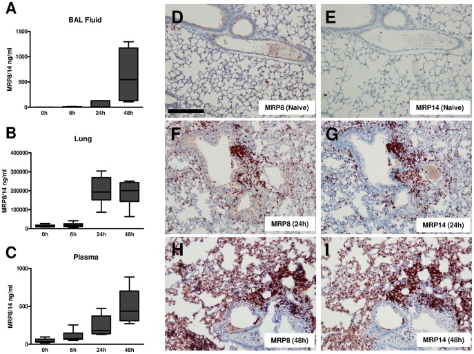
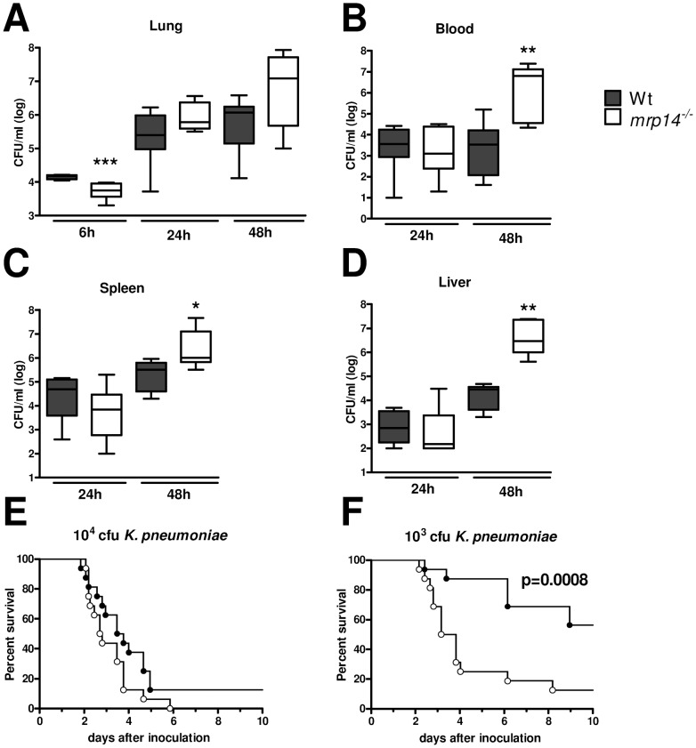
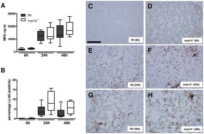
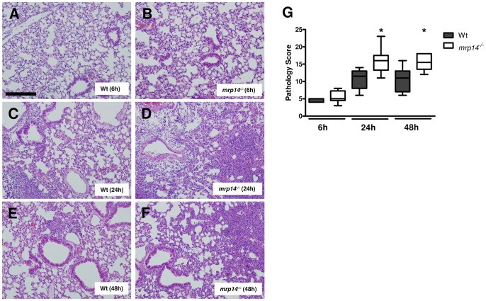
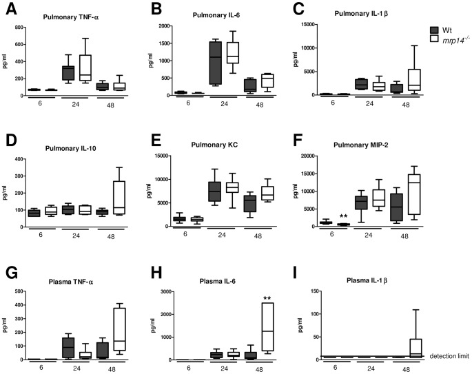
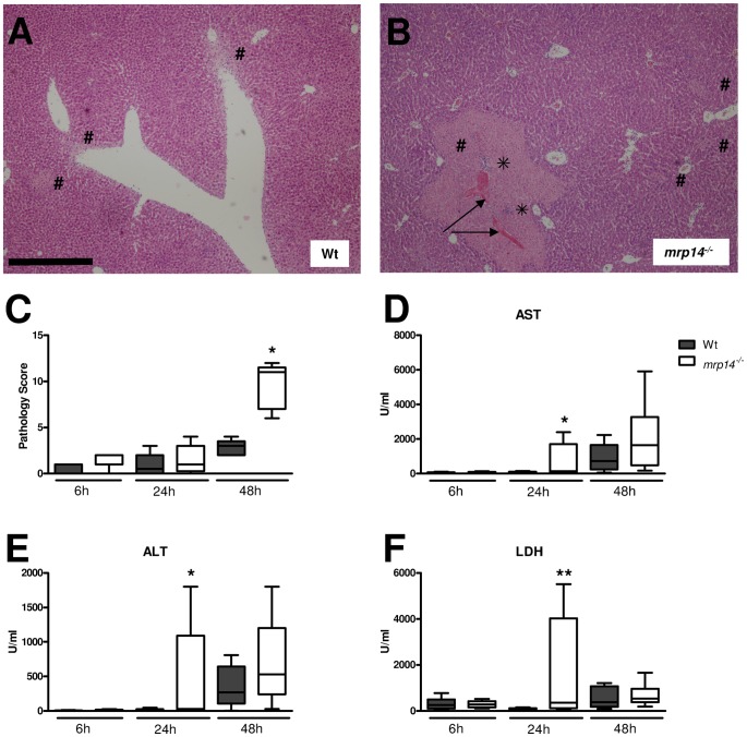
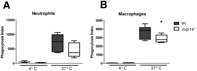
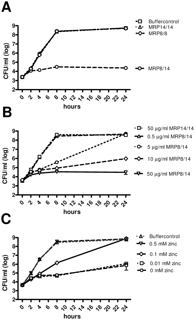
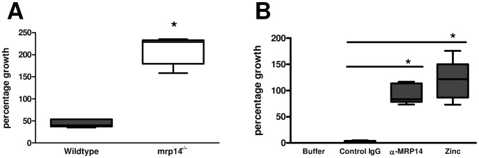
Klebsiella (K.) pneumoniae is a common cause of pneumonia-derived sepsis. Myeloid related protein 8 (MRP8, S100A8) and MRP14 (S100A9) are the most abundant cytoplasmic proteins in neutrophils. They can form MRP8/14 heterodimers that are released upon cell stress stimuli. MRP8/14 reportedly exerts antimicrobial activity, but in acute fulminant sepsis models MRP8/14 has been found to contribute to organ damage and death. We here determined the role of MRP8/14 in K. pneumoniae sepsis originating from the lungs, using an established model characterized by gradual growth of bacteria with subsequent dissemination. Infection resulted in gradually increasing MRP8/14 levels in lungs and plasma. Mrp14 deficient (mrp14(-/-)) mice, unable to form MRP8/14 heterodimers, showed enhanced bacterial dissemination accompanied by increased organ damage and a reduced survival. Mrp14(-/-) macrophages were reduced in their capacity to phagocytose Klebsiella. In addition, recombinant MRP8/14 heterodimers, but not MRP8 or MRP14 alone, prevented growth of Klebsiella in vitro through chelation of divalent cations. Neutrophil extracellular traps (NETs) prepared from wildtype but not from mrp14(-/-) neutrophils inhibited Klebsiella growth; in accordance, the capacity of human NETs to kill Klebsiella was strongly impaired by an anti-MRP14 antibody or the addition of zinc. These results identify MRP8/14 as key player in protective innate immunity during Klebsiella pneumonia.


Conflict of interest statement

The authors have declared that no competing interests exist.

Figures

Figure 1
Figure 1. K. pneumoniae pneumonia results in an increase of endogenous MRP8/14 levels and enhanced MRP8 and MRP14 expression in lungs.
MRP8/14 levels in BAL fluid (A), whole lung homogenates (B), and plasma (C) were determined in naive mice (n = 4) and 6, 24 and 48 hours after intranasal K. pneumoniae infection (n = 6–8). Data are expressed as box-and-whisker diagrams depicting the smallest observation, lower quartile, median, upper quartile and largest observation. MRP8 and MRP14 were stained in lungs of naive and infected mice. Shown here are representative slides of MRP8 (D) and MRP14 (E) staining of naive mice, MRP8 (F) and MRP14 (G) staining 24 hours and MRP8 (H) and MRP14 (I) staining 48 hours after infection. Scalebar indicates 200 µm.
Figure 2
Figure 2. Mrp14−/− mice show enhanced bacterial dissemination and increased mortality during pneumonia derived sepsis caused by K. pneumoniae.
Bacterial loads in the lung (A), blood (B), spleen (C) and liver (D) of K. pneumoniae in Wt (grey) and mrp14−/− mice (white). Data are expressed as box-and-whisker diagrams depicting the smallest observation, lower quartile, median, upper quartile and largest observation (8 mice per group at each time point). * p<0.05, ** p<0.01 versus Wt mice at the same time point. Survival of Wt and mrp14−/− mice after intranasal inoculation of 10.000 (E) or 1.000 cfu (F) (n = 12–16 per group in each experiment).
Figure 3
Figure 3. Neutrophil influx in Klebsiella pneumonia is not influenced by MRP14 deficiency.
MPO levels in whole lung homogenates (A) and quantitation of pulmonary Ly-6G positivity 6, 24 and 48 hours after infection (B) in Wt (grey) and mrp14−/− mice (white). Data are expressed as box-and-whisker diagrams depicting the smallest observation, lower quartile, median, upper quartile and largest observation (8 mice per group at each time point). There were no statistically significant differences between the groups. Representative neutrophil stainings (brown) of Wt (C) and mrp14−/− mice (D) 6 hours, Wt (E) and mrp14−/− mice (F) 24 hours and Wt (G) and mrp14−/− mice (H) 48 hours after induction of K. pneumoniae pneumonia.. Scalebar indicates 200 µm.
Figure 4
Figure 4. Mrp14−/− mice show enhanced lung pathology during K. pneumoniae pneumonia.
Representative slides of lung haematoxylin and eosin (HE) staining of Wt (A) and mrp14−/− mice (B) 6 hours, Wt (C) and mrp14−/− mice (D) 24 hours and Wt (E) and mrp14−/− mice (F) 48 hours after intranasal K. pneumoniae infection. Scalebar indicates 200 µm. Total pathology score at indicated time points post infection in Wt (grey) and mrp14−/− mice (white) was determined according to the scoring system described in the Methods section (G). Data are expressed as box-and-whisker diagrams depicting the smallest observation, lower quartile, median, upper quartile and largest observation (8 mice per group at each time point). * p<0.05 versus Wt mice at the same time point.
Figure 5
Figure 5. Cytokine and chemokine levels in lungs and plasma.
Lung cytokine (TNF-α, L-1β, IL-6, IL-10) (A–D), chemokine (KC and MIP-2) (E–F) and plasma cytokine (TNF-α, L-1β, IL-6 ) levels (G–I), 6, 24 and 48 hours after intranasal K. pneumoniae infection in Wt (grey) and mrp14−/− mice (white). Data are expressed as box-and-whisker diagrams depicting the smallest observation, lower quartile, median, upper quartile and largest observation (8 mice per group at each time point). * p<0.05, ** p<0.01 versus Wt mice.
Figure 6
Figure 6. Mrp14−/− mice show enhanced hepatocellular injury during pneumonia derived sepsis caused by Klebsiella.
Representative slides of liver haematoxylin and eosin HE staining of Wt (A) and mrp14−/− mice (B) 48 hours post infection. Livers from mrp14−/− mice displayed more advanced liver necrosis (#) accompanied with (micro) abscesses (<$>\vskip -1pt\scale 70%\raster="rg1"<$>). Arrows indicate fibrin deposits as a sign of thrombosis. Scalebar indicates 0.5 mm. Total pathology score was determined at indicated time points in Wt (grey) and mrp14−/− mice (white) according to the scoring system described in the Methods section (C). Aspartate aminotransferase (AST) (D), alanine aminotransferase (ALT) (E), and lactate dehydrogenase (LDH) (F) were measured in plasma. Data are expressed as box-and-whisker diagrams depicting the smallest observation, lower quartile, median, upper quartile and largest observation (8 mice per group at each time point). *p<0.05, **p<0.01 versus Wt mice at the same time point.
Figure 7
Figure 7. Phagocytosis is impaired in MRP14 deficient macrophages.
Growth arrested, CFSE-labelled K. pneumoniae were incubated with peripheral blood neutrophils (A) or macrophages (B) from Wt (grey) and mrp14−/− mice (white) at 4°C (n = 3–4 per mouse strain) or 37°C (n = 6–8 per mouse strain) for 20 and 60 minutes respectively after which phagocytosis was quantified (see Materials and Methods). Data are expressed as box-and-whisker diagrams depicting the smallest observation, lower quartile, median, upper quartile and largest observation (8 mice per group at each time point). *p<0.05.
Figure 8
Figure 8. MRP8/14 reduces bacterial growth of K. pneumoniae through metal chelation.
Growth of K. pneumoniae was assessed for a maximum of 24 hours in the presence of recombinant MRP8/14, MRP8 homodimer or MRP14 homodimer (50 µg/ml) (A). Bacterial growth was dose dependently inhibited by MRP8/14 (B); the growth inhibiting effect of MRP8/14 (10 µg/ml) was reversed by the addition of zinc (C). Data are means ± SEs of at least 3 replicates and representative of triplicate experiments.
Figure 9
Figure 9. Growth inhibition of Klebsiella by mouse and human NETs is MRP8/14 dependent.
3×10⁵ mouse neutrophils isolated from Wt and mrp14−/− mice were induced to make neutrophil extracellular traps (NETs) and subsequently infected with 5000 cfu K. pneumoniae. Cfu counts were determined after incubation of 7 hours at 37°C (A). 5×10⁵ human neutrophils were induced to make NETs and pretreated with a rabbit polyclonal anti-MRP14 antibody (α-MRP14), an unspecific rabbit polyclonal control antibody (control IgG) or an excess of zinc and then infected with 100 cfu Klebsiella. Cfu counts were determined after incubation of 10 hours (B). Percentage bacterial growth in the presence of NETs was calculated based on bacterial counts relative to medium controls without NETs. Data are expressed as box-and-whisker diagrams depicting the smallest observation, lower quartile, median, upper quartile and largest observation of at least 5 replicates. *p<0.05 versus controls.

Similar articles

Cited by

References

    1. Kollef MH, Kollef KE (2005) Antibiotic utilization and outcomes for patients with clinically suspected ventilator-associated pneumonia and negative quantitative BAL culture results. Chest 128: 2706–2713. - PubMed
    1. Podschun R, Ullmann U (1998) Klebsiella spp. as nosocomial pathogens: epidemiology, taxonomy, typing methods, and pathogenicity factors. Clin Microbiol Rev 11: 589–603. - PMC - PubMed
    1. Kang CI, Song JH, Chung DR, Peck KR, Ko KS, et al. (2011) Risk factors and pathogenic significance of severe sepsis and septic shock in 2286 patients with gram-negative bacteremia. J Infect 62: 26–33. - PubMed
    1. Shorr AF, Tabak YP, Killian AD, Gupta V, Liu LZ, et al. (2006) Healthcare-associated bloodstream infection: A distinct entity? Insights from a large U.S. database. Crit Care Med 34: 2588–2595. - PubMed
    1. Giamarellou H (2005) Multidrug resistance in Gram-negative bacteria that produce extended-spectrum beta-lactamases (ESBLs). Clin Microbiol Infect 11 Suppl 4: 1–16. - PubMed

Publication types

MeSH terms