. 2012;8(11):e1002985.
doi: 10.1371/journal.ppat.1002985. Epub 2012 Nov 1.

Prion pathogenesis is faithfully reproduced in cerebellar organotypic slice cultures

Affiliations

Prion pathogenesis is faithfully reproduced in cerebellar organotypic slice cultures

Jeppe Falsig et al. PLoS Pathog. 2012.

Abstract

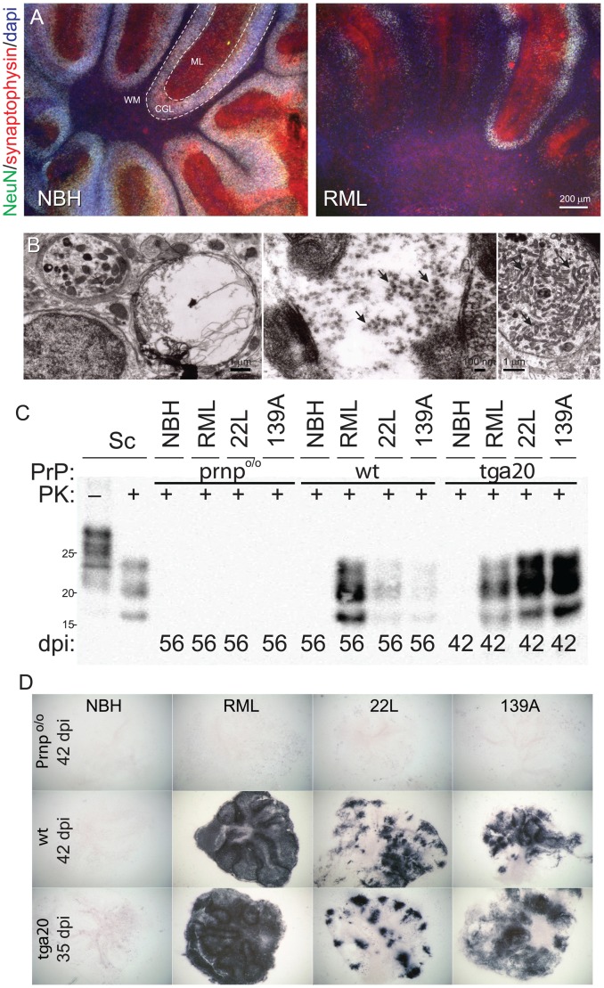
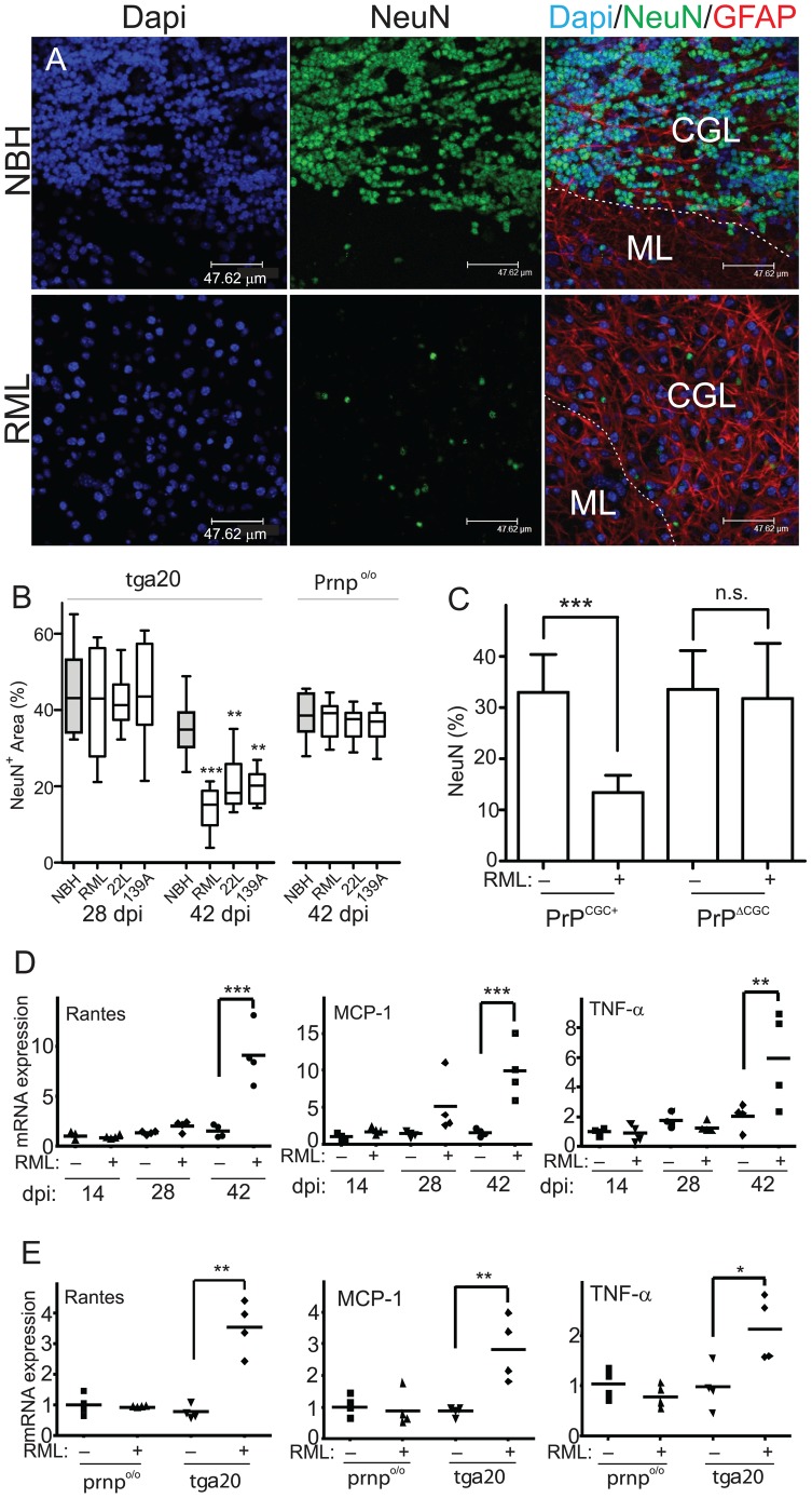
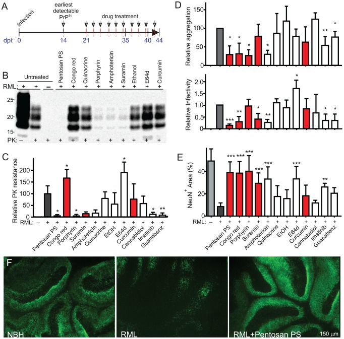
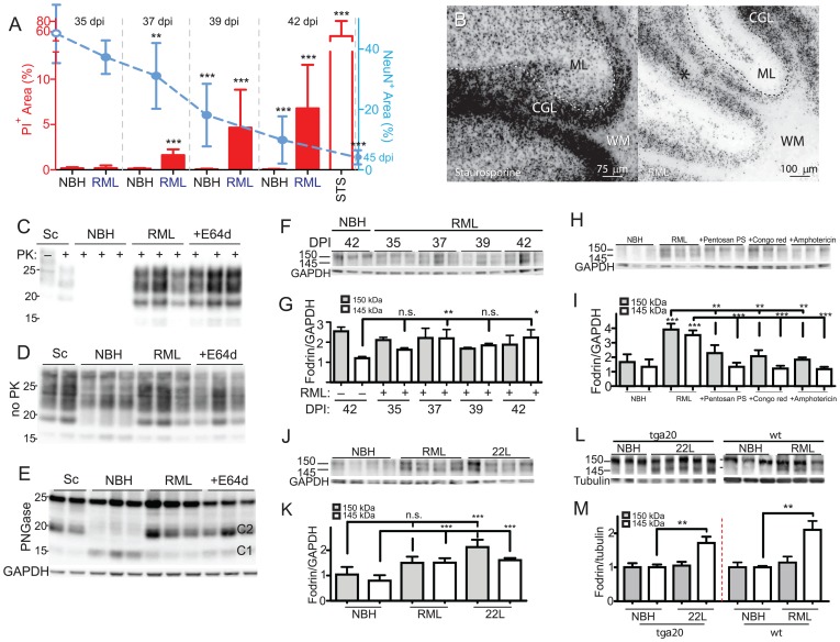
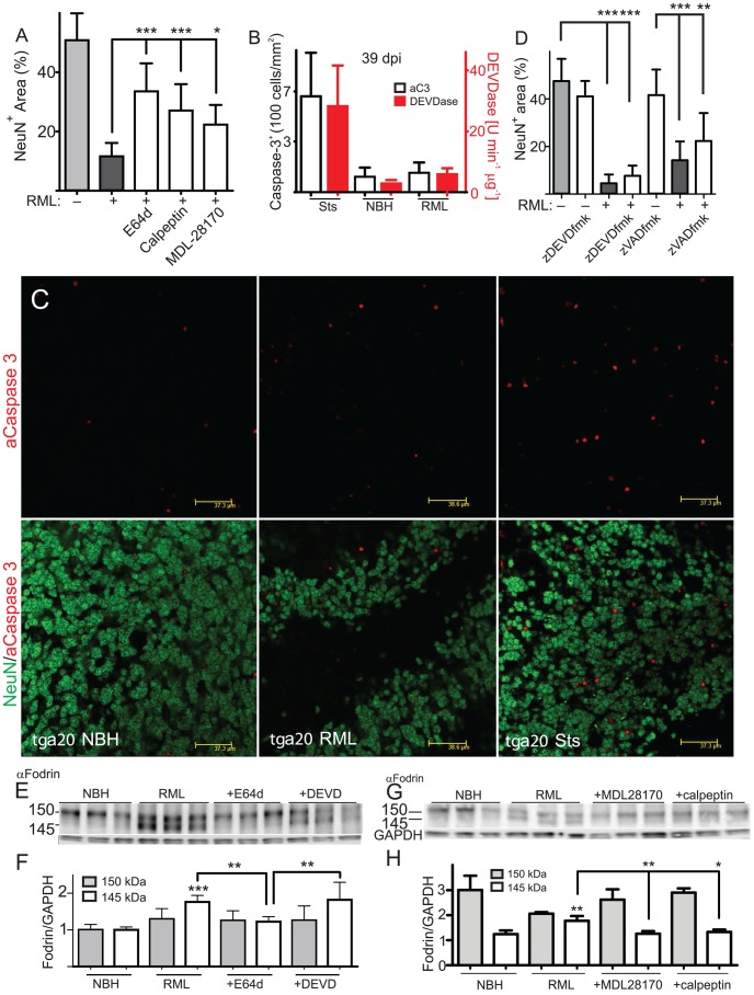
Prions cause neurodegeneration in vivo, yet prion-infected cultured cells do not show cytotoxicity. This has hampered mechanistic studies of prion-induced neurodegeneration. Here we report that prion-infected cultured organotypic cerebellar slices (COCS) experienced progressive spongiform neurodegeneration closely reproducing prion disease, with three different prion strains giving rise to three distinct patterns of prion protein deposition. Neurodegeneration did not occur when PrP was genetically removed from neurons, and a comprehensive pharmacological screen indicated that neurodegeneration was abrogated by compounds known to antagonize prion replication. Prion infection of COCS and mice led to enhanced fodrin cleavage, suggesting the involvement of calpains or caspases in pathogenesis. Accordingly, neurotoxicity and fodrin cleavage were prevented by calpain inhibitors but not by caspase inhibitors, whereas prion replication proceeded unimpeded. Hence calpain inhibition can uncouple prion replication from its neurotoxic sequelae. These data validate COCS as a powerful model system that faithfully reproduces most morphological hallmarks of prion infections. The exquisite accessibility of COCS to pharmacological manipulations was instrumental in recognizing the role of calpains in neurotoxicity, and significantly extends the collection of tools necessary for rigorously dissecting prion pathogenesis.


Conflict of interest statement

The authors have declared that no competing interests exist.

Figures

Figure 1
Figure 1. Localization and consequences of prion replication in COCS.
(A) Fluorescence micrographs showing profound loss of NeuN+ cerebellar granule cells and of synaptophysin in wt COCS challenged with RML prions (56 dpi, right panel). No neuronal damage was detected in COCS exposed to non-infectious brain homogenate (NBH; left panel). WM: white matter. ML: molecular layer. CGL: cerebellar granule cell layer. (B) Electron microscopy showing membrane-enclosed intraneuronal spongiform vacuoles (left), tubulovesicular structures (PrP-deficient spheres measuring between 20 and 40 nm in diameter, arrow, middle), and degenerating axons accumulating intra-cellular organelles including mitochondria (arrow, right) in RML-infected wt slices at 56 dpi. (C) Immunoblots showing PrPSc in tga20 and wt, but not Prnpo/o COCS exposed to prions (RML, 22L, 139A) or NBH. Sc; scrapie-sick wt mouse brain homogenate, used as positive control. (D) Histoblots showing strain-specific differences in PrPSc deposition patterns of tga20 and wt COCS. No PrPSc signal was observed in Prnpo/o COCS and in PrP-expressing COCS exposed to NBH.
Figure 2
Figure 2. Prion-induced neurodegeneration ex vivo.
(A) COCS were stained with IgG1 antibodies to NeuN (green) and GFAP (red) and counterstained with DAPI (blue). Representative images were recorded by confocal laser-scanning microscopy approximately 5 µm below the tissue surface using a 40× oil-immersion lens. Prion infection elicited severe NeuN+ cell loss. (B) Morphometric quantification of NeuN coverage in COCS prepared from tga20 and Prnpo/o mice. The loss of NeuN immunoreactivity was progressive over time (left graph). No NeuN loss was seen in PrPC-deficient COCS (right graph). Statistical shorthand here and henceforth: *: p<0.05; **: p<0.01; ***: p<0.001. (C) In contrast to control COCS (PrPCGC+), COCS prepared from mice with conditional PrP-ablation in CGCs (PrPΔCGC) showed no prion toxicity at 56 dpi. (D) Tga20 cultures exposed to NBH (−) or RML (+) were analyzed at various time points by quantitative reverse-transcription PCR (qPCR) for Rantes, MCP-1 and TNFα. ΔCt values were normalized to actin, with 1 being the ratio in uninfected cultures at 14 dpi ± s.d. Each data point is the average of 4 biological replicas. (E) Tga20 and Prnpo/o cultures treated with NBH (−) or RML (+) were harvested at 42 dpi and analyzed by qPCR for Rantes, MCP-1 and TNFα. ΔCt values were normalized to actin, with 1 being the ratio in untreated Prnpo/o COCS.
Figure 3
Figure 3. Anti-prion compounds.
(A) Timeline of the pharmacological experiments in tga20 COCS. Drug treatments were initiated at 21 dpi and re-added with each medium change (arrows). Longitudinal analyses revealed no cell loss at <35 dpi. Prion replication was assessed at 35 dpi, actively ongoing cell death at 39–42 dpi (PI; fodrin; TUNEL) and severe neuronal loss at 42–45 dpi (NeuN). (B) Representative PrPSc immunoblots of wt COCS challenged with RML prions (+) or NBH (−) and treated with compounds at 21–35 dpi. Blots were probed with POM1. (C) Total PrPSc (all PrPSc bands) was quantified densitometrically and compared to untreated RML-infected COCS (grey). Red: compounds which were reported to interact with PrPC or PrPSc. (D) Misfolded protein assay (upper panel) and scrapie cell assay (lower panel) of all wt samples from C. Relative aggregation and infectivity: values were compared to untreated RML-infected COCS ± s.d. (EF) NeuN morphometry (lower panel) of tga20 slices exposed to RML or NBH and treated with compounds from 21–42 dpi. All compounds reducing aggregation and/or infectivity were neuroprotective except guanabenz. In contrast, E64d was neuroprotective although it enhanced prion infectivity. (F) NeuN staining showing CGL damage by RML infection (middle) and its prevention by pentosan polysulphate (right).
Figure 4
Figure 4. Temporal aspects of prion toxicity.
(AB) Tga20 slices were exposed to RML or NBH, cultured and scored for PI incorporation (red bars) and NeuN+ morphometry (cyan dashed line) at 35, 37, 39, 42 and 45 dpi (RML; closed circles, NBH 45 dpi; open circle). The progressive appearance of PI+ cells coincided with NeuN degradation, suggesting that dying cells were efficiently removed. Sts: staurosporine treatment (48 hrs, 5 µM). (B) Fluorescent images of PI-stained COCS (42 dpi). For better contrast, a non-linearly modified negative of the image is shown. PI+ cells are mostly located in the CGL. (CE) Treatment of tga20 RML cultures with tool compounds was initiated at 21 dpi. COCS were harvested at 39 dpi (n = 3). Homogenates were treated with PK (C), left untreated (D) or treated with PNGase (E), and Western blots were probed with POM1 to detect PrPSc, total PrP, or total unglycosylated PrP (full-length and C1/C2 proteolytic fragments). The GAPDH band in panel E is representative for D, and samples were loaded as in panel C. (F) Western blots of infected tga20 COCS (35–42 dpi; n = 3) showing increased 145 kDa α-fodrin cleavage. (G) Densitometric quantitation of α-fodrin cleavage fragments shown in panel F (ordinate: ratio of α-fodrin/GAPDH). α-Fodrin cleavage peaked at 37–42 dpi. (HI) Western blots (H) and densitometric analysis (I) of infected tga20 COCS treated with anti-prion compounds (21–40 dpi) showing increasing 150/145 kDa α-fodrin cleavage in RML samples and reversal by anti-prion compounds. Densitometry readings were normalized against GAPDH. (JK) Western blots (J) and densitometric analysis (K) of NBH, RML and 22L treated tga20 COCS at 42 dpi, showing significantly increased 145 kDa α-fodrin cleavage in RML and 22L samples. Densitometry readings were normalized against GAPDH. (LM) Western blots (L) and densitometric analysis (M) of brain tissue from terminally sick 22L-infected tga20 mice and NBH-injected mice (left half of graph) or RML and NBH-treated wt mice (right half), showing significantly increased 145 kDa α-fodrin cleavage in infected samples. The left panel of consist of two cropped strips, cropped from the same blot at the same exposure. Densitometry readings were normalized against tubulin and normalized to NBH samples.
Figure 5
Figure 5. Calpain-mediated prion toxicity.
(A) Treatment of RML infected tga20 COCS with three calpain inhibitors from 21–44 dpi significantly antagonized neuronal loss in prion-infected tga20 COCS. Grey bars: untreated NBH (−) and RML (+) samples. (B, C) Tga20 cultures were harvested at 39 dpi and stained for activated caspase-3 (white bars) or homogenized and assayed for DEVDase activity (red bars). In contrast to staurosporine treatment (STS; 24 h), prion infection did not significantly increase DEVDase and aC3+ cells. Average DEVDase activity in COCS (4 pools of 20 slices each) normalized to the protein amount ± s.d. (C) Representative images of B. (D) Tga20 slices were treated with z-DEVD-fmk or zVAD-fmk from 21–44 dpi and analyzed by morphometric analysis. Grey bars: untreated. (EF) Tga20 cultures were treated with calpain and caspase inhibitors from 21–41 dpi, and probed for α-fodrin (n = 6–7). GAPDH: loading control. (F) Densitometric quantification showing that α-fodrin cleavage was enhanced by RML prion infection, and suppressed by E64d, but not by caspase inhibition (DEVD). (GH) Tga20 cultures were treated with calpain inhibitors as in E, and probed for α-fodrin (n = 3). GAPDH: loading control. (H) Densitometric quantification showing that α-fodrin cleavage was enhanced by RML prion infection, and suppressed by MDL-28170 and calpeptin.

Similar articles

Cited by

References

    1. Prusiner SB (1982) Novel proteinaceous infectious particles cause scrapie. Science 216: 136–144. - PubMed
    1. Büeler HR, Aguzzi A, Sailer A, Greiner RA, Autenried P, et al. (1993) Mice devoid of PrP are resistant to scrapie. Cell 73: 1339–1347. - PubMed
    1. Knowles TP, Waudby CA, Devlin GL, Cohen SI, Aguzzi A, et al. (2009) An analytical solution to the kinetics of breakable filament assembly. Science 326: 1533–1537. - PubMed
    1. Bremer J, Baumann F, Tiberi C, Wessig C, Fischer H, et al. (2010) Axonal prion protein is required for peripheral myelin maintenance. Nat Neurosci 13: 310–318. - PubMed
    1. Büeler HR, Fischer M, Lang Y, Bluethmann H, Lipp HP, et al. (1992) Normal development and behaviour of mice lacking the neuronal cell-surface PrP protein. Nature 356: 577–582. - PubMed

Publication types

MeSH terms