. 2012;8(11):e1002986.
doi: 10.1371/journal.ppat.1002986. Epub 2012 Nov 1.

A heparan-dependent herpesvirus targets the olfactory neuroepithelium for host entry

Affiliations

A heparan-dependent herpesvirus targets the olfactory neuroepithelium for host entry

Ricardo Milho et al. PLoS Pathog. 2012.

Abstract

Herpesviruses are ubiquitous pathogens that cause much disease. The difficulty of clearing their established infections makes host entry an important target for control. However, while herpesviruses have been studied extensively in vitro, how they cross differentiated mucus-covered epithelia in vivo is unclear. To establish general principles we tracked host entry by Murid Herpesvirus-4 (MuHV-4), a lymphotropic rhadinovirus related to the Kaposi's Sarcoma-associated Herpesvirus. Spontaneously acquired virions targeted the olfactory neuroepithelium. Like many herpesviruses, MuHV-4 binds to heparan sulfate (HS), and virions unable to bind HS showed poor host entry. While the respiratory epithelium expressed only basolateral HS and was bound poorly by incoming virions, the neuroepithelium also displayed HS on its apical neuronal cilia and was bound strongly. Incoming virions tracked down the neuronal cilia, and either infected neurons or reached the underlying microvilli of the adjacent glial (sustentacular) cells and infected them. Thus the olfactory neuroepithelium provides an important and complex site of HS-dependent herpesvirus uptake.


Conflict of interest statement

The authors have declared that no competing interests exist.

Figures

Figure 1
Figure 1. MuHV-4 infection localizes to the nasal septum and tubinates.
a. BALB/c mice were allowed to inhale spontaneously a 5 µl droplet containing 104 p.f.u. luciferase+ MuHV-4. 3 days later luciferase expression was identified by luciferin injection and CCD camera scanning of emitted light. The images shown are typical of >100 infections. Dissection localized the luciferase signal to a region occupied by the nasal septum/turbinates. b. BALB/c mice were infected and imaged as in a. Removing the septum/turbinates demonstrated that they accounted for all of the signal observed. Further dissection established that the vomeronasal organ lacked luciferase expression. Equivalent results were obtained in 10 mice. c. Luciferase+ MuHV-4 (106 p.f.u. in 5 µl) was applied with a small brush to the external genitalia of female mice. Over the following 2 weeks, 8/12 co-caged male and female mice acquired nasal luciferase signals. Dissection again localized the luciferase signal to a region occupied by the nasal septum/turbinates. Representative images are shown.
Figure 2
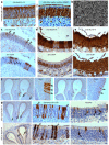
Figure 2. MuHV-4 infection localizes to the olfactory neuroepithelium.
a. BALB/c mice were allowed to inhale a 5 µl droplet containing 106 p.f.u. eGFP+ MuHV-4 and 3 days later analysed by immunostaining with a MuHV-4-specific polyclonal rabbit serum (brown) and counterstained with Mayer's Hemalum. The multi-layered main olfactory neuroepithelium and single-layered respiratory epithelium are indicated, as is the neuroepithelium of the vomeronasal organ (VNO). Equivalent staining was seen in >10 mice. Images enlarged ×5 in the right-hand panels show the sharp divide between virus+ neuroepithelium and virus respiratory epithelium; a representative area of (virus) squamous epithelium; and background staining of sections from naive mice. b An adjacent section was stained for viral eGFP (brown), which is expressed independently of lytic cycle genes. Again the main olfactory neuroepithelium was virus+ and other sites virus. c. BALB/c mice were infected with MuHV-4 (104 p.f.u. in 5 µl) and 3 days later analysed for viral tRNA/miRNA expression by in situ hybridization of nose sections with a digoxigenin-labelled riboprobe. The arrows show examples of positive neuroepithelial staining. The respiratory epithelium remained negative. Similar results were obtained in 3 mice. d. BALB/c mice were infected with MuHV-4 (106 p.f.u. in 5 µl) and 4 days later analysed either for viral tRNA/miRNA expression as in c, or for viral lytic antigen expression as in a. Arrows show examples of positive staining. e. BALB/c mice were allowed to inhale a 5 µl droplet containing 106 p.f.u. influenza A/PR/8/34. 1 day later they were analysed by immunostaining with a polyclonal influenza-specific rabbit serum that recognizes predominantly the viral hemagglutinin. Only the respiratory epithelium was virus+. 5 more mice gave similar results.
Figure 3
Figure 3. Both olfactory neurons and sustentacular cells are direct infection targets.
a. The neuroepithelium of a naive BALB/c mouse was stained for cytokeratin-18 to reveal sustentacular cells (brown) and for olfactory marker protein to reveal neurons plus apical neuronal cilia (brown). Sections were counter-stained with Mayer's Hemalum. b. A scanning electron micrograph of the murine neuroepithelium showing the dense apical network of neuronal cilia. c. BALB/c mice were allowed to inhale eGFP+ MuHV-4 (106 p.f.u.) and 1 day later analysed for viral lytic antigen (anti-MuHV-4) and eGFP expression with polyclonal sera (brown). The sections were counter-stained with Mayer's Hemalum. Filled arrowheads show examples of virus+ nuclei in the sustentacular cell layer; open arrowheads show examples of eGFP+ nuclei in the neuronal layer. d. At day 3 after infection as in c, the neuroepithelium was stained for MuHV-4 lytic antigens, or for OMP to show the distribution of neurons. Most virus+ nuclei were in the sustentacular cell layer between the neuronal nuclei and neuronal cilia. e. At day 3 after infection as in c, adjacent neuroepithelial sections were stained for MuHV-4 lytic antigens or virus-expressed eGFP. The former again showed positive nuclei mainly in the sustentacular cell layer, while the latter also stained nuclei throughout the neuronal layer. f. At day 1 after infection with eGFP+ MuHV-4 as in c, adjacent sections were stained for virus-expressed eGFP or lytic antigens. Each zoomed image shows the corresponding boxed region. The number of eGFP+ cells consistently exceeded by at least 3-fold the number of lytic antigen+ cells. The arrows show examples of positive staining. g. BALB/c mice were infected as in c, either with wild-type eGFP+ MuHV-4 or with an equivalent dose of ORF50 (replication-deficient) eGFP+ MuHV-4. Samples taken 1 day later were stained for virus-expressed eGFP with a polyclonal serum. The arrows show examples of positive staining, which conformed mainly to the distribution of sustentacular cells. To the right, arrowheads show examples of sustentacular and neuronal staining at higher magnification.
Figure 4
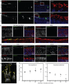
Figure 4. Direct neuronal and sustentacular infections also occur in infant mice.
a. 1 week old C57BL/6 mice - BALB/c and C57BL/6 strains gave equivalent data in both adults and juveniles - were allowed to inhale luciferase+ MuHV-4 (104 p.f.u. in 2 µl), and imaged for light emission 5 and 14 days later. The scale shows maximum radiance (photons sec−1 cm−2 steradian−1). The images are representative of >30 infections. b. Mice were infected as in a, then monitored serially by luciferin infection and CCD camera scanning of emitted light. Each point shows the result for 1 mouse. SCLN = superficial cervical lymph nodes (neck signal). The dashed line shows the lower limit of assay sensitivity. c. 1 week old C57BL/6 mice were allowed to inhale wild-type eGFP+ MuHV-4 (106 p.f.u. in 2 µl) and 4 days later analysed by immunostaining for virus-expressed eGFP. The zoomed image shows the boxed region. The lower image shows a further magnified region of neuroepithelium with both sustentacular and neuronal eGFP expression. Equivalent results were obtained in 6 mice. d. 1 week old C57BL/6 mice were infected as in c but with replication-defective ORF50eGFP+ MuHV-4 (106 p.f.u., assayed on complementing cells). Infection was identified 1 day later by immunostaining for virus-expressed eGFP. The zoomed image shows the boxed region, with arrowheads indicating examples of positive (sustentacular cell) staining. The lower image shows a further magnified region of neuroepithelium with eGFP+ neurons. Equivalent results were obtained in 6 mice. e. Immunofluorescent staining of naive 1 week old C57BL/6 mice shows the characteristic distributions of OMP (neuronal axons, dendrites and cell bodies), α-tubulin (broadly expressed, including neuronal cilia) and cytokeratin-18 (sustentacular cells). Nuclei were counterstained with DAPI. The merged image shows distinct layers of neuronal cell bodies, sustentacular cells and neuronal cilia. f. 1 week old C57BL/6 mice were allowed to inhale ORF50eGFP+ (106 p.f.u. with complementation) and 1 day later analysed by immunofluorescent staining for eGFP, cytokeratin-18 and OMP. Nuclei were counterstained with DAPI. Co-localization appears yellow in the merged images. Equivalent results were obtained in 3 mice.
Figure 5
Figure 5. Neuroepithelial infection is HS-dependent.
Adult C57BL/6 mice were allowed to inhale either wild-type, gLgp70 or gLgp70gp150 virions in 5 µl. As gLgp70 MuHV-4 plaques poorly, the wild-type virus was titrated by plaque assay and all other virus stocks normalized to this by immunoblotting with MuHV-4-specific mAbs (Fig.S2). Groups of 8 mice were then exposed to virus and 1 month later scored as infected or not based on ELISA for virus-specific serum IgG. At 101 and 102 p.f.u. equivalents, gLgp70 MuHV-4 infected significantly fewer mice than either the wild-type or the HS-independent triple mutant (p<0.03 by 2-tailed Fisher's exact test). The graph on the right illustrates ELISA results for 3 mice per group from the 102 p.f.u. equivalent dose, plus 3 naive controls. Note that the one mouse infected with gLgp70 virus shows an antibody response comparable to those infected with wild-type virus.
Figure 6
Figure 6. Identification of neuroepithelial HS.
a. The neuroepithelia of infant and adult mice were analysed for HS expression by immunostaining with mAbs F58-10E4 (HS, sulfated) and NAH46 (HS, non-sulfated). Counter-staining was with Mayer's Hemalum. The arrows show areas of positive staining (brown). Arrowheads show the junction between neuroepithelium and respiratory epithelium. b. Tissues from naive adult mice were immunostained for HS using mAb NAH46 (brown) and counterstained with Mayer's hemalum (blue). c. Naive 1 week old mice were analysed for neuroepithelial HS expression by immunofluorescence with mAbs NAH46 and F58-10E4, and for α-tubulin to show the neuronal cilia. Nuclei were counter-stained with DAPI. The zoomed image showed the boxed region of the merge. NAH46 staining occupied the same region as the cilia, although their co-localization (yellow) was only partial.
Figure 7
Figure 7. Neuroepithelial binding by virion glycoproteins.
The virion HS binding proteins gp70 and gH/gL were expressed as IgG Fc fusions and used to stain neuroepithelial and respiratory epithelial sections from 1 week old naive mice. α-tubulin staining was used to identify the neuronal cilia. The zoomed image shows the boxed region of the merge. Colocalisation appears yellow. Similar results were obtained with 3 mice. The arrows show apical epithelial staining by gHgL-Fc.
Figure 8
Figure 8. Virus binding to the olfactory neuroepithelium.
a. 1 week old C57BL/6 mice were allowed to inhale wild-type eGFP+ MuHV-4 (106 pf.u. in 2 µl). The next day neuroepithelial sections were analysed for viral lytic antigens and virus-expressed eGFP by immunofluorescent staining with polyclonal sera. Nuclei were counter-stained with DAPI. The zoomed image shows the boxed region of the merge. b. The same analysis was applied to ORF50eGFP+ MuHV-4, which without complementation expresses eGFP but not new lytic antigens. c. 1 week old C57BL/6 mice were allowed to inhale MuHV-4 (106 pf.u. in 2 µl) and 1 min later the same volume of PBS. They were then analysed by immunofluorescent staining of neuroepithelial sections for viral antigens. Neuronal cilia were visualised by staining for α-tubulin. Nuclei were counter-stained with DAPI. An equivalent section from a naive mouse is shown for comparison. d. Mice were infected as in c and examined for virus binding to the neuroepithelium and respiratory epithelium after 5 min or 45 min. The arrows at 45 min show viral antigen+ neurons. e. In a higher power image of the neuroepithelium 45 min after virus inhalation (106 pf.u. in 2 µl), the dashed yellow lines outline neurons, as defined by α-tubulin staining, and the arrows show aggregated virion antigens along neuronal dendrites. f. After infection and staining as in d, virus binding was quantitated by counting MuHV-4+ pixels over a fixed area of apical epithelium and then normalizing by the α-tubulin signal of the same area. Each point shows the result for 3 sections from 1 mouse. The horizontal bars show medians. Comparison by Student's 2 tailed unpaired t test showed that neuroepithelial and respiratory epithelial binding were not significantly different at 1 min post-inoculation (p = 0.08) but at all subsequent time points neuroepithelial binding was significantly greater (p<0.05).
Figure 9
Figure 9. Virus uptake at the olfactory neuroepithelium.
a. 1 week old C57BL/6 mice were allowed to inhale ORF50eGFP+ MuHV-4 (106 p.f.u. in 2 µl). The next day neuroepithelial sections were analysed for viral antigens and virus-expressed eGFP by immunofluorescent staining with polyclonal sera. Nuclei were counter-stained with DAPI. The zoomed image shows the boxed regions of the merge, with viral antigen+ neuronal cilia close to eGFP+ sustentacular cells. b. Mice were infected and analysed as in a. The zoomed images show eGFP+ sustentacular cells with viral antigen+ apical microvilli. The data are representative of >12 mice examined.
Figure 10
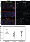
Figure 10. HS-dependent virion binding to the olfactory neuroepithelium.
a. 1 week old mice were allowed to inhale wild-type, gLgp70, or gLgp70gp150 virions (106 p.f.u. equivalents in 2 µl). 45 min later the nasal passages were rinsed by PBS inhalation and neuroepithelial sections examined for viral antigens by immunofluorescent staining with a polyclonal immune serum. α-tubulin staining was used to visualise the neuroepithelial surface. Nuclei were counter-stained with DAPI. The images are representative of data from 3 mice per group. b. Viral antigen staining, as in a, was quantitated by counting MuHV-4 antigen+ pixels over representative fixed areas, and dividing by the α-tubulin+ pixel number. Each point shows the result for 1 section. Sections were pooled from 3 mice per group. The horizontal bars show medians. By Student's 2-tailed t test, gLgp70 virion binding was significantly reduced relative to wild-type (p<0.005) whereas gLgp70gp150 virion binding was not (p = 0.3).

Similar articles

Cited by

References

    1. Bagni R, Whitby D (2009) Kaposi's sarcoma-associated herpesvirus transmission and primary infection. Curr Opin HIV AIDS 4: 22–26. - PubMed
    1. Faulkner GC, Krajewski AS, Crawford DH (2000) The ins and outs of EBV infection. Trends Microbiol 8: 185–189. - PubMed
    1. Hoagland RJ (1964) The incubation period of infectious mononucleosis. Am J Public Health Nations Health 54: 1699–1705. - PMC - PubMed
    1. Nash AA, Dutia BM, Stewart JP, Davison AJ (2001) Natural history of murine gamma-herpesvirus infection. Philos Trans R Soc Lond 356: 569–579. - PMC - PubMed
    1. Flaño E, Woodland DL, Blackman MA (2002) A mouse model for infectious mononucleosis. Immunol Res 25: 201–217. - PubMed

Publication types

Substances