. 2012;8(11):e1003003.
doi: 10.1371/journal.ppat.1003003. Epub 2012 Nov 1.

Expression of a cryptic secondary sigma factor gene unveils natural competence for DNA transformation in Staphylococcus aureus

Affiliations

Expression of a cryptic secondary sigma factor gene unveils natural competence for DNA transformation in Staphylococcus aureus

Kazuya Morikawa et al. PLoS Pathog. 2012.

Abstract

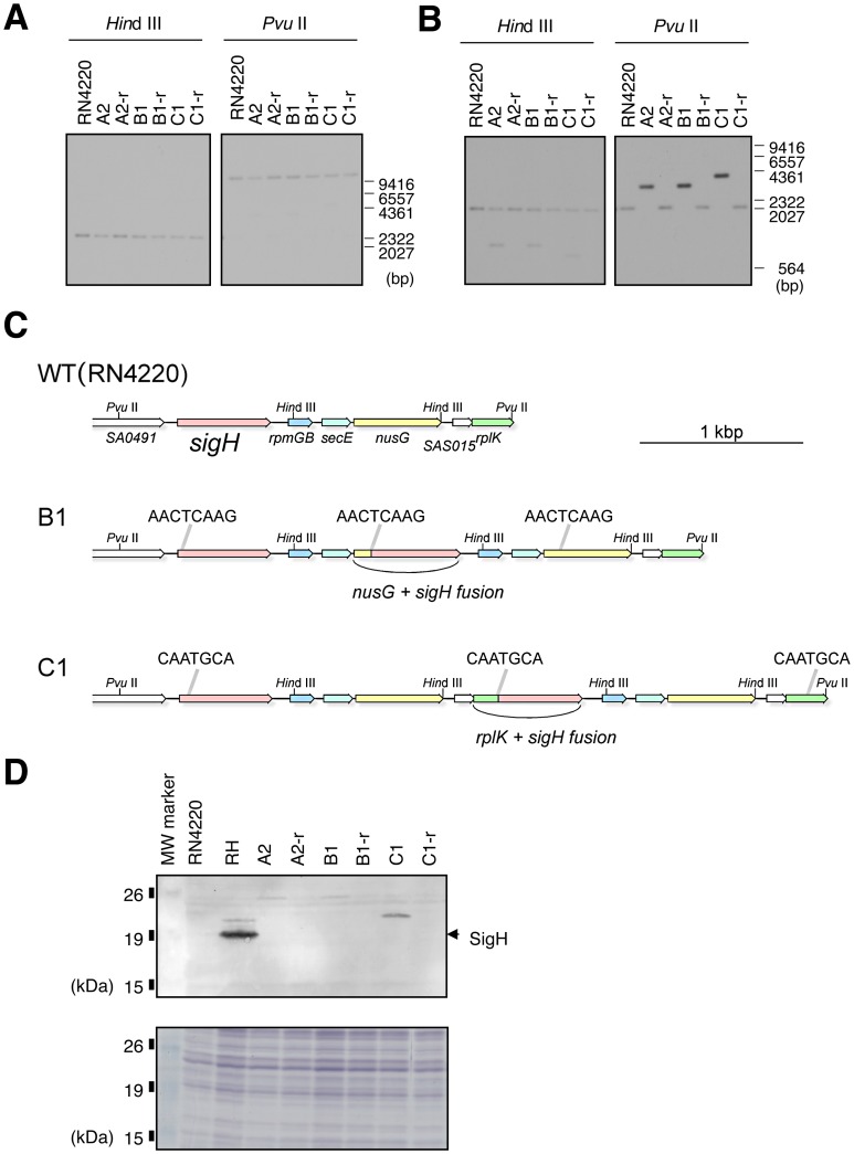
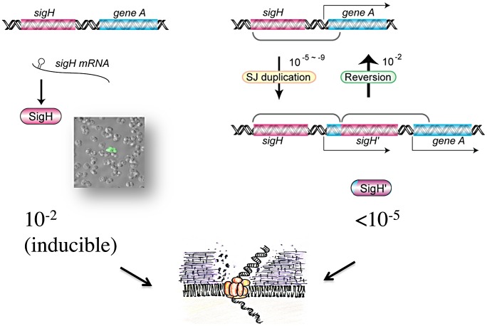
It has long been a question whether Staphylococcus aureus, a major human pathogen, is able to develop natural competence for transformation by DNA. We previously showed that a novel staphylococcal secondary sigma factor, SigH, was a likely key component for competence development, but the corresponding gene appeared to be cryptic as its expression could not be detected during growth under standard laboratory conditions. Here, we have uncovered two distinct mechanisms allowing activation of SigH production in a minor fraction of the bacterial cell population. The first is a chromosomal gene duplication rearrangement occurring spontaneously at a low frequency [≤10(-5)], generating expression of a new chimeric sigH gene. The second involves post-transcriptional regulation through an upstream inverted repeat sequence, effectively suppressing expression of the sigH gene. Importantly, we have demonstrated for the first time that S. aureus cells producing active SigH become competent for transformation by plasmid or chromosomal DNA, which requires the expression of SigH-controlled competence genes. Additionally, using DNA from the N315 MRSA strain, we successfully transferred the full length SCCmecII element through natural transformation to a methicillin-sensitive strain, conferring methicillin resistance to the resulting S. aureus transformants. Taken together, we propose a unique model for staphylococcal competence regulation by SigH that could help explain the acquisition of antibiotic resistance genes through horizontal gene transfer in this important pathogen.


Conflict of interest statement

The authors have declared that no competing interests exist.

Figures

Figure 1
Figure 1. Positive selection for Staphylococcus aureus cells with active SigH.
A) Positive selection tetracycline resistance (tet) reporter plasmid (pTet-rep). Reporter strains carrying pTet-rep were selected with 5 µg/ml tetracycline. cat: chloramphenicol resistance gene. bgaB: β-galactosidase gene. PcomG: promoter region of comG operon. PcomE: promoter region of comE operon. B) Fluctuation tests indicate that SigH activation occurs spontaneously. The y-axis represents the numbers of SigH active colonies detected from 109 cfu. Cells were grown in drug-free TSB and then selected for tetracycline resistance. Open symbols: aliquots from a single flask. Closed symbols: independent cultures in separate test tubes. Diamonds: N315 pTet-rep, Triangles: RNtet-rep (RN4220 pTet-rep). C) SigH activity can be stably maintained through generations without selection pressure. pTet-rep was cured from SigH active cells, and pRIT-PcomE-bgaB was introduced. Cells were grown on TSA plates containing 100 µg/ml X-gal and 12.5 µg/ml chloramphenicol. Rare white cells (about 1%) that lost SigH activity were also observed (see text).
Figure 2
Figure 2. Activation of sigH expression involves a chromosomal rearrangement with a tandem gene duplication/fusion.
A) and B) Southern blot analysis of the sigH locus for the parental strain (RN4220), SigH active mutants (A2, B1, C1) and revertants (A2-r, B1-r, C1-r). Genomic DNA was digested with HindIII or PvuII. A) Blots were probed with a H1p-H2p PCR fragment (upstream region of sigH). B) Probed with H5p-SA0492Rp PCR fragment (sigH coding sequence). Positions of the DNA molecular mass marker (λ HindIII) are indicated in base pairs on the right. C) Schematic representations of the sigH loci of RN4220 and SigH active derivatives B1 and C1. Arrows represent coding sequences, and are colored by gene, e.g. red represents sigH, yellow represents nusG etc. The direct repeat sequences encompassing duplication units are indicated above the gene maps. These direct repeats originally exist in the RN4220 sequence, and the duplication event increases their number from two (in RN4220) to three (in B1/C1). Gene maps are illustrated to scale and the bar represents 1 kbp. D) Western blot analysis showing that SigH is expressed as a fusion protein encoded by the new chimeric genes. A2, B1, and C1 are SigH active derivatives. A2-r, B1-r, and C1-r are the revertants that have lost SigH activity. R: RN4220, RH: RN4220 + pRITsigH. The position of full-length native SigH (calculated molecular weight 23.0 kDa) is indicated by an arrow-head. Signals in B1 and C1 were observed at higher molecular weight positions, in agreement with the calculated molecular weights of the SigH chimeric proteins in strains B1 and C1 (28.3 kDa and 25.5 kDa, respectively). Sizes and positions of the Bench Mark pre-stained protein ladder (Promega) are indicated on the left. The lower panel is a loading control SDS-PAGE gel stained with Coomassie Brilliant Blue staining.
Figure 3
Figure 3. SigH is activated under specific growth conditions.
A) Western blot analyses directed against GFP showing that SigH is not active in standard culture conditions. Strains N315aspGFP (asp23 promoter), N315comGFP (comG promoter) and N315-GFP (promoter-less) were grown aerobically in BHI or RPMI 1640 medium. B) Western blot against GFP showing that SigH activity is induced in CS2 medium. Incubation times (hours) are indicated above the panel. Positive control cells of N315aspGFP were grown in TSB for 10.5 h. C) Northern blot analysis confirming SigH-dependent expression of SA1365, one of the comG operon genes. Strains N315, NKSH (sigH inactivation mutant), and NKSHh (complemented strain) were grown in anaerobic and static conditions in -GS medium.
Figure 4
Figure 4. SigH is activated in a minor fraction of the cell population.
Confocal microscopy reveals that SigH-dependent GFP expression is limited to a minor fraction of the cell population. In N315comGFP, the GFP-positive frequency was 1.6+−0.6% (mean ± SD, n = 10; Panel A), while 100% of N315aspGFP cells expressed GFP (Panel B). Left panels: Fluorescent GFP signals detected by confocal microscopy. Right panels: Phase contrast images. Middle: merged images. A) N315comGFP grown in CS2 medium. GFP expression is observed only in a minor fraction of the cell population. The maximum frequency of GFP-positive cells was about 1%. B) N315aspGFP grown in BHI medium. All cells express GFP.
Figure 5
Figure 5. Two distinct mechanisms are responsible for SigH activation.
The translation initiation codon of sigH is required for SigH activation in CS2 medium, but not for SJ-dependent SigH activation. A) Nucleotide sequence of the sigH translation initiation site region. The expected translation initiation codon, and the downstream in-frame TTG codon are underlined. Sequences for the wild type strain (N315ex; top), and the constructed mutant (N315ex-sigH*; bottom) are shown and the mutated residues are shown in lower case. B) Western blot analysis of SigH-dependent GFP production in WT (N315ex) and N315ex-sigH* carrying pMK3-com-gfp. Cells were grown in CS2 medium for the indicated times. No GFP signal was detected in the translation initiation codon mutant strain (N315ex-sigH*). C) Rare GFP expressing cells are still detectable in N315ex-sigH* carrying pMK3-com-gfp.
Figure 6
Figure 6. SigH activity is restricted to a minor fraction of the cell population through post-transcriptional regulation.
A) Overexpression of sigH mRNA is not sufficient for SigH activation. N315ex carrying pMKcomGFP and pRIT-sigHNH7 was grown in CS2, LB, or BHI medium aerobically at 37°C for indicated time periods, and analyzed by Western blot. Only growth in CS2 medium led to GFP production. B–C) An inverted repeat sequence in the sigH 5′-UTR negatively controls its expression. B) Sequences of the 5′-UTR region in each sigH-expressing plasmid. Arrows show the 13 bp inverted repeat, and the translation initiation codon is underlined. PRIT-sigHNH7 carries the native nucleotide sequence. The inverted repeat sequence was partially deleted in pRIT-sigHIRd, whereas it was entirely removed and the SD sequence replaced with a consensus ribosome binding site in pRIT-sigH. C) Deletion of one half of the inverted repeat allows all cells to produce active SigH. N315ex derivatives carrying the plasmids indicated on the left were grown in CS2 or TSB at 37°C with shaking. Note that all cells of N315ex carrying pRIT-sigHIRd show GFP signals, even in TSB. D) Northern blot analysis confirmed the accumulation of sigH mRNA in cells carrying all of the different sigH expression plasmids. N315ex cells carrying the designated plasmid together with pMKcomGFP, were grown in TSB medium. Exponentially growing cells (OD600 = 0.5) were used for Northern blot analysis as described in Materials and Methods. The lower panel shows the EtBr stained agarose gel as a loading control.
Figure 7
Figure 7. Competence development in Staphylococcus aureus involves two distinct mechanisms.
A rare SJ-duplication mechanism generates a new chimeric sigH gene, and SigH is produced as a fusion protein. The duplication is cured at a high frequency, concomitant with the loss of gene duplication. Under the specific culture conditions used, SigH was expressed stochastically at a frequency of ca. 10−2, through a post-transcriptional regulatory mechanism. The inverted repeat sequence upstream of the translation initiation site prevents sigH expression, likely forming a secondary structure trapping the ribosome binding site, and/or serving as a post-transcriptional regulatory target, restricting SigH activation to a minor fraction of the cell population. SigH active cells express genes for DNA-binding and uptake machinery and become competent for DNA transformation.

Similar articles

Cited by

References

    1. Pasteur L (1880) De l'extension de la théorie des germes à l'étiologie de quelques maladies communes. C R Acad Sci 90: 1033–1044.
    1. Ogston A (1882) Micrococcus Poisoning. J Anat Physiol 17: 24–58. - PMC - PubMed
    1. Wertheim HF, Melles DC, Vos MC, van Leeuwen W, van Belkum A, et al. (2005) The role of nasal carriage in Staphylococcus aureus infections. Lancet Infect Dis 5: 751–762. - PubMed
    1. Lowy FD (1998) Staphylococcus aureus infections. N Engl J Med 339: 520–532. - PubMed
    1. Lowy FD (2003) Antimicrobial resistance: the example of Staphylococcus aureus . J Clin Invest 111: 1265–1273. - PMC - PubMed

MeSH terms