. 2012;8(11):e1003002.
doi: 10.1371/journal.pgen.1003002. Epub 2012 Nov 1.

Trps1 and its target gene Sox9 regulate epithelial proliferation in the developing hair follicle and are associated with hypertrichosis

Affiliations

Trps1 and its target gene Sox9 regulate epithelial proliferation in the developing hair follicle and are associated with hypertrichosis

Katherine A Fantauzzo et al. PLoS Genet. 2012.

Abstract

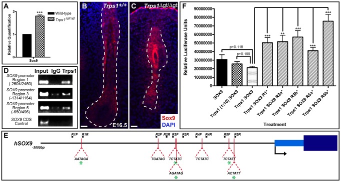
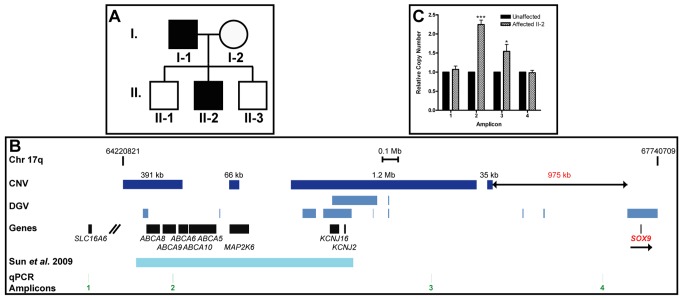
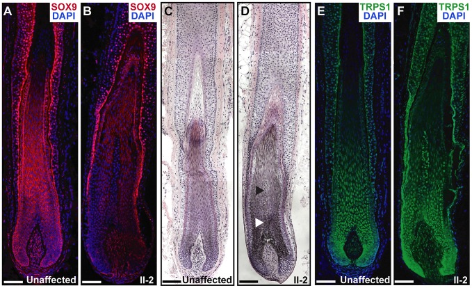
Hereditary hypertrichoses are a group of hair overgrowth syndromes that are extremely rare in humans. We have previously demonstrated that a position effect on TRPS1 is associated with hypertrichosis in humans and mice. To gain insight into the functional role of Trps1, we analyzed the late morphogenesis vibrissae phenotype of Trps1(Δgt) mutant mice, which is characterized by follicle degeneration after peg downgrowth has been initiated. We found that Trps1 directly represses expression of the hair follicle stem cell regulator Sox9 to control proliferation of the follicle epithelium. Furthermore, we identified a copy number variation upstream of SOX9 in a family with hypertrichosis that significantly decreases expression of the gene in the hair follicle, providing new insights into the long-range regulation of SOX9. Our findings uncover a novel transcriptional hierarchy that regulates epithelial proliferation in the developing hair follicle and contributes to the pathology of hypertrichosis.


Conflict of interest statement

The authors have declared that no competing interests exist.

Figures

Figure 1
Figure 1. Late morphogenesis vibrissa follicle abnormalities in Trps1Δgt/Δgt embryos.
(A) Trps1+/+ and Trps1Δgt/Δgt embryos at E16.5. Note the reduced number of vibrissae follicles and decreased size of the maxillary region (white brackets) in mutant embryos. (B) Trps1+/+ embryos have five rows of vibrissae (rows A–E), whereas Trps1Δgt/Δgt embryos have four rows due to a convergence of rows C and D. (C–H) Hematoxylin and eosin staining of transverse Trps1+/+, Trps1+/Δgt and Trps1Δgt/Δgt whisker pad sections at E16.5–E18.5 revealed small and irregularly spaced vibrissae follicles in heterozygous and homozygous mutant embryos. (I–L) Hematoxylin and eosin staining of sagittal Trps1+/+ and Trps1Δgt/Δgt head and dorsal skin sections at P0. Note the absence of hair follicles in the upper and lower jaws (black arrows) (I,J) and reduction in pelage follicle density (K,L) in mutant mice. Peg, epithelial peg; DC, dermal condensate. Scale bars, 100 µm.
Figure 2
Figure 2. Trps1Δgt/Δgt vibrissae follicles exhibit increased levels of proliferation.
(A,B) Keratin 14 staining (red) was consistent in the epithelial compartment of Trps1+/+ and Trps1Δgt/Δgt vibrissae follicles at E16.5. (C,D) Alkaline phosphatase staining (purple) revealed an intact dermal papilla and reduced collagen capsule surrounding Trps1Δgt/Δgt mutant vibrissae. (E,F) Collagen type I staining (red) was consistent in the glassy membrane of Trps1+/+ and Trps1Δgt/Δgt vibrissae follicles. (G,H) Ki67 staining (red) revealed a marked increase in proliferation throughout Trps1Δgt/Δgt mutant vibrissae follicles. (I,J) TUNEL staining (red) indicated similar levels of apoptosis between Trps1+/+ and Trps1Δgt/Δgt vibrissae. (K,L) Lef1 staining (red) was consistent in the dermal papillae and matrix cells of Trps1+/+ and Trps1Δgt/Δgt vibrissae follicles. Nuclei were stained with DAPI (blue). Scale bars, 100 µm.
Figure 3
Figure 3. Trps1 directly represses the expression of Sox9 in the vibrissa follicle.
(A) Bar graph depicting quantitative RT-PCR values revealing increased expression of Sox9 in E12.5 Trps1Δgt/Δgt whisker pad samples as compared to wild-type expression levels. Data are represented as mean ± standard deviation. *** = p<0.001. (B,C) Immunofluorescence analyses revealed increased Sox9 expression (red) throughout the epithelial compartment of Trps1Δgt/Δgt vibrissae follicles and surrounding collagen capsule at E16.5. Nuclei were stained with DAPI (blue). Scale bars, 100 µm. (D) Trps1 bound up to five sites in the SOX9 promoter in endogenous chromatin immunoprecipitation experiments in HEK 293T cells. No binding was observed in a coding sequence negative control region. (E) Identification of canonical GATA-binding sites within 3 kb of the transcriptional start site of SOX9. Green asterisks represent sites to which Trps1 was shown to bind. (F) Bar graph depicting relative luciferase units in luciferase reporter promoter assays demonstrating dose-dependent repression of SOX9 transcription by Trps1. Mutation of each of the Trps1-binding sites from WGATAR to WGATCR (*) alleviated Trps1-mediated repression of SOX9. ** = p<0.01; *** = p<0.001.
Figure 4
Figure 4. A Shh null allele can rescue the vibrissae phenotype of Trps1+/Δgt embryos.
(A–P) Hematoxylin and eosin staining of transverse Trps1+/+;Shh+/+, Trps1+/Δgt;Shh+/+, Trps1+/+;Shh+/GFP-cre, and Trps1+/Δgt;Shh+/GFP-cre whisker pad sections at E12.5–E18.5 revealed that Trps1+/+;Shh+/GFP-cre vibrissae (C,G,K,O) developed similarly to wild-type follicles (A,E,I,M), while Trps1+/Δgt;Shh+/+ embryos exhibited a reduction in vibrissae placode number at E12.5 (B) and displayed follicles that were reduced in number, irregularly spaced and slightly smaller than wild-type vibrissae throughout the remainder of embryogenesis (F,J,N). The vibrissae phenotype of Trps1+/Δgt;Shh+/+ embryos was completely rescued at all timepoints in Trps1+/Δgt;Shh+/GFP-cre compound heterozygous mice (D,H,L,P). (Q) Bar graph depicting quantitative RT-PCR values revealing increased expression of Sox9 in E16.5 Trps1+/Δgt;Shh+/+ whisker pad samples as compared to wild-type expression levels. Sox9 expression was recovered to wild-type levels in Trps1+/Δgt;Shh+/GFP-cre whisker pad samples. Data are represented as mean ± standard deviation. * = p<0.05. (R,S) Sox9 expression (red) was recovered to wild-type levels throughout the epithelial compartment of the vibrissae follicles in Trps1+/Δgt;Shh+/GFP-cre embryos as detected by immunofluorescence analyses. Nuclei were stained with DAPI (blue). Scale bars, 100 µm.
Figure 5
Figure 5. Copy number variations upstream of SOX9 associated with hypertrichosis.
(A) Pedigree of family in which the father (patient I-1) and proband (patient II-2) exhibited CGHT. (B) Map of human chromosome 17q spanning base pairs 64,220,821–67,740,709 according to build hg18. Copy number variations (CNV) detected in our analyses are represented by dark blue boxes. The telomeric end of the duplication region identified here lies 975 kb upstream of SOX9. Duplications present in the Database of Genomic Variations (DGV) or previously identified by Sun et al. are represented by lighter blue boxes. The locations of amplicons used in quantitative PCR analysis are represented by green lines. Scale bar, 0.1 Mb. (C) Bar graph depicting quantitative PCR values revealing significant increases in relative copy number of two amplicons in the proband within the duplication region identified here as compared to an unaffected control individual. Data are represented as mean ± standard deviation. * = p<0.05; *** = p<0.001.
Figure 6
Figure 6. Expression and histological analyses of CGHT follicles.
(A,B) SOX9 expression (red) was decreased in the hair follicle of the proband (B) as compared to an unaffected control follicle (A), particularly in the proliferative epithelial cells at the base of the follicle, as detected by immunofluorescence analyses. Nuclei were stained with DAPI (blue). (C,D) Hematoxylin and eosin staining of hair follicles from an unaffected control individual (C) and patient II-2 (D), revealing increased pigmentation and diameter in the CGHT follicle, particularly in the medulla layer (arrowheads) at the center of the hair shaft. (E,F) Consistent TRPS1 expression (green) between control (E) and CGHT follicles (F). Nuclei were stained with DAPI (blue). Scale bars, 100 µm.

References

    1. Garcia-Cruz D, Figuera LE, Cantu JM (2002) Inherited hypertrichoses. Clin Genet 61: 321–329. - PubMed
    1. Beighton P (1970) Congenital hypertrichosis lanuginosa. Arch Dermatol 101: 669–672. - PubMed
    1. Baumeister FA, Egger J, Schildhauer MT, Stengel-Rutkowski S (1993) Ambras syndrome: delineation of a unique hypertrichosis universalis congenita and association with a balanced pericentric inversion (8) (p11.2; q22). Clin Genet 44: 121–128. - PubMed
    1. Macías-Flores MA, García-Cruz D, Rivera H, Escobar-Luján M, Melendrez-Vega A, et al. (1984) A new form of hypertrichosis inherited as an X-linked dominant trait. Hum Genet 66: 66–70. - PubMed
    1. Canun S, Guevara-Sangines EG, Elvira-Morales A, Sierra-Romero Mdel C, Rodriguez-Asbun H (2003) Hypertrichosis terminalis, gingival hyperplasia, and a characteristic face: a new distinct entity. Am J Med Genet A 116A: 278–283. - PubMed

Publication types

MeSH terms