. 2012;7(10):e47796.
doi: 10.1371/journal.pone.0047796. Epub 2012 Oct 25.

Gatifloxacin induces S and G2-phase cell cycle arrest in pancreatic cancer cells via p21/p27/p53

Affiliations

Gatifloxacin induces S and G2-phase cell cycle arrest in pancreatic cancer cells via p21/p27/p53

Vikas Yadav et al. PLoS One. 2012.

Abstract

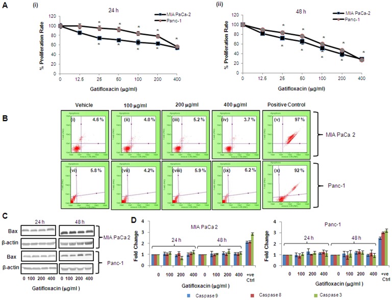
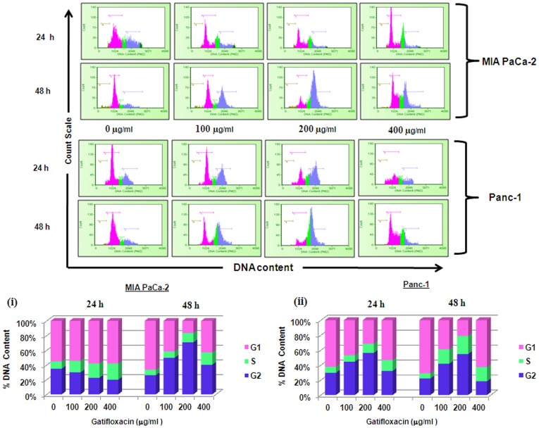
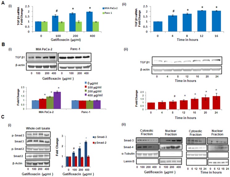
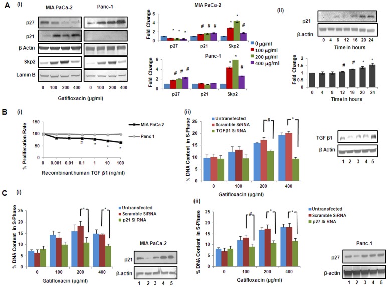
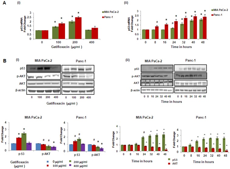
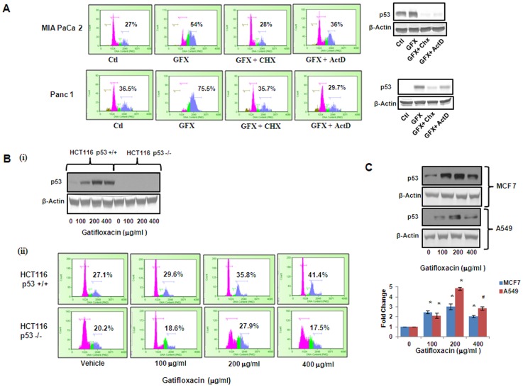
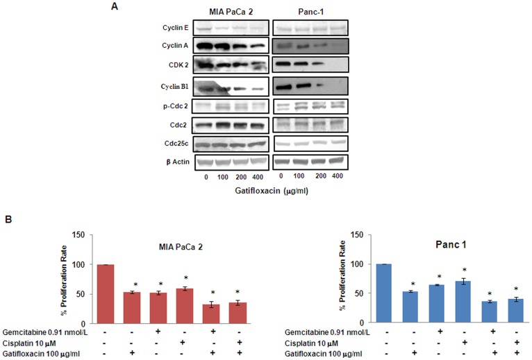
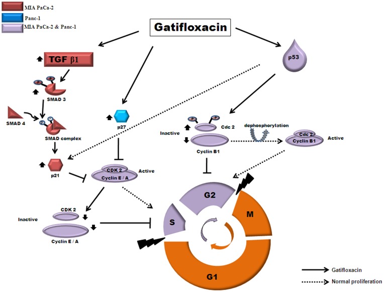
Pancreatic cancer, despite being the most dreadful among gastrointestinal cancers, is poorly diagnosed, and further, the situation has been aggravated owing to acquired drug resistance against the single known drug therapy. While previous studies have highlighted the growth inhibitory effects of older generation fluoroquinolones, the current study aims to evaluate the growth inhibitory effects of newer generation fluoroquinolone, Gatifloxacin, on pancreatic cancer cell lines MIA PaCa-2 and Panc-1 as well as to elucidate the underlying molecular mechanisms. Herein, we report that Gatifloxacin suppresses the proliferation of MIA PaCa-2 and Panc-1 cells by causing S and G(2)-phase cell cycle arrest without induction of apoptosis. Blockade in S-phase of the cell cycle was associated with increased TGF-β1 expression and translocation of Smad3-4 complex to the nucleus with subsequent activation of p21 in MIA PaCa-2 cells, whereas TGF-β signalling attenuated Panc-1 cells showed S-phase arrest by direct activation of p27. However, Gatifloxacin mediated G(2)-phase cell cycle arrest was found to be p53 dependent in both the cell lines. Our study is of interest because fluoroquinolones have the ability to penetrate pancreatic tissue which can be very effective in combating pancreatic cancers that are usually associated with loss or downregulation of CDK inhibitors p21/p27 as well as mutational inactivation of p53. Additionally, Gatifloxacin was also found to synergize the effect of Gemcitabine, the only known drug against pancreatic cancer, as well as the broad spectrum anticancer drug cisplatin. Taken together our results suggest that Gatifloxacin possesses anticancer activities against pancreatic cancer and is a promising candidate to be repositioned from broad spectrum antibiotics to anticancer agent.


Conflict of interest statement

Competing Interests: The authors have declared that no competing interests exist.

Figures

Figure 1
Figure 1. Gatifloxacin inhibits proliferation of cultured pancreatic cancer cells without induction of Apoptosis.
MTT assay of MIA PaCa-2 and Panc-1 cells after treatment with Gatifloxacin (0–400 µg/ml) for 24 h (Ai) and 48 h (Aii). Cells were seeded in 96 well plates (1×10⁴ cells/well) which were allowed to adhere overnight and were subsequently treated with increasing concentration of Gatifloxacin for 24 and 48 h. Vertical axis represents % proliferation rate whereas Horizontal axis represents increasing concentration of Gatifloxacin in µg/ml. Data are mean ± SEM three independent experiments performed in triplicate. * p<0.01 compared to vehicle control. (B) Annexin V-PE binding in MIA PaCa-2 (i-v) and Panc-1 (vi-x) after treatment with Gatifloxacin for 48 h as evaluated by 7-AAD and AnnexinV staining. (i) and (vi) Vehicle treated control cells, (ii) and (vii) cells treated with 100 µg/ml, (iii) and (viii) cells treated with 200 µg/ml, (iv) and (ix) cells treated with 400 µg/ml of Gatifloxacin. (v) MIA PaCa-2 cells and (x) Panc-1 cells treated with curcumin 60 µM for 24 h as positive control. Vertical axis represents 7-AAD positive cells whereas horizontal axis represents Annexin V-PE positive cells. Representative of three independent experiments has been shown with similar results. (C) Western blot analysis of the expression of Bax protein under the effect of Gatifloxacin in a time (24, 48 h) and dose (0, 100, 200, 400 µg/ml) dependent manner. β-Actin was used as a loading control. (D) Caspase 3, 8, 9 activity of MIA PaCa-2 and Panc-1 cells treated with Gatifloxacin in time (24, 48 h) and dose (0, 100, 200, 400 µg/ml) dependent manner. Bar graph represents mean ± SEM from three independent experiments.
Figure 2
Figure 2. Gatifloxacin induces S and G2 phase cell cycle arrest in pancreatic cancer cells.
Effects of Gatifloxacin on cell cycle were investigated using PI (Propidium Iodide) staining. Cells were treated with (0–400 µg/ml) Gatifloxacin for 24 and 48 h, collected and stained with PI. Here Pink peak represents G1-phase, Green peak represents S-phase and Blue peak represents G2-phase respectively. Upper panel shows representative of three independent experiments with similar results and lower panel represents the bar diagram of cells in different phases. Bar graph represents mean ± SEM from three independent experiments. (i) Representative bar graph for MIA PaCa-2, (ii) Representative bar graph for Panc-1.
Figure 3
Figure 3. Gatifloxacin causes activation of TGF-β1 and Smad Complex in MIA PaCa-2.
(A) (i) Real Time PCR analysis of TGF-β1 expression in MIA PaCa-2 and Panc-1 cells treated with Gatifloxacin in a dose dependent manner, (ii) Real Time PCR analysis of TGF-β1 expression in MIA PaCa-2 cells treated with 400 µg/ml of Gatifloxacin in a time dependent manner. 18S rRNA was used to normalize the results. (B) Western blot analysis of TGF-β1 expression in MIA PaCa-2 and Panc-1 cells treated with Gatifloxacin in a dose dependent manner (i) western blot analysis of TGF-β1 expression in MIA PaCa-2 cells treated with 400 µg/ml of Gatifloxacin in a time dependent manner (ii). Data are representative of typical experiment repeated three times with similar results. Bar Graph represents the mean ± SEM. (C) (i) Effect of Gatifloxacin on receptor mediated Smads (pSmad-2 and pSmad-3) when assessed in whole cell lysate in MIA PaCa-2 cells. β-actin was used as a loading control. (ii) Translocation of Smad 3–4 complex from cytoplasm to nucleus under the effect of Gatifloxacin in a dose (0, 100, 200, 400 µg/ml) and time dependent manner (0, 6, 12, 18, 24 h) was assessed by western blotting. Nuclear and Cytoplasmic fractions were separated as described in the “Material and methods” section. Lamin B (Nuclear specific protein) and α-Tubulin (Cytoplasmic specific protein) were used as loading controls.
Figure 4
Figure 4. Gatifloxacin causes activation of p21 in MIA PaCa-2 and p27 in Panc-1 cells, but Skp2 in both cell lines.
(A) (i) Western blot analysis of p21, p27 and Skp2 expression in MIA PaCa-2 and Panc-1 cells treated with Gatifloxacin in a dose dependent manner. β-Actin was used as loading control for p21 and p27, whereas Lamin B was used as loading control for Skp2, (A) (ii) Western blot analysis of p21 expression in MIA PaCa-2 cells treated with 400 µg/ml of Gatifloxacin in a time dependent manner. Data are representative of typical experiment repeated three times with similar results. Bar Graph represents the mean ± SEM. *p<0.01, #p<0.05 versus control. Gatifloxacin mediated S phase arrest is TGF-β1 dependent in MIA PaCa-2 (B) (i) MTT assay of MIA PaCa-2 and Panc-1 cells in presence of recombinant-TGF-β1 for 48 h. Vertical axis represents % proliferation rate whereas Horizontal axis represents increasing concentration of recombinant-TGF-β1 in ng/ml. Data are mean ± SEM three independent experiments performed in triplicate. *p<0.01, #p<0.05 versus control. (B) (ii) Abolishment of S-phase arrest in MIA PaCa-2 cells as assessed by PI (Propidium Iodide) staining. Cells transfected with TGF-β1 siRNA (48 h) and scrambled siRNA alongwith untransfected cells were treated from 100 µg/ml to 400 µg/ml Gatifloxacin (24 h) after 24 h of transfection, collected and stained with PI. Left panel represents the bar graph where vertical axis represents %DNA content in S-phase and Horizontal axis represents Gatifloxacin concentration in µg/ml. Bar graph represents mean ± SEM from three independent experiments. *p<0.01, #p<0.05 versus control. Right panel shows western blot for the knockdown efficiency of TGF-β1 siRNA. (1) represents untransfected control cells, (2) TGF-β1 siRNA transfected cells, (3) TGF-β1 siRNA transfected cells with 400 µg/ml of Gatifloxacin, (4) siRNA transfected cells, (5) siRNA transfected cells with 400 µg/ml of Gatifloxacin. Gatifloxacin mediated S phase arrest is p21 dependent in MIA PaCa-2 and p27 dependent in Panc-1 cells. (C) Abolishment of S-phase arrest in MIA PaCa-2 cells transfected with p21 siRNA (i) and Panc-1 cells transfected with p27 siRNA (ii), respectively as assessed by PI (Propidium Iodide) staining. Cells transfected with p21/p27 siRNA and scrambled siRNA (48 h) alongwith untransfected cells were treated with 100 µg/ml to 400 µg/ml of Gatifloxacin (24 h) after 24 h of transfection, collected and stained with PI. Left panel represents the bar graph where vertical axis represents %DNA content in S-phase and Horizontal axis represents Gatifloxacin concentration in µg/ml. Right panel shows western blot for the knockdown efficiency of p21 or p27 siRNA. (1) represents untransfected control cells, (2) p21/p27 siRNA transfected cells, (3) p21/p27 siRNA transfected cells treated with 400 µg/ml of Gatifloxacin, (4) scrambled siRNA transfected cells, (5) scrambled siRNA transfected cells treated with 400 µg/ml of Gatifloxacin. Bar graph represents mean ± SEM from three independent experiments. *p<0.01, #p<0.05 versus control.
Figure 5
Figure 5. Gatifloxacin Causes activation of p53 and inhibits pAKT.
(A) (i) Real Time PCR analysis of p53 expression in MIA PaCa-2 and Panc-1 cells treated with Gatifloxacin in a dose dependent manner, (ii) Real Time PCR analysis of p53 expression in MIA PaCa-2 and Panc-1 cells treated with 200µg/ml of Gatifloxacin in a time dependent manner. 18S rRNA was used to normalize the results. (B) (i) Western blot analysis of p53, AKT and pAKT expression in MIA PaCa-2 and Panc-1 cells treated with Gatifloxacin in a dose dependent manner, (ii) Western blot analysis of p53, AKT and pAKT expression in MIA PaCa-2 and Panc-1 cells treated with Gatifloxacin in a time dependent manner. β-actin was used as loading control. Data are representative of typical experiment repeated three times with similar results. Bar Graph represents the mean ± SEM. *p<0.01, #p<0.05 compared to control.
Figure 6
Figure 6. Gatifloxacin-induced G2-phase arrest is p53 dependent.
(A) Left Panel shows the Cell cycle analysis of MIA PaCa-2 and Panc-1 cells when grown in the presence or absence of p53 transcriptional inhibitor ActinomycinD (1 µg/ml) or Translational inhibitor cycloheximide (2 µg/ml) along with 200 µg/ml Gatifloxacin for 48 h and right panel shows the p53 protein expression in MIA PaCa-2 and Panc-1 cells in presence or absence of 200 µg/ml Gatifloxacin with or without 2 µg/ml CHX or 1 µg/ml ActD. % here indicates percentage of G2 phase subpopulation. (B) (i) Western blot analysis for p53 expression in HCT116 p53+/+ and P53−/− cell lines treated with Gatifloxacin in a dose dependent manner for 24 h. (ii) Cell cycle analysis of HCT116 p53+/+ and P53−/− treated with Gatifloxacin (0–400 µg/ml) for 24 h. % here indicates percentage of G2 phase subpopulation. (C) Western blot analysis of p53 protein expression in MCF 7 and A549 cells treated with Gatifloxacin in a dose dependent manner. Bar Graph represents the mean ± SEM. *p<0.01, compared to control.
Figure 7
Figure 7. Gatifloxacin affects cell cycle regulatory proteins and synergizes the antiproliferative effects of other broad spectrum anticancer drugs.
(A) Effect of Gatifloxacin on S and G2-phase regulatory Cyclins and CDKs as assessed by western blot analysis in MIA PaCa-2 and Panc-1 cells (B) Gatifloxacin (100 µg/ml) synergizes the antiproliferative effect of Gemcitabine (0.91nM/L) and Cisplatin (10 µM) in MIA PaCa-2 and Panc-1 cells as assessed by MTT assay. Vertical axis represents % proliferation rate whereas Horizontal axis represents presence or absence of above mentioned drugs. Data are mean ± SEM of three independent experiments performed in triplicate. *p<0.02 as compared to untreated control.
Figure 8
Figure 8. Schematic representation of proposed mechanism of action of Gatifloxacin induced S and G2 phase arrest in pancreatic cancer cells.
Gatifloxacin leads to activation of TGF-β1 only in MIA PaCa-2 cell line which than activates Smad-3 but not Smad-2 via its phosphorylation and forms complex with Smad-4 in cytoplasm. Complex than translocates to the nucleus where it activates p21, however in case of Panc-1 Gatifloxacin directly activates p27. p21 (waf1/cip1) and p27 (kip1) both being cyclin dependent kinase inhibitor, than inhibits and down regulates Cyclin A, Cyclin E and CDK-2 which than causes S-phase arrest in both the cell lines. Gatifloxacin also activates p53 in both MIA PaCa-2 and Panc-1 cell lines which than down regulates Cyclin B1, upregulates inhibitory pCdc2 and Cdc2 which than leads to G2 arrest. However, activation of p53 doesn’t affect the levels of Cdc25c. pAKT, an inhibitor of Smad complex, p21, p27 and p53 was found to be downregulated in both cell lines under the effect of Gatifloxacin.

Similar articles

Cited by

References

    1. Lillemoe KD (1995) Current management of pancreatic carcinoma. Ann Surg 221: 133–148. - PMC - PubMed
    1. Duffy JP, Eibl G, Reber HA, Hines OJ (2003) Influence of hypoxia and neoangiogenesis on the growth of pancreatic cancer. Mol.Cancer 2: 12. - PMC - PubMed
    1. Apte MV, Park S, Phillips PA, Santucci N, Goldstein D, et al. (2004) Desmoplastic reaction in pancreatic cancer: role of pancreatic stellate cells. Pancreas 29: 179–187. - PubMed
    1. Riall TS, Cameron JL, Lillemoe KD, Winter JM, Campbell KA, et al. (2006) Resected periampullary adenocarcinoma: 5-year survivors and their 6- to 10-year follow-up. Surgery 140: 764–772. - PubMed
    1. Bao PQ, Ramanathan RK, Krasinkas A, Bahary N, Lembersky BC, et al... (2011) III, Erratum to: Phase II Study of Gemcitabine and Erlotinib as Adjuvant Therapy for Patients with Resected Pancreatic Cancer Ann Surg Oncol. - PubMed

Publication types

MeSH terms