. 2012;7(10):e48049.
doi: 10.1371/journal.pone.0048049. Epub 2012 Oct 25.

Mechanical disruption of tumors by iron particles and magnetic field application results in increased anti-tumor immune responses

Affiliations

Mechanical disruption of tumors by iron particles and magnetic field application results in increased anti-tumor immune responses

Myriam N Bouchlaka et al. PLoS One. 2012.

Abstract

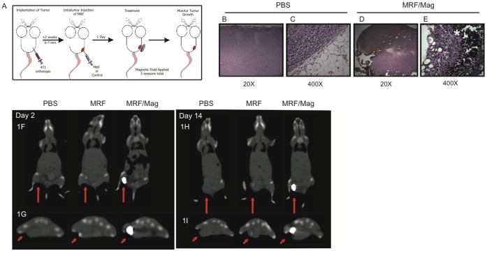
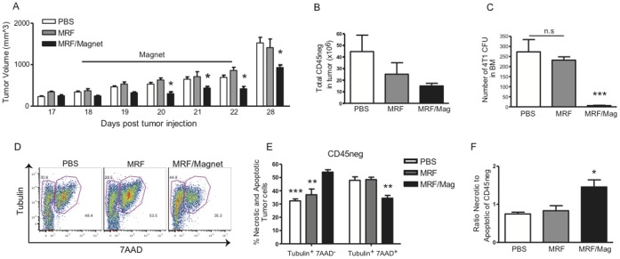
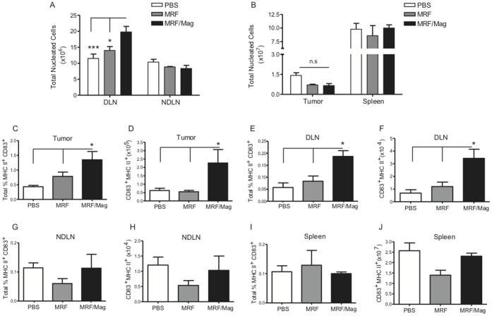
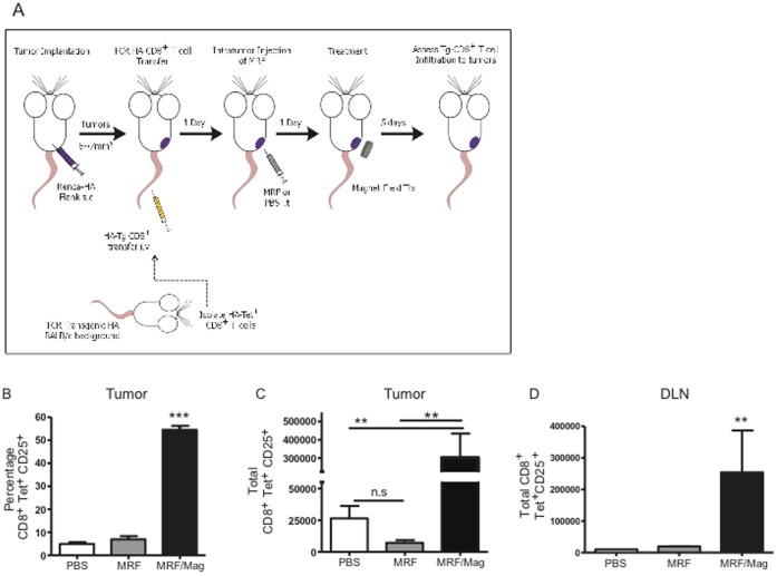
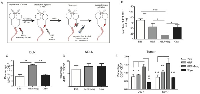
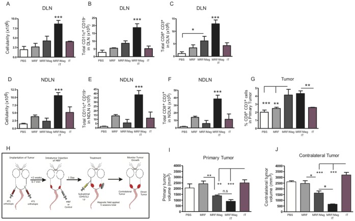
The primary tumor represents a potential source of antigens for priming immune responses for disseminated disease. Current means of debulking tumors involves the use of cytoreductive conditioning that impairs immune cells or removal by surgery. We hypothesized that activation of the immune system could occur through the localized release of tumor antigens and induction of tumor death due to physical disruption of tumor architecture and destruction of the primary tumor in situ. This was accomplished by intratumor injection of magneto-rheological fluid (MRF) consisting of iron microparticles, in Balb/c mice bearing orthotopic 4T1 breast cancer, followed by local application of a magnetic field resulting in immediate coalescence of the particles, tumor cell death, slower growth of primary tumors as well as decreased tumor progression in distant sites and metastatic spread. This treatment was associated with increased activation of DCs in the draining lymph nodes and recruitment of both DCs and CD8(+)T cells to the tumor. The particles remained within the tumor and no toxicities were observed. The immune induction observed was significantly greater compared to cryoablation. Further anti-tumor effects were observed when MRF/magnet therapy was combined with systemic low dose immunotherapy. Thus, mechanical disruption of the primary tumor with MRF/magnetic field application represents a novel means to induce systemic immune activation in cancer.


Conflict of interest statement

Competing Interests: The authors have declared that no competing interests exist.

Figures

Figure 1
Figure 1. Histological assessment and biodistribution of iron particles following intratumoral MRF/magnetic field application.
(A) Experimental design and schedule of treatment: 4T1 cells were injected s.c into the mammary fat pad of female BALB/c mice. When tumors reached 6–7 mm, 100 µL of 60% MRF w/v in PBS was injected into the tumor (i.t) for treatment groups or 100 µL PBS in control group. Iron particles are dispersed randomly in the tumor. One group received magnetic field treatments by direct application of permanent magnets on the primary tumor. Mice received 5 min/session of magnet treatments using a 0.4 Tesla magnet starting 24 hours after MRF injection for 5 consecutive days. Another group received MRF i.t and no further treatments. During magnetic field treatment, the iron particles aggregate and lead to (B–E) At the end of MRF/magnet treatment, tumors were collected, fixed and sectioned in formalin and then stained with H&E. (B–C) Images of PBS or (D–E) MRF/magnet treated groups (20X or 400X magnification). Iron particles depicted by the brownish particles (1D) or black particles (white star) (1E). One representative experiment of three independent experiments. (n = 3 mice/group). (F–I) Trafficking of iron particles to distant sites by in vivo imaging 2 days (F–G) and 14 days (H–I) post iron injections as depicted by CT-scan images of one representative animal per group. Groups include PBS, MRF and MRF/magnet treatment showing sagittal CT-scanning (F, H) and coronal CT-scanning of the lower hind area where tumor was inoculated (G, I). Red arrows point at tumor and site of iron particles (white area). n = 3 mice/group from one experiment.
Figure 2
Figure 2. Inhibition of local and systemic tumor growth as a result of increased primary tumor necrosis after MRF implantation and magnetic field treatment.
4T1 tumors were established into the mammary fat pad of female Balb/c mice as illustrated in Fig. 1A. When tumors reached 6–7 mm, 100 µL of 60% MRF was injected i.t (day 17) for treatment groups or 100 µl PBS in control group. One group was treated 24 hours after MRF injection with direct magnetic field application for 5 consecutive days (days 18–22). (A) Tumor volume during and after MRF/Magnet treatments. Data is representative of one of six experiments (n = 9 mice/group). (B) Total number of tumor cells by flow cytometry after 5 days of magnet treatment showing smaller tumor load after magnet treatment and (C) tumor CFUs of bone marrow. MRF/magnet treatments result in inhibition of growth of metastatic disease. (D) Representative flow staining for tubulin and 7AAD in the tumor. (E) Percentage of tubulin+7AAD (necrotic) cells in the tumor gated on CD45 cells. (B–E) Data representative of one of two experiments with similar results (n = 3–4 mice/group). One-way or Two-way ANOVA. * P<0.05, ** P<0.01, *** P<0.001.
Figure 3
Figure 3. DC expansion and activation after MRF and magnetic field treatment.
4T1 tumors were injected into the mammary fat pad of female Balb/c mice as illustrated in Fig. 1A. Briefly, primary tumors were injected with 100 µL of 60% MRF i.t in the treatment groups, some mice received no further treatments and some mice received direct magnet application over the tumor for a total of 5 days. Control group received 100 µL PBS i.t. Five days after magnet treatments, DLN, NDLN, tumor and spleen were excised and analyzed by flow cytometry for (A–B) total number of nucleated cells in (A) DLN and NDLN or (B) tumor and spleen. All four organs were analyzed for CD83, MHC II expression gating on CD45+ CD11c+ CD19 as follow: (C–D) represents both the total percentage and numbers of activated DCs in the tumor, (E–F) in the DLN, (G–H) in the NDLN and (I–J) in the spleen. Data (mean ± SEM) representative of one of four experiments (n = 3–4 mice/group). One-way ANOVA. * P<0.05, *** P<0.001. n.s: not significant.
Figure 4
Figure 4. MRF and magnetic field application results in antigen-specific T cell accumulation.
(A) Experimental model for antigen-specific responses following MRF and magnet treatments: BALB/c mice received Renca-HA on the right flank s.c. When tumors reached the desired size, mice first received 2×10⁶ CD8(+)-HA-Tet+ cells i.v. One day following Tg-CD8(+) T cell transfer, mice were administered MRF or PBS intratumor. 24 h later, some groups are left untreated or receive magnetic treatments for 5 min a day for 5 consecutive days. After 5 days of daily magnetic field treatments, tumors or tumor-DLNs were collected and assayed by flow cytometry. (A) Percentage and (B) total number of transgenic HA-Tg-CD8(+)-CD25+ T cells in Renca-HA tumor. (C) DLNs from the same mice in (A–B) were analyzed for homing of HA-Tg-CD8(+)CD25+ T cells. Data representative of one single experiment (n = 3 mice/group in PBS or MRF group and n = 5 mice in the MRF/magnet group). One-way ANOVA. ** P<0.01, *** P<0.001, n.s: not significant.
Figure 5
Figure 5. Augmented immune activation and inhibition of metastasis with MRF/magnetic treatments in comparison to cryoablation.
(A) Experimental design of MRF and magnet treatments and cryoablation. BALB/c mice received 4T1 orthotopically on one side of the mammary fat pad. When tumors reached 6–7 mm in size, some mice received one of the following: PBS i.t, MRF i.t, MRF i.t and magnet treatment (5 min/day for 5 days) or cryoablation once. (B) Lung cells were collected and plated for tumor CFUs (after 4 days of magnet application). (C–D) Percentage of activated DCs (MHC II+ CD83+ of CD45+ CD11c+ CD19) in the DLNs (B) and NDLNs (C). (E) Percentage of CD8(+) T cells (gated on CD3+ CD45+) in primary tumor in each group relative to 4 or 7 days of magnet treatments. Values represent the mean ± SEM of one of two experiments with similar results (n = 3–5 mice/group). One-way or Two-way ANOVA. * P<0.05, ** P<0.01, *** P<0.001, n.s: not significant.
Figure 6
Figure 6. Combination of MRF/magnetic field application with immunotherapy results in heightened systemic anti-tumor responses.
4TI breast cancer cells were implanted into the mammary fat pad of BALB/c mice. Treatment was initiated when tumors reached an average volume of 6–7 mm. Mice received one of the following 5 treatments: 1) PBS i.t, 2) MRF i.t alone, 3) MRF and magnet, 4) MRF+ magnet + anti-CD40 (25 ug) and recombinant human IL2 (2.5 ×10⁵ IU) i.p or 5) anti-CD40 and IL2 alone. Anti-CD40 was given for 5 consecutive days starting the same day as the magnetic field treatments and IL2 was administered on days 2, 5, 9 and 11 post MRF injections. (A–G) Immune effects of combination of low dose anti-CD40/IL2 with MRF/magnet after 5 days of magnet treatment and anti-CD40/IL2. (A, D) Total nucleated cells in the DLN and NDLNs, respectively. (B) Total percentage of DCs (CD11c+ CD19 CD45+) in DLNs and (E) NDLNs. (C) Total CD3+ CD8(+) T cells in DLNs or (F) NDLNs or (G) in the tumor. Data representative of one of three experiments with similar results (n = 3–4 mice/group). (H) Experimental model for systemic anti-tumor effects: 4TI breast cancer cells were implanted s.c. on the right and left side of the mammary fat pad of BALB/c mice. Only the right tumors were injected with MRF or PBS. Some groups were further treated for 5 consecutive days with magnetic field. Other groups received anti-CD40 (25 ug) on the same days of magnet treatment and rh-IL2 (2.5×10⁵ IU) (days 2, 5, 9 and 11 post MRF injections). (I-J) Tumor volumes after 28 days post tumor inoculation or equivalent to 12 days after start of magnet treatments are represented for (J) primary tumors that received MRF or (J) the contralateral untreated tumors. Data representative of one of five experiments with similar results (n = 5–7 mice/group). One-way ANOVA. * P<0.05, ** P<0.01, *** P<0.001. n.s: not significant. IT: anti-CD40/IL2.

References

    1. Curley SA (2003) Radiofrequency ablation of malignant liver tumors. Ann Surg Oncol 10: 338–347. - PubMed
    1. Raj GV, Reddan DJ, Hoey MF, Polascik TJ (2003) Management of small renal tumors with radiofrequency ablation. Urology 61: 23–29. - PubMed
    1. Zhao Z, Wu F (2010) Minimally-invasive thermal ablation of early-stage breast cancer: a systemic review. Eur J Surg Oncol 36: 1149–1155. - PubMed
    1. Garcea G, Lloyd TD, Aylott C, Maddern G, Berry DP (2003) The emergent role of focal liver ablation techniques in the treatment of primary and secondary liver tumours. Eur J Cancer 39: 2150–2164. - PubMed
    1. Pawlik TM, Izzo F, Cohen DS, Morris JS, Curley SA (2003) Combined resection and radiofrequency ablation for advanced hepatic malignancies: results in 172 patients. Ann Surg Oncol 10: 1059–1069. - PMC - PubMed

Publication types

MeSH terms