

Self-assembling complexes of quantum dots and scFv antibodies for cancer cell targeting and imaging
- PMID: 23133578
- PMCID: PMC3484990
- DOI: 10.1371/journal.pone.0048248
Self-assembling complexes of quantum dots and scFv antibodies for cancer cell targeting and imaging
Abstract
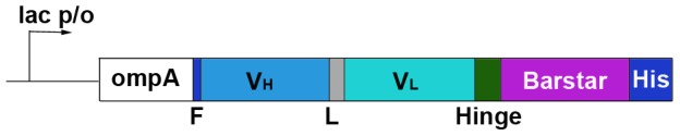
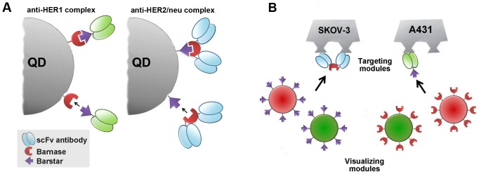
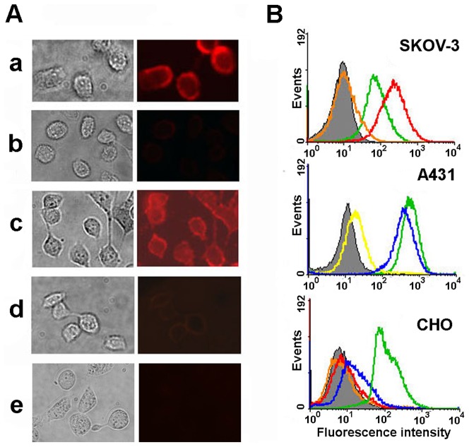
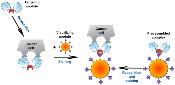
Semiconductor quantum dots represent a novel class of fluorophores with unique physical and chemical properties which could enable a remarkable broadening of the current applications of fluorescent imaging and optical diagnostics. Complexes of quantum dots and antibodies are promising visualising agents for fluorescent detection of selective biomarkers overexpressed in tumor tissues. Here we describe the construction of self-assembling fluorescent complexes of quantum dots and anti-HER1 or anti-HER2/neu scFv antibodies and their interactions with cultured tumor cells. A binding strategy based on a very specific non-covalent interaction between two proteins, barnase and barstar, was used to connect quantum dots and the targeting antibodies. Such a strategy allows combining the targeting and visualization functions simply by varying the corresponding modules of the fluorescent complex.
Conflict of interest statement
Figures
Similar articles
-
Fluorescent immunolabeling of cancer cells by quantum dots and antibody scFv fragment.J Biomed Opt. 2009 Mar-Apr;14(2):021004. doi: 10.1117/1.3122775. J Biomed Opt. 2009. PMID: 19405717
-
[Targeted Delivery of Quantum Dots to HER2-Expressing Tumor Using Recombinant Antibodies].Bioorg Khim. 2015 Sep-Oct;41(5):599-605. doi: 10.1134/s1068162015050040. Bioorg Khim. 2015. PMID: 26762098 Russian.
-
Near-infrared quantum dots for HER2 localization and imaging of cancer cells.Int J Nanomedicine. 2014 Mar 11;9:1323-37. doi: 10.2147/IJN.S51535. eCollection 2014. Int J Nanomedicine. 2014. PMID: 24648731 Free PMC article.
-
Bioconjugated quantum dots as fluorescent probes for biomedical imaging.J Nanosci Nanotechnol. 2011 Sep;11(9):7521-36. doi: 10.1166/jnn.2011.5122. J Nanosci Nanotechnol. 2011. PMID: 22097457 Review.
-
Molecular profiling of single cancer cells and clinical tissue specimens with semiconductor quantum dots.Int J Nanomedicine. 2006;1(4):473-81. doi: 10.2147/nano.2006.1.4.473. Int J Nanomedicine. 2006. PMID: 17722280 Free PMC article. Review.
Cited by
-
HER2-specific recombinant immunotoxin 4D5scFv-PE40 passes through retrograde trafficking route and forces cells to enter apoptosis.Oncotarget. 2017 Mar 28;8(13):22048-22058. doi: 10.18632/oncotarget.15833. Oncotarget. 2017. PMID: 28423549 Free PMC article.
-
Specific visualization of tumor cells using upconversion nanophosphors.Acta Naturae. 2014 Oct;6(4):48-53. Acta Naturae. 2014. PMID: 25558394 Free PMC article.
-
Experimental Evaluation of Quantum Dots and Antibodies Conjugation by Surface Plasmon Resonance Spectroscopy.Int J Mol Sci. 2022 Oct 20;23(20):12626. doi: 10.3390/ijms232012626. Int J Mol Sci. 2022. PMID: 36293491 Free PMC article.
-
Two-Step Targeted Drug Delivery via Proteinaceous Barnase-Barstar Interface and Doxorubicin-Loaded Nano-PLGA Outperforms One-Step Strategy for Targeted Delivery to HER2-Overexpressing Cells.Pharmaceutics. 2022 Dec 24;15(1):52. doi: 10.3390/pharmaceutics15010052. Pharmaceutics. 2022. PMID: 36678681 Free PMC article.
-
Genetically Encoded Self-Assembling Protein Nanoparticles for the Targeted Delivery In Vitro and In Vivo.Pharmaceutics. 2023 Jan 10;15(1):231. doi: 10.3390/pharmaceutics15010231. Pharmaceutics. 2023. PMID: 36678860 Free PMC article. Review.
References
-
- Hanash S (2004) Integrated global profiling of cancer. Nat. Rev. Cancer 4: 638–644. - PubMed
-
- Bird RE, Hardman KD, Jacobson JW, Johnson S, Kaufman BM, et al. (1988) Single-chain antigen-binding proteins. Science 242: 423–426. - PubMed
-
- Müller KM, Arndt KM, Strittmatter W (1998) Plückthun (1998) A The first constant domain (C(H)1 and C(L)) of an antibody used as heterodimerization domain for bispecific miniantibodies. FEBS Lett. 422: 259–264. - PubMed
-
- Eigenbrot C, Randal M, Presta L, Carter P, Kossiakoff AA (1993) X-ray structures of the antigen-binding domains from three variants of humanized anti-p185HER2 antibody 4D5 and comparison with molecular modeling. J. Mol. Biol. 229: 969–995. - PubMed
Publication types
MeSH terms
Substances
LinkOut - more resources
Full Text Sources
Other Literature Sources
Research Materials
Miscellaneous
