

IgG expression in human colorectal cancer and its relationship to cancer cell behaviors
- PMID: 23133595
- PMCID: PMC3486799
- DOI: 10.1371/journal.pone.0047362
IgG expression in human colorectal cancer and its relationship to cancer cell behaviors
Abstract
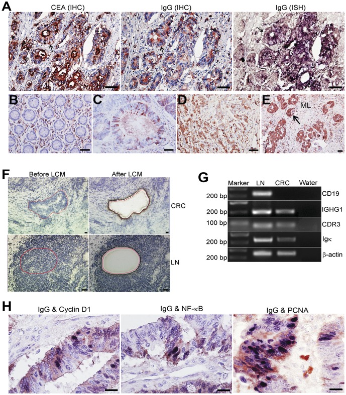
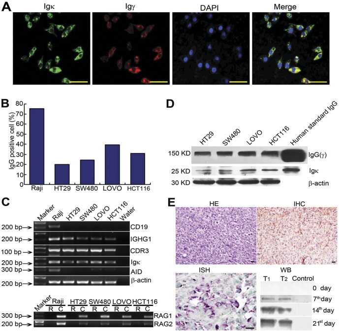
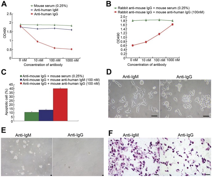
Increasing evidence indicates that various cancer cell types are capable of producing IgG. The exact function of cancer-derived IgG has, however, not been elucidated. Here we demonstrated the expression of IgG genes with V(D)J recombination in 80 cases of colorectal cancers, 4 colon cancer cell lines and a tumor bearing immune deficient mouse model. IgG expression was associated with tumor differentiation, pTNM stage, lymph node involvement and inflammatory infiltration and positively correlated with the expressions of Cyclin D1, NF-κB and PCNA. Furthermore, we investigated the effect of cancer-derived IgG on the malignant behaviors of colorectal cancer cells and showed that blockage of IgG resulted in increased apoptosis and negatively affected the potential for anchor-independent colony formation and cancer cell invasion. These findings suggest that IgG synthesized by colorectal cancer cells is involved in the development and growth of colorectal cancer and blockage of IgG may be a potential therapy in treating this cancer.
Conflict of interest statement
Figures
References
-
- Lee G, Chu RA, Ting HH (2009) Preclinical assessment of anti-cancer drugs by using RP215 monoclonal antibody. Cancer Biol Ther 8: 161–166. - PubMed
-
- Kimoto Y (1998) Expression of heavy-chain constant region of immunoglobulin and T-cell receptor gene transcripts in human non-hematopoietic tumor cell lines. Genes Chromosomes Cancer 22: 83–86. - PubMed
-
- Qiu X, Zhu X, Zhang L, Mao Y, Zhang J, et al. (2003) Human epithelial cancers secrete immunoglobulin g with unidentified specificity to promote growth and survival of tumor cells. Cancer Res 63: 6488–6495. - PubMed
-
- Chen Z, Gu J (2007) Immunoglobulin G expression in carcinomas and cancer cell lines. Faseb J 21: 2931–2938. - PubMed
-
- Lee G, Laflamme E, Chien CH, Ting HH (2008) Molecular identity of a pan cancer marker, CA215. Cancer Biol Ther 7: 2007–2014. - PubMed
Publication types
MeSH terms
Substances
LinkOut - more resources
Full Text Sources
Medical
Research Materials
Miscellaneous
