

Genome-wide comparative analysis of annexin superfamily in plants
- PMID: 23133603
- PMCID: PMC3487801
- DOI: 10.1371/journal.pone.0047801
Genome-wide comparative analysis of annexin superfamily in plants
Abstract
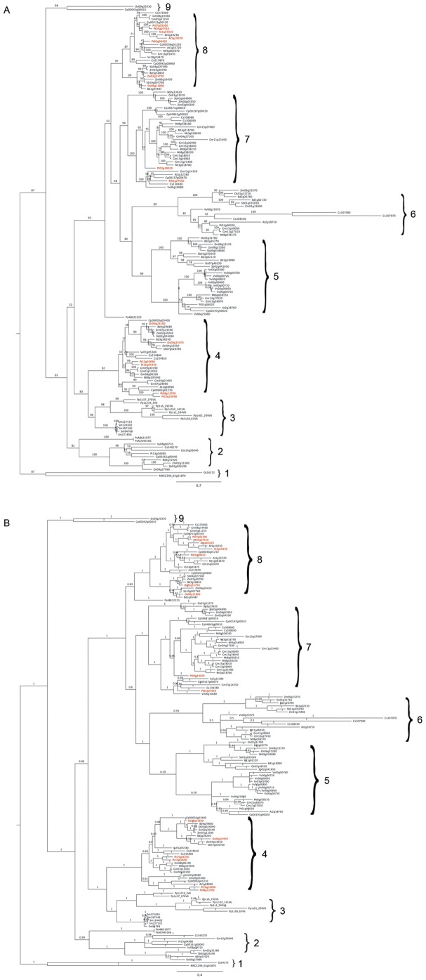
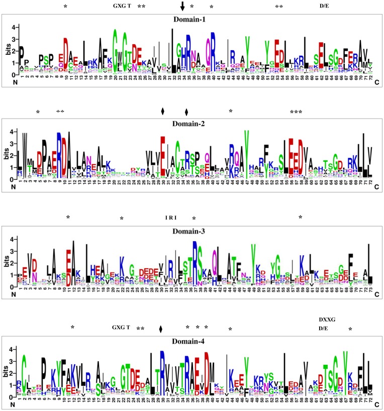
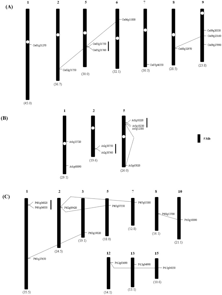
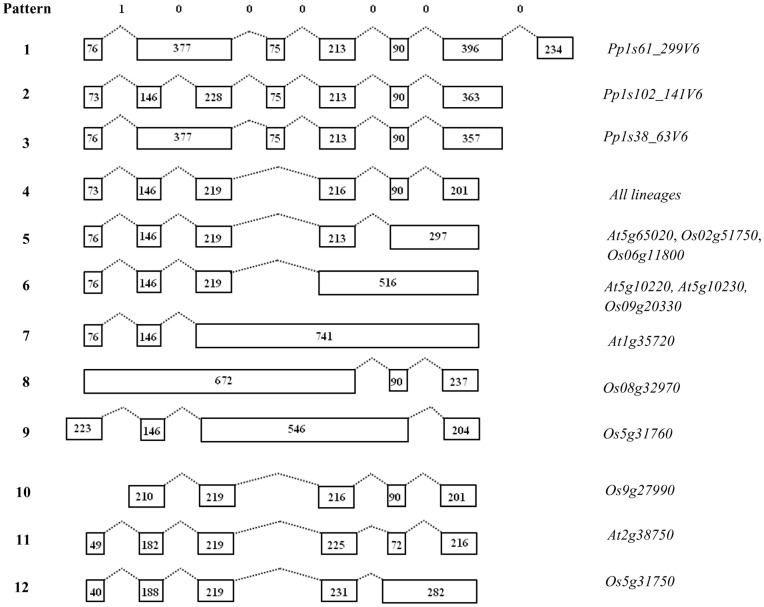
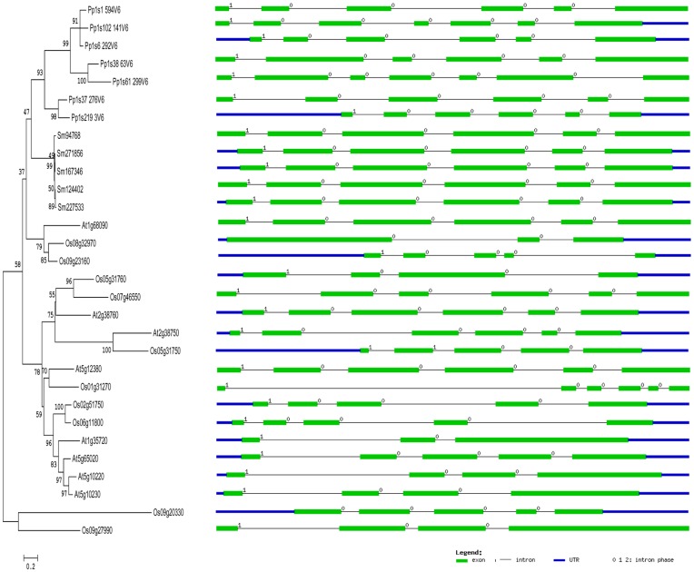
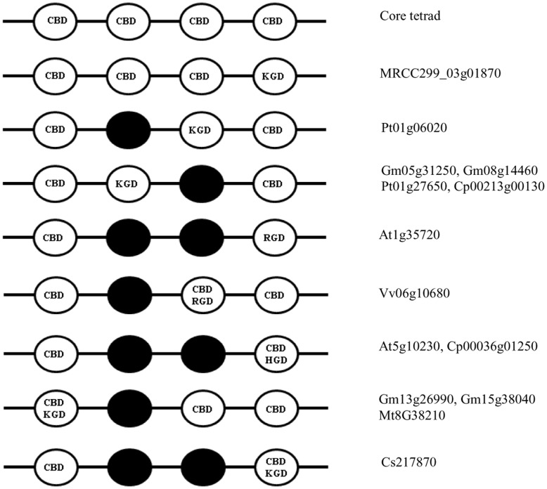
Most annexins are calcium-dependent, phospholipid-binding proteins with suggested functions in response to environmental stresses and signaling during plant growth and development. They have previously been identified and characterized in Arabidopsis and rice, and constitute a multigene family in plants. In this study, we performed a comparative analysis of annexin gene families in the sequenced genomes of Viridiplantae ranging from unicellular green algae to multicellular plants, and identified 149 genes. Phylogenetic studies of these deduced annexins classified them into nine different arbitrary groups. The occurrence and distribution of bona fide type II calcium binding sites within the four annexin domains were found to be different in each of these groups. Analysis of chromosomal distribution of annexin genes in rice, Arabidopsis and poplar revealed their localization on various chromosomes with some members also found on duplicated chromosomal segments leading to gene family expansion. Analysis of gene structure suggests sequential or differential loss of introns during the evolution of land plant annexin genes. Intron positions and phases are well conserved in annexin genes from representative genomes ranging from Physcomitrella to higher plants. The occurrence of alternative motifs such as K/R/HGD was found to be overlapping or at the mutated regions of the type II calcium binding sites indicating potential functional divergence in certain plant annexins. This study provides a basis for further functional analysis and characterization of annexin multigene families in the plant lineage.
Conflict of interest statement
Figures
Similar articles
-
Analyses of phylogeny, evolution, conserved sequences and genome-wide expression of the ICK/KRP family of plant CDK inhibitors.Ann Bot. 2011 May;107(7):1141-57. doi: 10.1093/aob/mcr034. Epub 2011 Mar 7. Ann Bot. 2011. PMID: 21385782 Free PMC article.
-
Identification and characterization of annexin gene family in rice.Plant Cell Rep. 2012 May;31(5):813-25. doi: 10.1007/s00299-011-1201-0. Epub 2011 Dec 14. Plant Cell Rep. 2012. PMID: 22167239
-
Comprehensive analyses of the annexin gene family in wheat.BMC Genomics. 2016 May 28;17:415. doi: 10.1186/s12864-016-2750-y. BMC Genomics. 2016. PMID: 27236332 Free PMC article.
-
Evolutionary adaptation of plant annexins has diversified their molecular structures, interactions and functional roles.New Phytol. 2012 Nov;196(3):695-712. doi: 10.1111/j.1469-8137.2012.04308.x. Epub 2012 Sep 19. New Phytol. 2012. PMID: 22994944 Review.
-
Annexins: central regulators of plant growth and stress signaling.Acta Biochim Biophys Sin (Shanghai). 2025 Jan 15;57(4):507-520. doi: 10.3724/abbs.2024228. Acta Biochim Biophys Sin (Shanghai). 2025. PMID: 39821233 Free PMC article. Review.
Cited by
-
Overexpression of Arabidopsis AnnAt8 Alleviates Abiotic Stress in Transgenic Arabidopsis and Tobacco.Plants (Basel). 2016 Apr 14;5(2):18. doi: 10.3390/plants5020018. Plants (Basel). 2016. PMID: 27135239 Free PMC article.
-
Structure, Function, and Applications of Soybean Calcium Transporters.Int J Mol Sci. 2022 Nov 17;23(22):14220. doi: 10.3390/ijms232214220. Int J Mol Sci. 2022. PMID: 36430698 Free PMC article. Review.
-
Selenium-binding Protein 1 (SBD1): A stress response regulator in Chlamydomonas reinhardtii.Plant Physiol. 2022 Aug 1;189(4):2368-2381. doi: 10.1093/plphys/kiac230. Plant Physiol. 2022. PMID: 35579367 Free PMC article.
-
Genome-Wide Identification and Transcriptional Expression Analysis of Annexin Genes in Capsicum annuum and Characterization of CaAnn9 in Salt Tolerance.Int J Mol Sci. 2021 Aug 12;22(16):8667. doi: 10.3390/ijms22168667. Int J Mol Sci. 2021. PMID: 34445369 Free PMC article.
-
Spatial and temporal specificity of Ca2+ signalling in Chlamydomonas reinhardtii in response to osmotic stress.New Phytol. 2016 Dec;212(4):920-933. doi: 10.1111/nph.14128. Epub 2016 Aug 12. New Phytol. 2016. PMID: 27516045 Free PMC article.
References
-
- Gerke V, Moss SE (2002) Annexins: From structure to function. Physiol Rev 82: 331–371. - PubMed
-
- Morgan RO, Fernandez MP (1995) Molecular phylogeny of annexins and identification of a primitive homologue in Giardia lambia . Mol Biol Evol 12: 967–979. - PubMed
-
- Laohavisit A, Davies JM (2011) Annexins. New Phytol 189: 40–53. - PubMed
MeSH terms
Substances
LinkOut - more resources
Full Text Sources
