. 2012;7(11):e48228.
doi: 10.1371/journal.pone.0048228. Epub 2012 Nov 1.

Genomic comparison of Escherichia coli O104:H4 isolates from 2009 and 2011 reveals plasmid, and prophage heterogeneity, including shiga toxin encoding phage stx2

Affiliations

Genomic comparison of Escherichia coli O104:H4 isolates from 2009 and 2011 reveals plasmid, and prophage heterogeneity, including shiga toxin encoding phage stx2

Sanaa A Ahmed et al. PLoS One. 2012.

Abstract

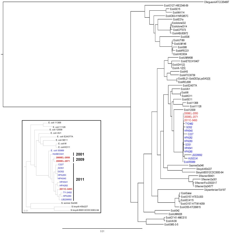
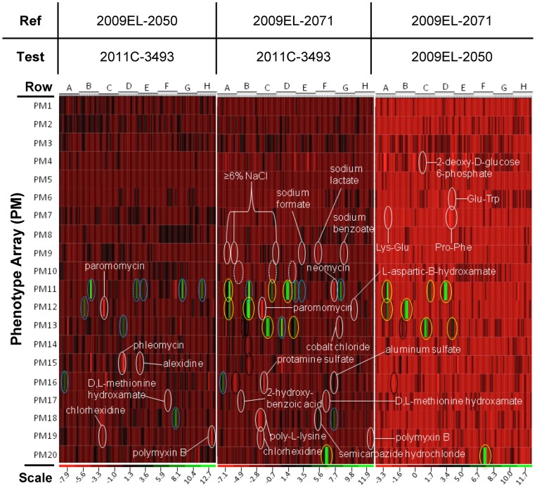
In May of 2011, an enteroaggregative Escherichia coli O104:H4 strain that had acquired a Shiga toxin 2-converting phage caused a large outbreak of bloody diarrhea in Europe which was notable for its high prevalence of hemolytic uremic syndrome cases. Several studies have described the genomic inventory and phylogenies of strains associated with the outbreak and a collection of historical E. coli O104:H4 isolates using draft genome assemblies. We present the complete, closed genome sequences of an isolate from the 2011 outbreak (2011C-3493) and two isolates from cases of bloody diarrhea that occurred in the Republic of Georgia in 2009 (2009EL-2050 and 2009EL-2071). Comparative genome analysis indicates that, while the Georgian strains are the nearest neighbors to the 2011 outbreak isolates sequenced to date, structural and nucleotide-level differences are evident in the Stx2 phage genomes, the mer/tet antibiotic resistance island, and in the prophage and plasmid profiles of the strains, including a previously undescribed plasmid with homology to the pMT virulence plasmid of Yersinia pestis. In addition, multiphenotype analysis showed that 2009EL-2071 possessed higher resistance to polymyxin and membrane-disrupting agents. Finally, we show evidence by electron microscopy of the presence of a common phage morphotype among the European and Georgian strains and a second phage morphotype among the Georgian strains. The presence of at least two stx2 phage genotypes in host genetic backgrounds that may derive from a recent common ancestor of the 2011 outbreak isolates indicates that the emergence of stx2 phage-containing E. coli O104:H4 strains probably occurred more than once, or that the current outbreak isolates may be the result of a recent transfer of a new stx2 phage element into a pre-existing stx2-positive genetic background.


Conflict of interest statement

Competing Interests: Aaron Kitzmiller and Brial Osborne are employees of Bioteam, Inc.; Kyle Hubbard, and Michael Krepps are employees of Excet Inc.; Susan Coyne and April Shea are employees of Team Ke’aki Tech. These companies provided contract labor and informatics support for this project. None of these companies have financial interests that would be influenced by the publication of this work. There are no patents, products in development or marketed products to declare. This does not alter the authors’ adherence to all the PLoS ONE policies on sharing data and materials.

Figures

Figure 1
Figure 1. Georgian strains and 2011 Outbreak Isolates have Different Plasmid Profiles. A)
Plasmid profiles of Georgian and 2011 Outbreak isolates. Left panel: PFGE of high-molecular weight plasmids. Right panel: conventional agarose gels showing small, HinDIII-resistant 1.5 kb plasmids. B) Structure of pAA-09EL50pAA-09EL50 and pAA-09EL71 and comparison to pAA/pAA-EA11 by MAUVE. The sequences are nearly identical but for the presence of IS element-associated sequence in pAA-09EL71 (arrow). C) Comparison of p09EL50 to the 108kb plasmid pLF82 from E. coli LF82 , Salmonella enteric subsp. enterica serovar Typhi typhi str. CT18 plasmid pHCM2, and Yersinia pestis CO92 plasmid pMT1. Elements common to all three plasmids are colored in light purple, whereas elements lacking in one or more strains are colored in dark purple.
Figure 2
Figure 2. The chromosomal architecture of 2009 and 2011 strains is similar. A)
Alignment of the chromosomes of the 2011 (2011C–3493) and 2009 outbreak genomes (2009EL–2050, 2009EL–2071) in MAUVE. Large regions of divergence are shown as white gaps. B) Locations of SNPs and small insertion/deletions in the 2009 and 2011 genomes. Most of the apparent differences between TY2482 and 2011EL–3493 in this map are due to sequence errors in the TY2482 sequence ; see Table S4. C) Location of insertion sequences (IS) and IS-like elements.
Figure 3
Figure 3. Georgian strains cluster with 2011 European outbreak strains.
Phylogenetic comparisons were carried using all core E. coliInset: Phylogenic analysis of subset of most closely related strains using all conserved orthologs. O104:H4 isolates are labeled in blue type; strains sequenced for this study are indicated in red type. Maximum likelihood phylogenies were constructed using RAxML (E. coli-wide tree) or Fasttree (inset) as described in Methods.
Figure 4
Figure 4. A unique repertoire of genomic islands is present in each strain. A)
Identification of chromosomal Regions of Divergence for each strain. Pair-wise BLASTp analysis of annotated protein sequences in RAST was used to determine reciprocal best-hits in each strain. Each closed, complete strain was used as a query against the other strains. RDs were defined as regions exhibiting 4 or more adjacent genes that were absent (red) or significantly divergent (<99% identical at the protein level) (yellow) in the target strains. *Annotation differences initially showed an RD3 locus but subsequent BLASTn alignments did not confirm. B) The Tet/Mer locus of the 2011 and 2009 outbreak strains. Genes encoding resistance are shown in red, transport/efflux functions are indicated in blue, regulatory functions in green, and transposon functions in dark red. Hypothetical proteins and proteins of unknown function are indicated in yellow. C) Loss of ascorbate/lyxose-metabolism genes from 2009EL-2071 due to insertion of an IS1B element.
Figure 5
Figure 5. Analysis of prophage content of O104:H4 strains.
A) Location of prophages in the genomes of the EAggEc strains analyzed in this study. Linear maps of the genomes and the location of prophages as boxes are shown. All genome sequences have the same starting position as described in methods. The prophage locations are drawn to scale. Phages are color-coded according to similarity; for example the red box indicates the stx2a phages. The exact genomic locations of the prophages in their respective genomes are given in Table 6. B) Architecture of individual prophages. Phage proteins are colored according to their predicted functions. The stx2ab genes are boxed in red; the island of pyrimidine biosynthesis genes identified as a part of this prophage by Phage_Finder is indicated by the blue box. In all cases the int genes are positioned on the left, regardless of the orientation of the prophage within the chromosome.
Figure 6
Figure 6. Genetic divergence of phage loci and induction of phage particles. A)
Differences in Shiga toxin phage genome organization and sequence. Top panel - Comparison of stx prophage region. stx2 prophage genomes were aligned in CLC Bio using the multiple alignment tool. Gaps in the regions of homology are indicated by pink spikes while areas of sequence divergence (including gaps) are indicated in red hashes. Red and green lettering indicate genes that are present in the Georgian strains but not in the European outbreak strain and vice versa, respectively. Bottom panel – Pairwise comparison of Stx2 phage protein orthologs in RAST relative to strain 2011C–3493. B) Deletion of small prophage region from Georgian isolates. C) Induction of infectious phage particles from 2011 and 2009 strains. D) Heterogeneous phage morphotypes are evident upon induction of phage from (a) 2011C–3493, induced with mitomycin C (b) 2009EL–2050, spontaneous (c) 2009EL–2071, induced with ciprofloxacin, (d) 2009EL–2071, induced with mitomycin C, (e) 2009EL–2050, induced with ciprofloxacin, (f) 2009EL–2071, induced with ciprofloxacin.
Figure 7
Figure 7. Pair-wise heat map phenotypic comparison of three E.coli strains.
Each strain was assayed for growth in the presence of various chemicals using OmniLog phenotypic microarrays, as detailed in Materials and Methods. Each well represents the average of three biological replicates. The columns represent the well position, and are denoted as Ai to Hi (i = 1 to 12) from the left to the right of the plot in each array along the x-axis (note that there were no values for wells H12). Each cell ratio value represents the average of three biological replicates. Plates PM01–PM10 contains single wells for each growth condition, while plates PM11–PM20 contain quadruplicate wells for each growth condition. Those wells which exhibited a two-fold or greater difference in growth and which were statistically significant, with P value less than 0.05 (See File S5), are indicated here. White dashed ovals indicate pH of 4.5, tetracyclines are indicated by a yellow oval, cephalosporins by a solid blue oval, lactams by a dashed blue oval, and chelators by a black oval. The apparent variation in the heat map in the comparison of 2009EL–2050 and 2009EL–2071 is a function of the reduced scale of the heat-map (−3.3 minimum value).
Figure 8
Figure 8. Model for evolution of the 2009 and 2011 E. coli O104:H4 strains.
An ancestor enteroaggregtaive strain similar to 55989 acquires a Stx2a phage (Stx2a-A), then an IncB plasmid to become the HUSEC041 strain, diverging from the lineage of strains which led to the European and Georgian strains. Those strains acquire the tet/mer resistance locus, replace the AAF/III plasmid with the AAF/I plasmid observed in all three strains, and acquire a cryptic 1.5 kb plasmid. 2009EL–2071 loses the tet/mer locus. 2009EL-2050 also acquires an IncF plasmid. 2011C–3493 acquires both an ESBL-expressing plasmid, and the Stx2a phage that initially infected the strain is displaced by a second Stx2a phage (Stx2a-B).

Similar articles

Cited by

References

    1. Kaper JB, Nataro JP, Mobley HL (2004) Pathogenic Escherichia coli. Nat Rev Microbiol 2: 123–140. - PubMed
    1. DuPont HL (2007) The growing threat of foodborne bacterial enteropathogens of animal origin. Clin Infect Dis 45: 1353–1361. - PubMed
    1. Barlow RS, Gobius KS, Desmarchelier PM (2006) Shiga toxin-producing Escherichia coli in ground beef and lamb cuts: results of a one-year study. Int J Food Microbiol 111: 1–5. - PubMed
    1. Berger CN, Sodha SV, Shaw RK, Griffin PM, Pink D, et al. (2010) Fresh fruit and vegetables as vehicles for the transmission of human pathogens. Environ Microbiol 12: 2385–2397. - PubMed
    1. Brandl MT (2006) Fitness of human enteric pathogens on plants and implications for food safety. Annu Rev Phytopathol 44: 367–392. - PubMed

Publication types

MeSH terms