. 2012;7(11):e48853.
doi: 10.1371/journal.pone.0048853. Epub 2012 Nov 1.

Endosomal maturation, Rab7 GTPase and phosphoinositides in African swine fever virus entry

Affiliations

Endosomal maturation, Rab7 GTPase and phosphoinositides in African swine fever virus entry

Miguel A Cuesta-Geijo et al. PLoS One. 2012.

Abstract

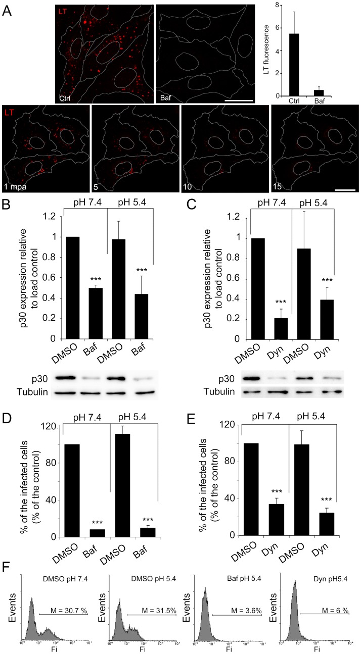
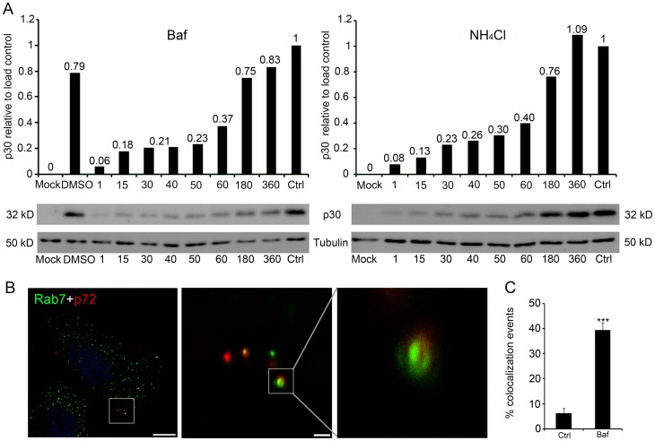
Here we analyzed the dependence of African swine fever virus (ASFV) infection on the integrity of the endosomal pathway. Using confocal immunofluorescence with antibodies against viral capsid proteins, we found colocalization of incoming viral particles with early endosomes (EE) during the first minutes of infection. Conversely, viral capsid protein was not detected in acidic late endosomal compartments, multivesicular bodies (MVBs), late endosomes (LEs) or lysosomes (LY). Using an antibody against a viral inner core protein, we found colocalization of viral cores with late compartments from 30 to 60 minutes postinfection. The absence of capsid protein staining in LEs and LYs suggested that virus desencapsidation would take place at the acid pH of these organelles. In fact, inhibitors of intraluminal acidification of endosomes caused retention of viral capsid staining virions in Rab7 expressing endosomes and more importantly, severely impaired subsequent viral protein production. Endosomal acidification in the first hour after virus entry was essential for successful infection but not thereafter. In addition, altering the balance of phosphoinositides (PIs) which are responsible of the maintenance of the endocytic pathway impaired ASFV infection. Early infection steps were dependent on the production of phosphatidylinositol 3-phosphate (PtdIns3P) which is involved in EE maturation and multivesicular body (MVB) biogenesis and on the interconversion of PtdIns3P to phosphatidylinositol 3, 5-biphosphate (PtdIns(3,5)P(2)). Likewise, GTPase Rab7 activity should remain intact, as well as processes related to LE compartment physiology, which are crucial during early infection. Our data demonstrate that the EE and LE compartments and the integrity of the endosomal maturation pathway orchestrated by Rab proteins and PIs play a central role during early stages of ASFV infection.


Conflict of interest statement

Competing Interests: The authors have declared that no competing interests exist.

Figures

Figure 1
Figure 1. Viral colocalization with endosomes at early infection.
Representative confocal micrographs of Vero cells infected with ASFV and immunostained for viral capsid proteins p72 and pE120R (shown in red), and in green, EE marker EEA1 (A), MVB marker CD63 (B), LE marker Rab7 (C) and LY marker Lamp1 (D) at 15 minutes postinfection (mpi). Scale bars, 10 µm. Cells were infected at a moi of 10 pfu/cell and adsorption was maintained at 4°C for 90 min. Unbound virus was then washed, cells were shifted to 37°C and infection was allowed to progress for indicated times. (E) Percentages of colocalization events of p72 capsid protein with EE or LE marker are expressed as means and relativized to the total cell-associated virus particles per individual cell at each time point in 10 cells in duplicates. (F) Percentages of colocalization events of p150 inner core protein with LE marker expressed as means and relativized to the total cell-associated virus particles per individual cell at each time point in 10 cells in duplicates. (G) Representative confocal micrograph of the colocalization of viral cores with Rab7 positive endosomes. Nuclei were stained with TOPRO3. (G1–4) Detail of colocalization between viral cores and LE in high magnification of the boxed areas in (G). (H) Colocalization of viral core protein p150 with Lamp1 marker.
Figure 2
Figure 2. Low intraluminal pH and Endocytosis are required for ASFV infectivity.
(A) Inhibition of intraluminal acidification of endosomes by Baf in a time dependent manner is shown using the pH sensitive dye lysotracker red. Bar 25 µm; mpa: minutes after Baf addition. (B–E) Inhibition of intraluminal acidification of endosomes with Baf and inhibition of endocytosis with Dyn impaired virus infectivity and neither Baf nor Dyn inhibition of early infection could be recovered with acid pH medium. (B) Early viral protein p30 expression in cells pretreated with 200 nM Baf or DMSO and pulsed for 1 h with ph 5.4 medium postadsorption or maintained at pH 7.4 for 6 hpi. Western blot with specific antibodies was quantified and normalized to protein load control values. Low early viral protein expression with Baf was not recovered by acid pH medium treatment. (C) Quantification of viral protein p30 expression at 6 hpi as determined by Western blot in cells pretreated with 80 μM Dyn or DMSO and maintained in presence of medium at pH 7.4 or pulsed at pH 5.4 for 1 h post-adsorption. (D) Flow cytometry of Vero cells pretreated with Baf and infected in medium at pH 7.4 or pulsed at pH 5.4 for 1 h post-adsorption. Infected cells were then detected by FACS and data normalized to infection rates in DMSO treated cells. (E) Flow cytometry of Vero cells pretreated with Dyn and infected in medium at pH 7.4 or pulsed at pH 5.4 for 1 h. Asterisks denote sadistically significant differences (*** P<0.001). (F) Representative FACS profiles obtained during the analysis are shown.
Figure 3
Figure 3. Acid pH of the late endosome is required at early stages of ASFV infection.
(A) Early viral protein p30 expression determined at 8 hpi by Western blotting with specific antibodies, quantified and normalized to protein load control values. Acid pH requirement was evidenced by the effect of lysosomotropic drug addition at any time point within the first hpi but not thereafter. (B) Representative confocal micrograph of Baf-pretreated cells fixed after 3 hpi and immunostained for major viral capsid protein p72 (red) and LE marker Rab7 (green); Bar 10 µm. Detail of colocalization between viral capsids and LEs in Baf-treated cells; Insets are magnifications of the boxed areas in the previous image, bar 1 µm. (C) Quantification of colocalization events relativized to the total number of cell-associated virions per individual cell, performed in 130 virions and expressed as means and standard deviations from two independent experiments. Asterisks denote statistically significant differences (***P<0.001).
Figure 4
Figure 4. Late endosomal compartment relevance for ASFV infection.
(A) Representative FACS profiles obtained during sorter analysis of COS-7 cells transfected with GFP-Rab7-wild type (Rab7 WT) and dominant negative mutant (GFP-Rab7-DN, T22N). R3 represents transfected cells expressing GFP to be sorted. (B) Representative confocal micrographs of transfected, sorted cells after isolation, infected with ASFV at a moi of 1 for 24 hpi and immunostained for major viral capsid protein p72 (red). Percentages of transfected infected cells decreased from 43.5% in cells expressing Rab7 WT to 1.65% in cells expressing Rab7 DN. Bar 25 µm.
Figure 5
Figure 5. ASFV entry depends on endosomal membrane phosphoinositides.
(A) Quantification of cell viability at 24 h by Trypan blue exclusion to determine the working concentration of PI3K inhibitor wortmannin. (B) Quantification of ASFV infectivity at 3 hpi (moi of 0.5 pfu/cell) in the presence of increasing concentrations of wortmannin. Data are expressed as percentages of infected cells from 30 random fields in triplicates and are means ± SD from three independent experiments. Asterisks denote statistically significant differences ***P<0.001. Representative confocal micrographs of cells immunostained for early viral protein p30 in red are shown in the right panels. Bar 20 µm. (C) Quantification of virus production in Vero cells untreated, treated with increasing concentrations of wortmannin from 2 h before adsorption during the whole infection cycle, or treated after 3 hpi. Cells were infected with ASFV at a moi of 0.5 pfu/cell for 24 hpi. Data are expressed as virus titers and are means ± SD from three independent experiments. Asterisks denote statistically significant differences ***P<0.001. (D) Viral protein expression at a range of post-infection times as determined by Western blot in cells to which 10 µM wortmannin was added 2 h before virus adsorption and maintained or left untreated.
Figure 6
Figure 6. Phosphoinositide interconversion and related late endosome fusion events in ASFV infection.
(A) Quantification of virus production in cells untreated, treated with 1 µM PIKfyve inhibitor YM201636 or treated with DMSO. Data are expressed as virus titers and are means ± SD from three independent experiments. Asterisks denote statistically significant differences **P<0.01; *P<0.05 (B) Infected cell numbers in cells treated with PIKfyve inhibitor (YM201636) at several time points or an equivalent volume of DMSO. Data are expressed as the number of infected cells at 6 hpi (moi of 1 pfu/cell) from 20 random fields and are means ± SD from two independent experiments. Asterisks denote statistically significant differences (***P<0.001 and **P<0.01). (C) Representative confocal micrographs of infected and non-infected PIKfyve-treated cells, immunostained for Rab7 (green) and viral protein p72 (red). The characteristic phenotype of cytoplasmic vacuoles due to impaired endosome fusion was readily found in uninfected cells. Infected cells are recognized in the image as those harboring viral factories in red and lacked cytoplasmic vacuolization phenotype. Bar 10 µm.
Figure 7
Figure 7. Model of ASFV infection progress through the endosomal pathway.
ASFV enters the host cell by clathrin-coated vesicles (CCVs) from clathrin-coated pits (CCPs) and clathrin molecules are recycled to the plasma membrane (PM) as the virus progresses to the endosomal pathway. First, virions gain access to EEs from PM. The EE compartment is characterized by the presence of Rab5 and EEA1. ASFV is then directed from the vacuolar domain of the EE to the acidic late compartments. Subsequently, the virions reach CD63 enriched membranes of MVBs. Under the acid intraluminal pH of these endosomes, viral capsid would be degraded and viral cores would reach LE which depends on the presence of Rab7. At this stage, viral cores could egress to the cytosol to reach their replication site at the perinuclear area. In this process, the PIs composition of the endosomal membrane seemed to be crucial. PtdIns3P is synthesized by PI3K and this process is inhibited by PI3K inhibitor wortmannin and PtdIns(3,5)P2, which is synthesized by the enzyme PIKfyve, a process blocked by the inhibitor YM201636. These PIs interconversions on the endosomal membrane are necessary for a successful infection.

Similar articles

Cited by

  • ASFV infection induces lipid metabolic disturbances and promotes viral replication.
    Chu X, Ge S, Li Y, Zhang Q, Cui X, Zuo Y, Li R, Sun H, Yin L, Wang Z, Li J, Xiao Y, Wang Z. Chu X, et al. Front Microbiol. 2025 Jan 7;15:1532678. doi: 10.3389/fmicb.2024.1532678. eCollection 2024. Front Microbiol. 2025. PMID: 39872814 Free PMC article.
  • Guidelines for the use and interpretation of assays for monitoring autophagy (4th edition)1.
    Klionsky DJ, Abdel-Aziz AK, Abdelfatah S, Abdellatif M, Abdoli A, Abel S, Abeliovich H, Abildgaard MH, Abudu YP, Acevedo-Arozena A, Adamopoulos IE, Adeli K, Adolph TE, Adornetto A, Aflaki E, Agam G, Agarwal A, Aggarwal BB, Agnello M, Agostinis P, Agrewala JN, Agrotis A, Aguilar PV, Ahmad ST, Ahmed ZM, Ahumada-Castro U, Aits S, Aizawa S, Akkoc Y, Akoumianaki T, Akpinar HA, Al-Abd AM, Al-Akra L, Al-Gharaibeh A, Alaoui-Jamali MA, Alberti S, Alcocer-Gómez E, Alessandri C, Ali M, Alim Al-Bari MA, Aliwaini S, Alizadeh J, Almacellas E, Almasan A, Alonso A, Alonso GD, Altan-Bonnet N, Altieri DC, Álvarez ÉMC, Alves S, Alves da Costa C, Alzaharna MM, Amadio M, Amantini C, Amaral C, Ambrosio S, Amer AO, Ammanathan V, An Z, Andersen SU, Andrabi SA, Andrade-Silva M, Andres AM, Angelini S, Ann D, Anozie UC, Ansari MY, Antas P, Antebi A, Antón Z, Anwar T, Apetoh L, Apostolova N, Araki T, Araki Y, Arasaki K, Araújo WL, Araya J, Arden C, Arévalo MA, Arguelles S, Arias E, Arikkath J, Arimoto H, Ariosa AR, Armstrong-James D, Arnauné-Pelloquin L, Aroca A, Arroyo DS, Arsov I, Artero R, Asaro DML, Aschner M, Ashrafizadeh M, Ashur-Fabian O, Atanasov AG, Au AK, Auberger P, Auner HW, Aurelian L, Autelli R… See abstract for full author list ➔ Klionsky DJ, et al. Autophagy. 2021 Jan;17(1):1-382. doi: 10.1080/15548627.2020.1797280. Epub 2021 Feb 8. Autophagy. 2021. PMID: 33634751 Free PMC article.
  • African Swine Fever Virus Exhibits Distinct Replication Defects in Different Cell Types.
    Gao Y, Xia T, Bai J, Zhang L, Jiang X, Yang X, Zhang K, Jiang P. Gao Y, et al. Viruses. 2022 Nov 26;14(12):2642. doi: 10.3390/v14122642. Viruses. 2022. PMID: 36560646 Free PMC article.
  • Feline herpesvirus 1 (FHV-1) enters the cell by receptor-mediated endocytosis.
    Synowiec A, Dąbrowska A, Pachota M, Baouche M, Owczarek K, Niżański W, Pyrc K. Synowiec A, et al. J Virol. 2023 Aug 31;97(8):e0068123. doi: 10.1128/jvi.00681-23. Epub 2023 Jul 26. J Virol. 2023. PMID: 37493545 Free PMC article.
  • The Potential of Disabled Infectious Single Cycle (DISC) Virus Platforms for Next Generation African Swine Fever Vaccine Development.
    Jia F, Lynch SE, Williams DT. Jia F, et al. Transbound Emerg Dis. 2025 Jul 14;2025:8573171. doi: 10.1155/tbed/8573171. eCollection 2025. Transbound Emerg Dis. 2025. PMID: 40692871 Free PMC article. Review.

References

    1. Dixon L, Costa JV, Escribano JM, Rock DL, Vinuela E, et al.. (2000) Asfarviridae. New York: Academic Press. 159–165 p.
    1. Andres G, Simon-Mateo C, Vinuela E (1997) Assembly of African swine fever virus: role of polyprotein pp220. J Virol 71: 2331–2341. - PMC - PubMed
    1. Andres G, Garcia-Escudero R, Simon-Mateo C, Vinuela E (1998) African swine fever virus is enveloped by a two-membraned collapsed cisterna derived from the endoplasmic reticulum. J Virol 72: 8988–9001. - PMC - PubMed
    1. Carrascosa AL, del Val M, Santaren JF, Vinuela E (1985) Purification and properties of African swine fever virus. J Virol 54: 337–344. - PMC - PubMed
    1. Breese SS Jr, DeBoer CJ (1966) Electron microscope observations of African swine fever virus in tissue culture cells. Virology 28: 420–428. - PubMed

Publication types

Substances