. 2012;7(11):e49127.
doi: 10.1371/journal.pone.0049127. Epub 2012 Nov 1.

Fibroblast growth factor 10-fibroblast growth factor receptor 2b mediated signaling is not required for adult glandular stomach homeostasis

Affiliations

Fibroblast growth factor 10-fibroblast growth factor receptor 2b mediated signaling is not required for adult glandular stomach homeostasis

Allison L Speer et al. PLoS One. 2012.

Abstract

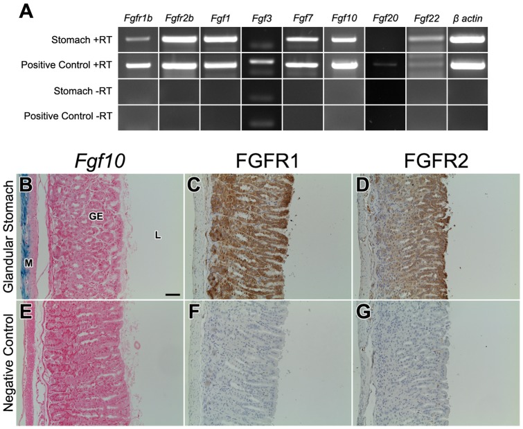
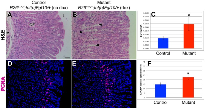
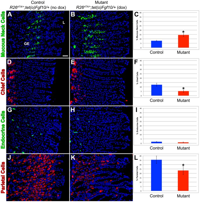
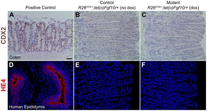
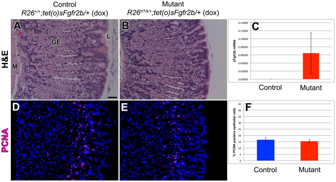
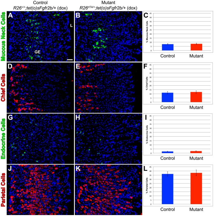
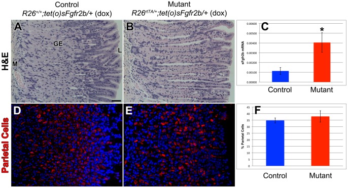
The signaling pathways that are essential for gastric organogenesis have been studied in some detail; however, those that regulate the maintenance of the gastric epithelium during adult homeostasis remain unclear. In this study, we investigated the role of Fibroblast growth factor 10 (FGF10) and its main receptor, Fibroblast growth factor receptor 2b (FGFR2b), in adult glandular stomach homeostasis. We first showed that mouse adult glandular stomach expressed Fgf10, its receptors, Fgfr1b and Fgfr2b, and most of the other FGFR2b ligands (Fgf1, Fgf7, Fgf22) except for Fgf3 and Fgf20. Fgf10 expression was mesenchymal whereas FGFR1 and FGFR2 expression were mostly epithelial. Studying double transgenic mice that allow inducible overexpression of Fgf10 in adult mice, we showed that Fgf10 overexpression in normal adult glandular stomach increased epithelial proliferation, drove mucous neck cell differentiation, and reduced parietal and chief cell differentiation. Although a similar phenotype can be associated with the development of metaplasia, we found that Fgf10 overexpression for a short duration does not cause metaplasia. Finally, investigating double transgenic mice that allow the expression of a soluble form of Fgfr2b, FGF10's main receptor, which acts as a dominant negative, we found no significant changes in gastric epithelial proliferation or differentiation in the mutants. Our work provides evidence, for the first time, that the FGF10-FGFR2b signaling pathway is not required for epithelial proliferation and differentiation during adult glandular stomach homeostasis.


Conflict of interest statement

Competing Interests: The authors have read the journal's policy and have the following conflict: SB currently serves as an editor of PLOS ONE. The authors would like to confirm that this does not alter the authors' adherence to all the PLOS ONE policies on sharing data and materials.

Figures

Figure 1
Figure 1. Normal expression pattern of Fgf10, its receptors, all other FGFR2b ligands in adult glandular stomach.
(A) PCR of adult glandular stomach and E14.5 wildtype whole embryo (positive control). RT negative controls for both are all negative. (B,E) β-galactosidase staining of adult glandular stomach in Fgf10LacZ/+ (B) and wildtype Fgf10+/+ (E) mice. Fgf10 expression occurs only in the mesenchyme. Immunohistochemical analysis of wildtype adult glandular stomach sections stained with anti-FGFR1 (C) or anti-FGFR2 (D). (F,G) Negative control stainings, in which the primary antibody was absent, show minimal to no signal. FGFR1 and FGFR2 staining is strong throughout the gastric epithelium whereas the mesenchymal staining is much weaker. The scale bar is 50 µm. M = mesenchyme, GE = gastric epithelium, L = lumen.
Figure 2
Figure 2. Fgf10 overexpression during homeostasis changes gastric gland histology but not epithelial proliferation in glandular stomach.
(A–B) Hematoxylin and eosin staining of sections of adult glandular stomach in control littermate (R26rtTA/+; tet(O)Fgf10/+ without dox) (A) and mutant Fgf10 overexpressing mice (R26rtTA/+; tet(O)Fgf10/+ with dox) (B). There is a visible decrease in the number of parietal cells and an obvious shift of the mucous neck cells from the luminal side towards the gastric gland base (black arrowheads). (C) qRT-PCR showing a significant increase in Fgf10 expression in the mutants vs controls (*p<0.05). (D-E) PCNA immunostaining of sections of adult glandular stomach in control littermate (R26rtTA/+; tet(O)Fgf10/+ without dox) (D) and mutant Fgf10 overexpressing mice (R26rtTA/+; tet(O)Fgf10/+ with dox) (E). (F) Quantification of the percentage of PCNA-positive epithelial cells showed a statistically significant increase in the percentage of PCNA positive epithelial cells indicating increased gastric epithelial proliferation in mutant Fgf10 overexpressing mice (R26rtTA/+; tet(O)Fgf10/+ with dox) compared to control littermates (R26rtTA/+; tet(O)Fgf10/+ without dox). Dox was given for 10 days. The scale bar is 50 µm. M = mesenchyme, GE = gastric epithelium, L = lumen.
Figure 3
Figure 3. Gastric epithelial differentiation is significantly altered by Fgf10 overexpression during homeostasis.
Immunostaining for markers of terminally differentiated gastric epithelial cells of sections of adult glandular stomach in control littermate (R26rtTA/+; tet(O)Fgf10/+ without dox) (A,D,G,J) and mutant Fgf10 overexpressing mice (R26rtTA/+; tet(O)Fgf10/+ with dox) (B,E,H,K). (A-B) Mucous neck cells are identified by GSII and (C) quantification reveals a significant increase in mutant Fgf10 overexpressing mice compared to control littermates (*p<0.05). (D,E) Chief cells are marked by intrinsic factor and (F) quantification shows a significant decrease in chief cell number (*p<0.05). (G,H) Endocrine cells are identified by chromogranin A and (I) quantification demonstrates no significant change in endocrine cell number. (J,K) Parietal cells are visualized with H/K ATPase immunostaining and (L) quantification demonstrates a significant reduction in mutant Fgf10 overexpressing mice compared to control littermates (*p<0.05). Dox was given for 10 days. The scale bar is 50 µm. GE = gastric epithelium, L = lumen.
Figure 4
Figure 4. Fgf10 overexpression during homeostasis does not cause metaplasia of the gastric epithelium.
(A–C) CDX2, a marker of intestinal metaplasia (IM), immunostaining in adult colon (positive control) (A), control littermate (R26rtTA/+; tet(O)Fgf10/+ without dox) (B) and mutant Fgf10 overexpressing mice (R26rtTA/+; tet(O)Fgf10/+ with dox) (C). (D-F) HE4, a marker of spasmolytic polypeptide expressing metaplasia (SPEM), immunostaining in human epididymis (positive control) (D), control littermate (R26rtTA/+; tet(O)Fgf10/+ without dox) (E) and mutant Fgf10 overexpressing mice (R26rtTA/+; tet(O)Fgf10/+) (F). Dox was given for 10 days. The scale bar is 50 µm.
Figure 5
Figure 5. FGF10-FGFR2b mediated signaling is not required for normal gastric histology and epithelial proliferation during homeostasis.
Hematoxylin and eosin staining of sections of adult glandular stomach in control littermate (R26+/+; tet(O)sFgfr2b/+ with dox) (A) and mutant mice (R26rtTA/+; tet(O)sFgfr2b/+ with dox) (B) showing normal stomach histology. (C) qRT-PCR confirming robust expression of sFgfr2b in the mutants as compared to controls where the level of sFgfr2b is nearly undetectable. PCNA immunostaining of sections of adult glandular stomach in control littermate (R26+/+; tet(O)sFgfr2b/+ with dox) (D) and mutant mice (R26rtTA/+; tet(O)sFgfr2b/+ with dox) (E) and (F) quantification of the percentage of PCNA-positive epithelial cells showing no significant change in gastric epithelial proliferation in mutant mice compared to control littermates. Dox was given for 1 month. The scale bar is 50 µm. M = mesenchyme, GE = gastric epithelium, L = lumen.
Figure 6
Figure 6. FGF10-FGFR2b mediated signaling is not required for gastric epithelial differentiation during homeostasis.
Mucous neck cells are marked by GSII immunostaining of sections of adult glandular stomach in control littermate (R26+/+; tet(O)sFgfr2b/+with dox) (A) and mutant mice (R26rtTA/+; tet(O)sFgfr2b/+ with dox) (B) and (C) quantification reveals no difference in mucous neck cell number. Chief cells are identified by intrinsic factor immunostaining of sections of adult glandular stomach in control littermate (R26+/+; tet(O)sFgfr2b/+ with dox) (D) and mutant mice (R26rtTA/+; tet(O)sFgfr2b/+ with dox) (E) and (F) quantification reveals no significant change in chief cell number. (G,H) Endocrine cells are identified by chromogranin A and (I) quantification shows no significant change in mutant sFgfr2b overexpressing mice compared to control littermates. Parietal cells are identified by H/K ATPase immunostaining of sections of adult glandular stomach in control littermate (R26+/+; tet(O)sFgfr2b/+with dox) (J) and mutant mice (R26rtTA/+; tet(O)sFgfr2b/+ with dox) (K) and (L) quantification reveals no significant change in parietal cell number. Dox was given for 1 month. The scale bar is 50 µm. GE = gastric epithelium, L = lumen.
Figure 7
Figure 7. FGF10-FGFR2b mediated signaling is not required for parietal cell differentiation during homeostasis.
Hematoxylin and eosin staining of sections of adult glandular stomach in control littermate (R26+/+; tet(O)sFgfr2b/+ with dox) (A) and mutant mice (R26rtTA/+; tet(O)sFgfr2b/+ with dox) (B) showing normal stomach histology. (C) qRT-PCR confirming a significant increase in the expression of sFgfr2b in the mutants as compared to controls (*p<0.05). Parietal cells are identified by H/K ATPase immunostaining of sections of adult glandular stomach in control littermate (R26+/+; tet(O)sFgfr2b/+ with dox) (D) and mutant mice (R26rtTA/+; tet(O)sFgfr2b/+ with dox) (E) and (F) quantification reveals no significant change in parietal cell number. Dox was given for 3 months. The scale bar is 50 µm. M = mesenchyme, GE = gastric epithelium, L = lumen.

Similar articles

Cited by

References

    1. Ferlay J, Shin HR, Bray F, Forman D, Mathers C, et al. (2010) Estimates of worldwide burden of cancer in 2008: GLOBOCAN 2008. Int J Cancer 127: 2893–2917. - PubMed
    1. Kamangar F, Dores GM, Anderson WF (2006) Patterns of cancer incidence, mortality, and prevalence across five continents: defining priorities to reduce cancer disparities in different geographic regions of the world. J Clin Oncol 24: 2137–2150. - PubMed
    1. Zabaleta J (2012) Multifactorial etiology of gastric cancer. Methods Mol Biol 863: 411–435. - PMC - PubMed
    1. Goldenring JR, Nam KT (2010) Oxyntic atrophy, metaplasia, and gastric cancer. Prog Mol Biol Transl Sci 96: 117–131. - PMC - PubMed
    1. Nozaki K, Weis V, Wang TC, Falus A, Goldenring JR (2009) Altered gastric chief cell lineage differentiation in histamine-deficient mice. Am J Physiol Gastrointest Liver Physiol 296: G1211–1220. - PMC - PubMed

Publication types

Substances