

Effect of dietary calcium on spinal bone fusion in an ovariectomized rat model
- PMID: 23133713
- PMCID: PMC3488633
- DOI: 10.3340/jkns.2012.52.4.281
Effect of dietary calcium on spinal bone fusion in an ovariectomized rat model
Abstract
Objective: To evaluate the effect of calcium supplementation on spinal bone fusion in ovariectomized (OVX) rats.
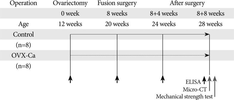
Methods: Sixteen female Sprague Dawley rats underwent bilateral ovariectomy at 12 weeks of age to induce osteoporosis and were randomly assigned to two groups : control group (n=8) and calcium-supplemented group (OVX-Ca, n=8). Autologous spinal bone fusion surgery was performed on both groups 8 weeks later. After fusion surgery, the OVX-Ca group was supplemented with calcium in drinking water for 8 weeks. Blood was obtained 4 and 8 weeks after fusion surgery. Eight weeks after fusion surgery, the rats were euthanized and the L4-5 spine removed. Bone fusion status and fusion volume were evaluated by manual palpation and three-dimensional computed tomography.
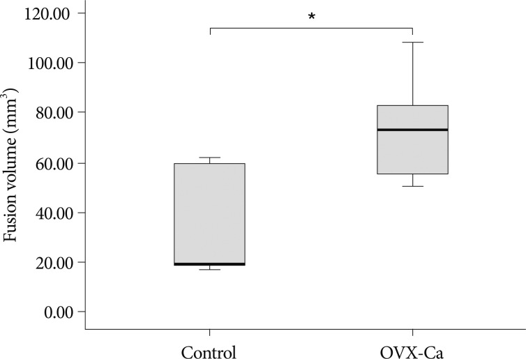
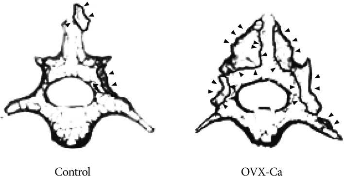
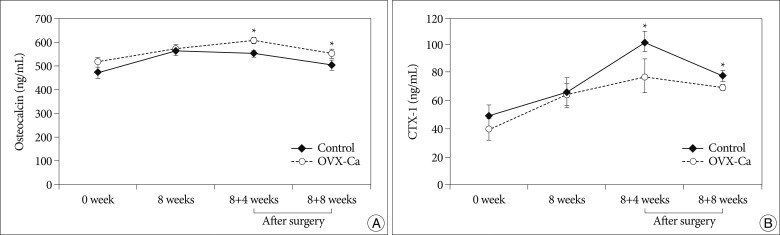
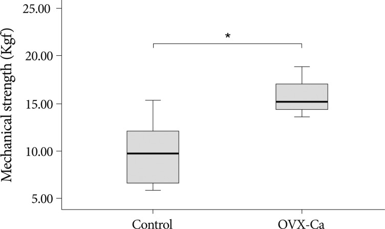
Results: The mean fusion volume in the L4-5 spine was significantly greater in the OVX-Ca group (71.80±8.06 mm(3)) than in controls (35.34±8.24 mm(3)) (p<0.01). The level of osteocalcin, a bone formation marker, was higher in OVX-Ca rats than in controls 4 weeks (610.08±10.41 vs. 551.61±12.34 ng/mL) and 8 weeks (552.05±19.67 vs. 502.98±22.76 ng/mL) after fusion surgery (p<0.05). The level of C-terminal telopeptide fragment of type I collagen, a bone resorption marker, was significantly lower in OVX-Ca rats than in controls 4 weeks (77.07±12.57 vs. 101.75±7.20 ng/mL) and 8 weeks (69.58±2.45 vs. 77.15±4.10 ng/mL) after fusion surgery (p<0.05). A mechanical strength test showed that the L4-5 vertebrae in the OVX-Ca group withstood a 50% higher maximal load compared with the controls (p<0.01).
Conclusion: Dietary calcium given to OVX rats after lumbar fusion surgery improved fusion volume and mechanical strength in an ovariectomized rat model.
Keywords: Calcium; Osteoporosis; Ovariectomized rat; Spinal bone fusion.
Figures
Similar articles
-
Effect of a selective estrogen receptor modulator on bone formation in osteoporotic spine fusion using an ovariectomized rat model.Spine J. 2016 Jan 1;16(1):72-81. doi: 10.1016/j.spinee.2015.08.061. Epub 2015 Sep 4. Spine J. 2016. PMID: 26343247
-
Short- and long-term effects of calcium and exercise on bone mineral density in ovariectomized rats.Br J Nutr. 2001 Oct;86(4):521-7. doi: 10.1079/bjn2001428. Br J Nutr. 2001. PMID: 11591240
-
Alendronate Prevents Intervertebral Disc Degeneration Adjacent to a Lumbar Fusion in Ovariectomized Rats.Spine (Phila Pa 1976). 2015 Oct 15;40(20):E1073-83. doi: 10.1097/BRS.0000000000001092. Spine (Phila Pa 1976). 2015. PMID: 26731708
-
BMP-2 induced early bone formation in spine fusion using rat ovariectomy osteoporosis model.Spine J. 2013 Oct;13(10):1273-80. doi: 10.1016/j.spinee.2013.06.010. Epub 2013 Aug 13. Spine J. 2013. PMID: 23953506
-
The time-dependent effect of ibandronate on bone graft remodeling in an ovariectomized rat spinal arthrodesis model.Spine J. 2014 Aug 1;14(8):1748-57. doi: 10.1016/j.spinee.2014.01.042. Epub 2014 Jan 30. Spine J. 2014. PMID: 24486470
Cited by
-
The effects of Cosmos caudatus (ulam raja) on dynamic and cellular bone histomorphometry in ovariectomized rats.BMC Res Notes. 2013 Jun 24;6:239. doi: 10.1186/1756-0500-6-239. BMC Res Notes. 2013. PMID: 23800238 Free PMC article.
-
Bone Health Optimization in Adult Spinal Deformity Patients: A Narrative Review.J Clin Med. 2024 Aug 19;13(16):4891. doi: 10.3390/jcm13164891. J Clin Med. 2024. PMID: 39201032 Free PMC article. Review.
-
Thiazide diuretic use is associated with fewer hardware complications after anterior cervical discectomy and fusion.Eur Spine J. 2025 Apr;34(4):1295-1300. doi: 10.1007/s00586-025-08756-6. Epub 2025 Mar 6. Eur Spine J. 2025. PMID: 40050514
-
Pre-Clinical Models in Implant Dentistry: Past, Present, Future.Biomedicines. 2021 Oct 26;9(11):1538. doi: 10.3390/biomedicines9111538. Biomedicines. 2021. PMID: 34829765 Free PMC article. Review.
-
Preventive effects of collagen Peptide from deer sinew on bone loss in ovariectomized rats.Evid Based Complement Alternat Med. 2014;2014:627285. doi: 10.1155/2014/627285. Epub 2014 Jul 1. Evid Based Complement Alternat Med. 2014. PMID: 25101135 Free PMC article.
References
-
- Abe Y, Takahata M, Ito M, Irie K, Abumi K, Minami A. Enhancement of graft bone healing by intermittent administration of human parathyroid hormone (1-34) in a rat spinal arthrodesis model. Bone. 2007;41:775–785. - PubMed
-
- Boden SD. Overview of the biology of lumbar spine fusion and principles for selecting a bone graft substitute. Spine (Phila Pa 1976) 2002;27:S26–S31. - PubMed
-
- Borgström F, Ström O, Marin F, Kutahov A, Ljunggren O. Cost effectiveness of teriparatide and PTH(1-84) in the treatment of postmenopausal osteoporosis. J Med Econ. 2010;13:381–392. - PubMed
-
- Bridwell KH, Sedgewick TA, O'Brien MF, Lenke LG, Baldus C. The role of fusion and instrumentation in the treatment of degenerative spondylolisthesis with spinal stenosis. J Spinal Disord. 1993;6:461–472. - PubMed
LinkOut - more resources
Full Text Sources
