

Locations and clinical significance of non-hemorrhagic brain lesions in diffuse axonal injuries
- PMID: 23133728
- PMCID: PMC3488648
- DOI: 10.3340/jkns.2012.52.4.377
Locations and clinical significance of non-hemorrhagic brain lesions in diffuse axonal injuries
Abstract
Objective: Detection of focal non-hemorrhagic lesion (NHL) has become more efficient in diffuse axonal injury (DAI) patients using an MRI. The aims of this study are to find out the radiological distribution, progress of NHL and its clinical significance.
Methods: Between September 2005 and October 2011, 32 individuals with NHLs on brain MRI were enrolled. NHLs were classified by brain location into 4 major districts and 13 detailed locations including cortical and subcortical, corpus callosum, deep nuclei and adjacent area, and brainstem. The severity of NHL was scored from grades 1 to 4, according to the number of districts involved. Fourteen patients with NHL were available for MRI follow-up and an investigation of the changes was conducted.
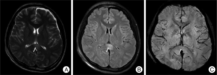
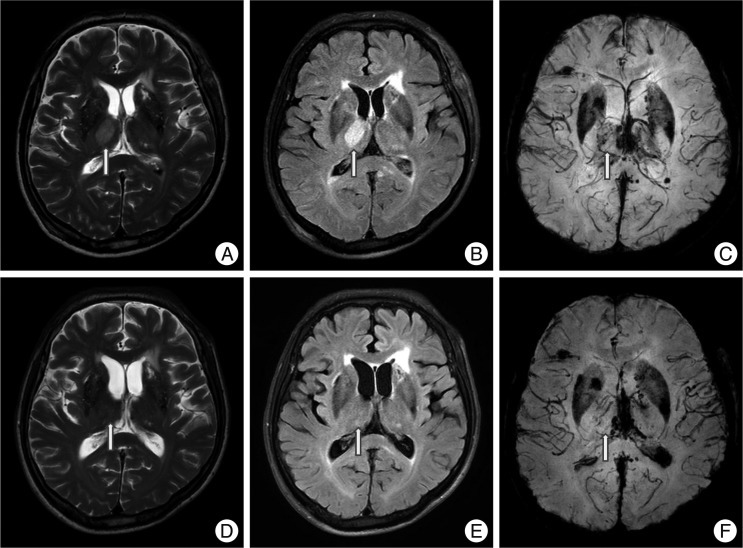
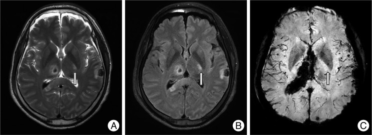
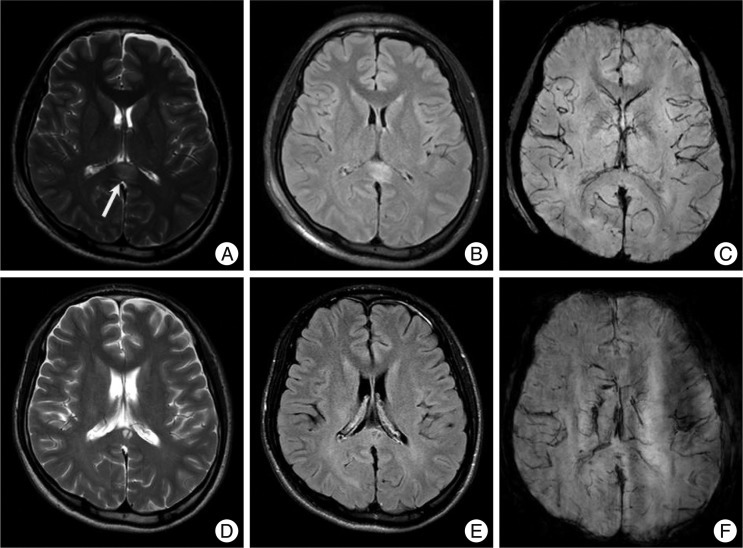
Results: Thirty-two patients had 59 NHLs. The most common district of NHL was cortical and subcortical area; 15 patients had 20 NHSs. However the most common specific location was the splenium of the corpus callosum; 14 patients had 14 lesions. The more lesions patients had, the lower the GCS, however, this was not a statistically meaningful difference. On follow-up MRI in 14 patients, out of 24 lesions, 13 NHLs resolved, 5 showed cystic change, and 6 showed atrophic changes.
Conclusion: NHLs were located most commonly in the splenium and occur frequently in the thalamus and the mesial temporal lobe. Because most NHS occur concomitantly with hemorrhagic lesions, it was difficult to determine their effects on prognosis. Since most NHLs resolve completely, they are probably less significant to prognosis than hemorrhagic lesions.
Keywords: Corpus callosum; Diffuse axonal injury; Magnetic resonance imaging; Non-hemorrhagic; Traumatic brain injury.
Figures

References
-
- Adams JH. Diffuse axonal injury in non-missile head injury. Injury. 1982;13:444–445. - PubMed
-
- Adams JH. The autopsy in fatal non-missile head injuries. Curr Top Pathol. 1988;76:1–22. - PubMed
-
- Adams JH, Doyle D, Ford I, Gennarelli TA, Graham DI, McLellan DR. Diffuse axonal injury in head injury : definition, diagnosis and grading. Histopathology. 1989;15:49–59. - PubMed
-
- Adams H, Mitchell DE, Graham DI, Doyle D. Diffuse brain damage of immediate impact type. Its relationship to 'primary brain-stem damage' in head injury. Brain. 1977;100:489–502. - PubMed
-
- Ezaki Y, Tsutsumi K, Morikawa M, Nagata I. Role of diffusion-weighted magnetic resonance imaging in diffuse axonal injury. Acta Radiol. 2006;47:733–740. - PubMed
LinkOut - more resources
Full Text Sources
