

Meningeal layers around anterior clinoid process as a delicate area in extradural anterior clinoidectomy : anatomical and clinical study
- PMID: 23133730
- PMCID: PMC3488650
- DOI: 10.3340/jkns.2012.52.4.391
Meningeal layers around anterior clinoid process as a delicate area in extradural anterior clinoidectomy : anatomical and clinical study
Abstract
Objective: Removal of the anterior clinoid process (ACP) is an essential process in the surgery of giant or complex aneurysms located near the proximal internal carotid artery or the distal basilar artery. An extradural clinoidectomy must be performed within the limits of the meningeal layers surrounding the ACP to prevent morbid complications. To identify the safest method of extradural exposure of the ACP, anatomical studies were done on cadaver heads.
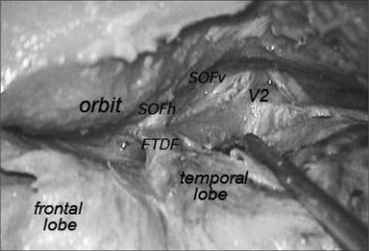
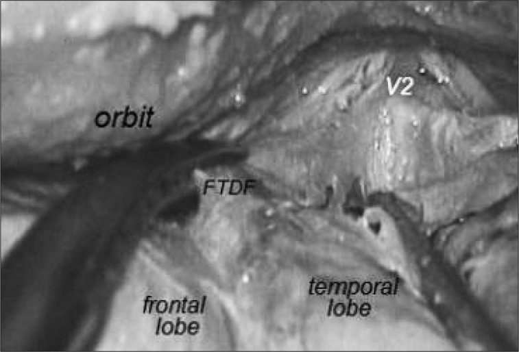
Methods: Anatomical dissections for extradural exposure of the ACP were performed on both sides of seven cadavers. Before dividing the frontotemporal dural fold (FTDF), we measured its length from the superomedial apex attached to the periorbita to the posterolateral apex which connects to the anterosuperior end of the cavernous sinus.
Results: The average length of the FTDF on cadaver dissections was 7 mm on the right side and 7.14 mm on the left side. Cranial nerves were usually exposed when cutting FTDF more than 7 mm of the FTDF.
Conclusion: The most delicate area in an extradural anterior clinoidectomy is the junction of the FTDF and the anterior triangular apex of the cavernous sinus. The FTDF must be cut from the anterior side of the triangle at the periorbital side rather than from the dural side. The length of the FTDF incision must not exceed 7 mm to avoid cranial nerve injury.
Keywords: Anatomical study; Extradural clinoidectomy; Frontotemporal dural fold; Superior orbital fissure.
Figures


References
-
- Avci E, Bademci G, Ozturk A. Microsurgical landmarks for safe removal of anterior clinoid process. Minim Invasive Neurosurg. 2005;48:268–272. - PubMed
-
- Bayassi S. [Meningo-orbital fold (MOF) as a guiding point in extradural approach to the anterior clinoid process] Neurol Neurochir Pol. 2005;39:49–55. - PubMed
-
- Collignon F, Link M. Paraclinoid and cavernous sinus regions : measurement of critical structures relevant for surgical procedure. Clin Anat. 2005;18:3–9. - PubMed
-
- Coscarella E, Başkaya MK, Morcos JJ. An alternative extradural exposure to the anterior clinoid process : the superior orbital fissure as a surgical corridor. Neurosurgery. 2003;53:162–166. discussion 166-167. - PubMed
-
- Day AL. Aneurysms of the ophthalmic segment. A clinical and anatomical analysis. J Neurosurg. 1990;72:677–691. - PubMed
LinkOut - more resources
Full Text Sources
Miscellaneous
