

Transorbital penetrating intracranial injury by a chopstick
- PMID: 23133735
- PMCID: PMC3488655
- DOI: 10.3340/jkns.2012.52.4.414
Transorbital penetrating intracranial injury by a chopstick
Abstract
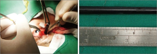
A 38-year-old man fell from a chair with a chopstick in his hand. The chopstick penetrated his left eye. He noticed pain, swelling, and numbness around his left eye. On physical examination, a linear wound was noted at the medial aspect of the left eyelid. Noncontrast computed tomography (CT) study showed a linear hypodense structure extending from the medial aspect of the left orbit to the occipital bone, suggesting a foreign body. This foreign body was hyperdense relative to normal parenchyma. From a CT scan with 3-dimensional reconstruction, the foreign body was found to be passing through the optic canal into the cranium. The clear plastic chopstick was withdrawn without difficulty. The patient was discharged home 3 weeks after his surgery. A treatment plan for a transorbital penetrating injury should be determined by a multidisciplinary team, with input from neurosurgeons and ophthalmologists.
Keywords: Craniocerebral trauma; Foreign body; Orbit; Penetrating.
Figures


Similar articles
-
A case of death of purulent meningitis caused by transorbital intracranial penetrating injury due to long-term residual bamboo chopstick.Leg Med (Tokyo). 2022 Mar;55:102012. doi: 10.1016/j.legalmed.2021.102012. Epub 2022 Jan 3. Leg Med (Tokyo). 2022. PMID: 34998200 Review.
-
Transorbital penetrating head injury by a wooden chopstick in the cavernous sinus: a case report and literature review.Nagoya J Med Sci. 2023 Feb;85(1):179-184. doi: 10.18999/nagjms.85.1.179. Nagoya J Med Sci. 2023. PMID: 36923619 Free PMC article. Review.
-
Transorbital penetrating injury by a chopstick--case report.Neurol Med Chir (Tokyo). 2001 Jul;41(7):345-8. doi: 10.2176/nmc.41.345. Neurol Med Chir (Tokyo). 2001. PMID: 11487998
-
The Role of Intraoperative Cerebral Angiography in Transorbital Intracranial Penetrating Trauma: A Case Report and Literature Review.World Neurosurg. 2017 Jan;97:761.e5-761.e10. doi: 10.1016/j.wneu.2016.09.083. Epub 2016 Sep 28. World Neurosurg. 2017. PMID: 27693768 Review.
-
Transorbital Penetrating Intracranial Injury by a Battery.J Craniofac Surg. 2018 Jan;29(1):e61-e64. doi: 10.1097/SCS.0000000000004054. J Craniofac Surg. 2018. PMID: 29065045
Cited by
-
Transorbital penetrating intracranial injury involving bilateral frontal lobes with evisceration of right eye: A case report.Clin Case Rep. 2024 May 31;12(6):e9018. doi: 10.1002/ccr3.9018. eCollection 2024 Jun. Clin Case Rep. 2024. PMID: 38827937 Free PMC article.
-
Transorbital stab injury with retained knife: a narrow escape.Case Rep Crit Care. 2014;2014:754053. doi: 10.1155/2014/754053. Epub 2014 Sep 23. Case Rep Crit Care. 2014. PMID: 25328717 Free PMC article.
-
Management of Penetrating Skull Base Injury: A Single Institutional Experience and Review of the Literature.Biomed Res Int. 2017;2017:2838167. doi: 10.1155/2017/2838167. Epub 2017 Jul 30. Biomed Res Int. 2017. PMID: 28828384 Free PMC article. Review.
-
Transorbital-penetrating intracranial injury due to a homemade metal arrow: A case report.Ann Med Surg (Lond). 2020 Jul 28;57:183-189. doi: 10.1016/j.amsu.2020.07.049. eCollection 2020 Sep. Ann Med Surg (Lond). 2020. PMID: 32774851 Free PMC article.
-
Childhood penetrating intracranial injury by non-metallic objects: a case report of three pediatric cases.Transl Pediatr. 2025 May 30;14(5):1039-1049. doi: 10.21037/tp-2024-550. Epub 2025 May 27. Transl Pediatr. 2025. PMID: 40519730 Free PMC article.
References
-
- Carothers A. Orbitofacial wounds and cerebral artery injuries caused by umbrella tips. JAMA. 1978;239:1151–1152. - PubMed
-
- Chibbaro S, Tacconi L. Orbito-cranial injuries caused by penetrating non-missile foreign bodies. Experience with eighteen patients. Acta Neurochir (Wien) 2006;148:937–941. discussion 941-942. - PubMed
-
- Di Roio C, Jourdan C, Mottolese C, Convert J, Artru F. Craniocerebral injury resulting from transorbital stick penetration in children. Childs Nerv Syst. 2000;16:503–506. discussion 507. - PubMed
-
- du Trevou MD, van Dellen JR. Penetrating stab wounds to the brain : the timing of angiography in patients presenting with the weapon already removed. Neurosurgery. 1992;31:905–911. discussion 911-912. - PubMed
Publication types
LinkOut - more resources
Full Text Sources
