

A tragical paediatric case history of intraorbital and intracranial epithelioid hemangioendothelioma
- PMID: 23133764
- PMCID: PMC3485861
- DOI: 10.1155/2012/396097
A tragical paediatric case history of intraorbital and intracranial epithelioid hemangioendothelioma
Abstract
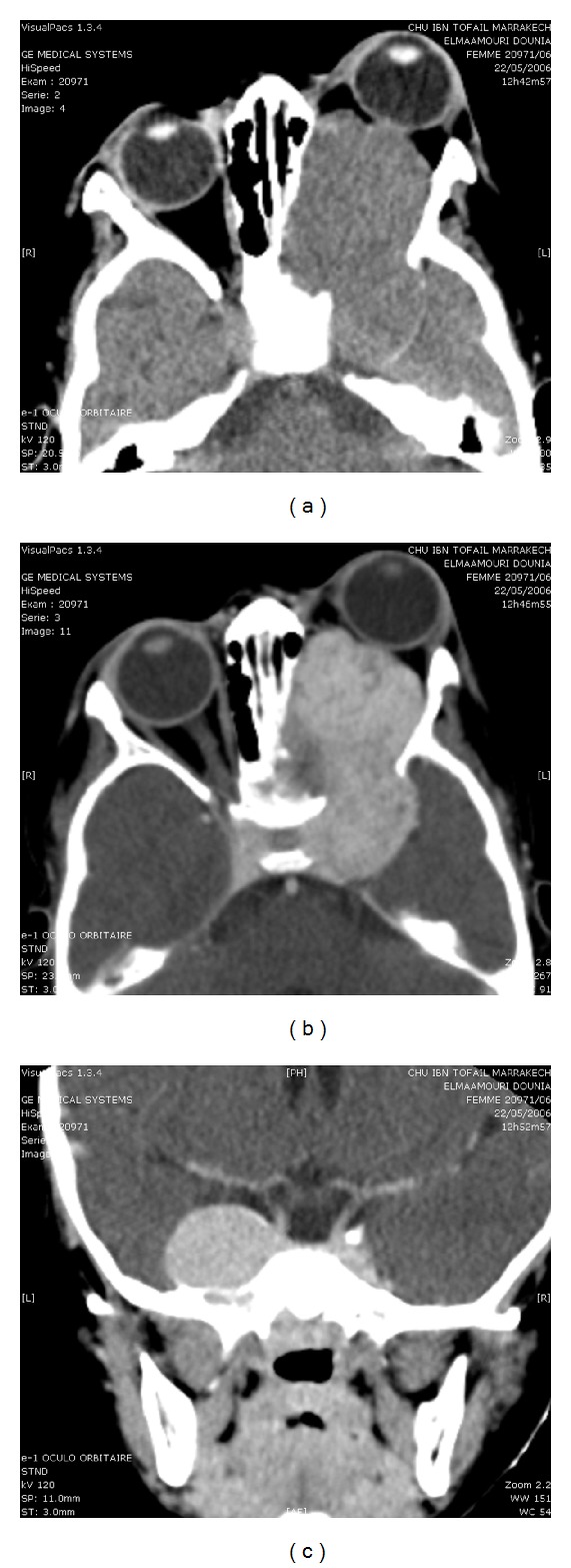
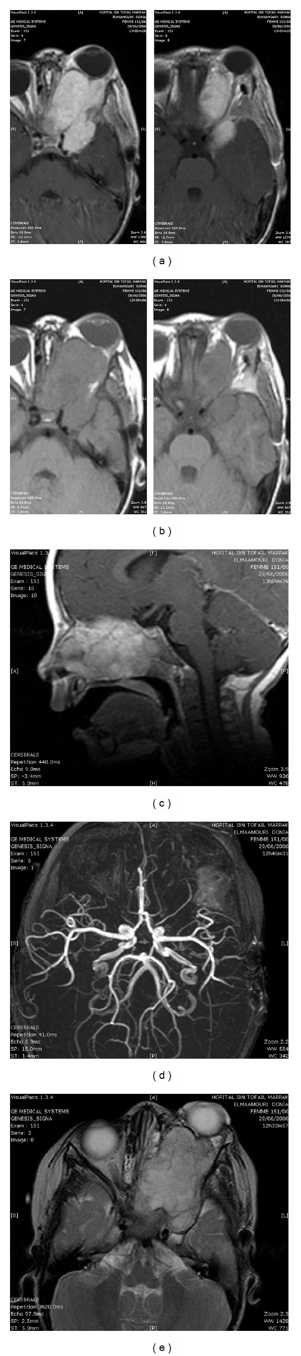
Epithelioid hemangioendothelioma (EHE) is a rare tumor of intermediate malignancy. We report a case of intracranial and intraorbitar EHE. A 3-year-old girl presented with a 3-month history of progressive left exophthalmia. Neuroradiologic imaging (CT scan and MRI) showed an intraorbitar process with an intense enhancement extending to temporal fossa, ethmoidal bone, nasal fossa, maxillary sinus, and cavernous sinus. The angiogram was normal. The tumor was operated through subfrontal approach but only a partial resection was performed. The histological diagnosis was epithelioid hemangioendothelioma. The patient was neurologically intact 2 months after surgery without exophtalmia. However 4 months after surgery he displayed a fall of the right eye vision with intense headache. Control CT scan showed persistence of important tumoral residue. Epithelioid hemangioendothelioma is a hemorrhagic tumor. Total removal must be possible. Otherwise, we recommend a complementary chemoradiotherapy and close followup. We propose this interesting case history of a tragical evolution of EHE in contradiction with what has already been reported.
Figures


Similar articles
-
Extensive epithelioid hemangioendothelioma of the maxillary sinus: A case report.Int J Surg Case Rep. 2019;58:70-73. doi: 10.1016/j.ijscr.2019.04.013. Epub 2019 Apr 16. Int J Surg Case Rep. 2019. PMID: 31015076 Free PMC article.
-
Epithelioid Hemangioendothelioma of the Mastoid Bone with Extension of Middle Cranial Fossa: A Case Report.Indian J Otolaryngol Head Neck Surg. 2023 Dec;75(4):4050-4053. doi: 10.1007/s12070-023-03954-4. Epub 2023 Jul 5. Indian J Otolaryngol Head Neck Surg. 2023. PMID: 37974704 Free PMC article.
-
Epithelioid Hemangioendothelioma of the Maxillary Sinus.Head Neck Pathol. 2016 Jun;10(2):229-32. doi: 10.1007/s12105-015-0633-1. Epub 2015 May 12. Head Neck Pathol. 2016. PMID: 25963905 Free PMC article.
-
Malignant intracranial epithelioid hemangioendothelioma presumably originating from the lung: case report.J Neurooncol. 2004 May;67(3):337-43. doi: 10.1023/b:neon.0000024215.79178.27. J Neurooncol. 2004. PMID: 15164990 Review.
-
Intracranial epithelioid hemangioendothelioma.Childs Nerv Syst. 2008 Jul;24(7):863-8. doi: 10.1007/s00381-008-0634-4. Epub 2008 May 14. Childs Nerv Syst. 2008. PMID: 18478237 Review.
Cited by
-
A Rare Case of Epithelioid Haemangioendothelioma of the Lateral Orbit in a 22-Year-Old Patient.Cureus. 2025 Jan 7;17(1):e77084. doi: 10.7759/cureus.77084. eCollection 2025 Jan. Cureus. 2025. PMID: 39777374 Free PMC article.
-
Dural composite hemangioendothelioma: The first intracranial case.Surg Neurol Int. 2024 Feb 23;15:55. doi: 10.25259/SNI_3_2024. eCollection 2024. Surg Neurol Int. 2024. PMID: 38468685 Free PMC article.
-
Surgical considerations in a paediatric case of a large skull-base epithelioid haemangioendothelioma.Childs Nerv Syst. 2019 Mar;35(3):559-563. doi: 10.1007/s00381-018-3988-2. Epub 2018 Oct 19. Childs Nerv Syst. 2019. PMID: 30341660
-
Multiple hemorrhagic intraparenchymal tumors presenting with fatal intracranial hypertension: A rare manifestation of systemic epithelioid hemangioendothelioma.Surg Neurol Int. 2015 Oct 6;6:156. doi: 10.4103/2152-7806.166799. eCollection 2015. Surg Neurol Int. 2015. PMID: 26539307 Free PMC article.
-
Suprasellar epithelioid hemangioendothelioma: Case report and review of the literature.Surg Neurol Int. 2016 Sep 1;7(Suppl 23):S596-602. doi: 10.4103/2152-7806.189729. eCollection 2016. Surg Neurol Int. 2016. PMID: 27656318 Free PMC article.
References
-
- Enzinger FM, Weiss SW. Hemangioendothelioma: vascular tumors of intermediate malignancy. In: Enzinger FM, Weiss SW, editors. Soft Tissue Tumors. 2 edition. St. Louis, Mo, USA: C.V. Mosby; 1988. pp. 533–544.
-
- Weiss SW, Enzinger FM. Epitheloid hemangioendothelioma. A vascular tumor often mistaken for a carcinoma. Cancer. 1982;50(5):970–981. - PubMed
-
- Puca A, Meglio M, Rollo M, Zannoni GF. Intracranial epithelioid hemangioendothelioma: case report. Neurosurgery. 1996;38(2):399–401. - PubMed
-
- Gray MH, Rosenberg AE, Dickersin GR, Bhan AK. Cytokeratin expression in epithelioid vascular neoplasms. Human Pathology. 1990;21(2):212–217. - PubMed
-
- Phookan G, Davis AT, Holmes B. Hemangioendothelioma of the cavernous sinus: case report. Neurosurgery. 1998;42(5):1153–1156. - PubMed
LinkOut - more resources
Full Text Sources
