

doi: 10.1155/2012/134603.
Epub 2012 Oct 22.
Imaging of postpartum ovarian vein thrombosis
Affiliations
- PMID: 23133765
- PMCID: PMC3485490
- DOI: 10.1155/2012/134603
Item in Clipboard
Imaging of postpartum ovarian vein thrombosis
Case Rep Obstet Gynecol.
2012.
Abstract
Postpartum ovarian vein thrombosis (OVT) is a rare but serious complication. Clinical findings of OVT are nonspecific. Postpartum OVT, which is a clinically difficultly diagnosed entity, must be thought of in differential diagnosis in cases of postpartum acute abdomen. OVT can be accurately diagnosed by appropriate noninvasive radiologic modalities to start early therapy with anticoagulants and intravenous antibiotics. In this paper, we review the imaging findings of a case with postpartum ovarian vein thrombosis that had been followed up for 6 months by ultrasonography (US), color Doppler US, computed tomography (CT), and magnetic resonance imaging (MRI).
Figures
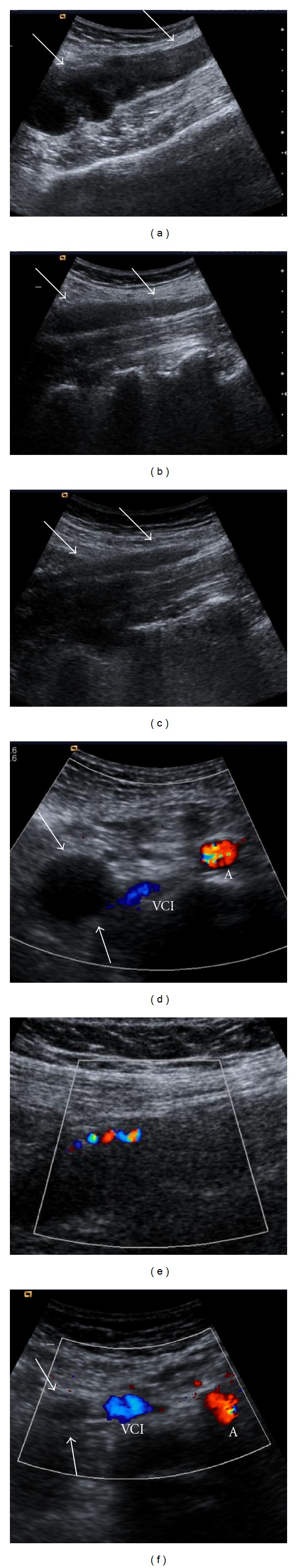
US and color Doppler US images obtained at first day ((a) and (d)), fourth month ((b) and (e)) and sixth month ((c) and (f)) show an enlarged, tortuous noncompressible tubular structure with hypoechoic material centrally, representing thrombosed right ovarian vein, extending superiorly from the right adnexa, lateral to the IVC (white arrows). With time, the width of the vein decreased. At fourth month, color Doppler US image shows flow with recanalization (e). VCI = vena cava inferior, A = abdominal aorta.
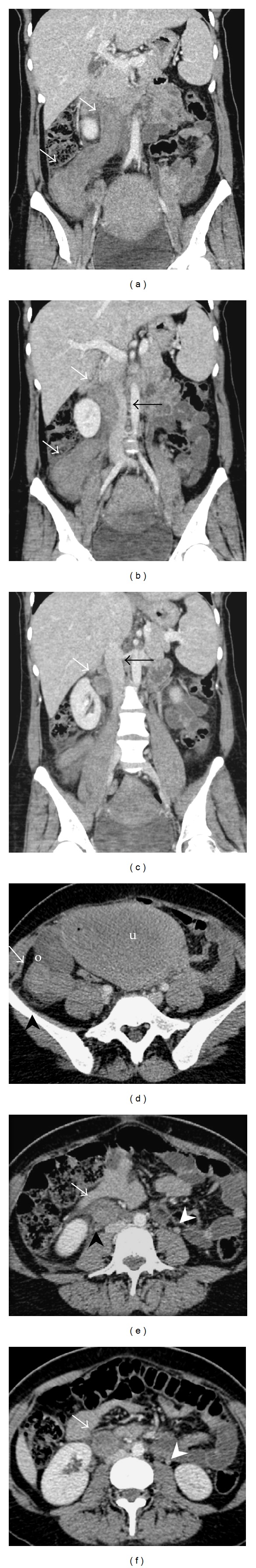
Coronal ((a)–(c)) and axial ((d)–(f)) contrast-enhanced multidetector computerized tomography (MDCT) images demonstrate an enlarged ovarian vein with central hypodensity, representing complete thrombosis (white arrows) extending up to the inferior vena cava, ending at the right renal hilus. There is also surrounding perivascular inflammatory reaction (black arrow heads). Note inflammatory changes in retroperitoneal fat around ovarian vein. Iliac vein is patent. The other ovarian vein is normal in calibration (white arrow heads).
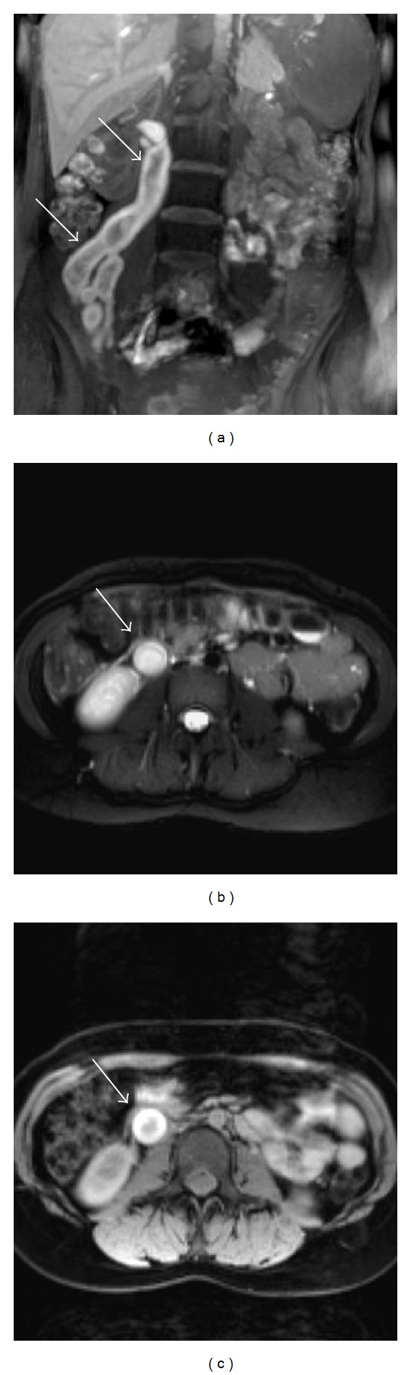
MR images show subacute right ovarian vein thrombosis. Coronal T1-weighted image (a) demonstrates an enlarged right ovarian vein showing increased signal (arrow) with an isointense central focus and axial T2-weighted image (b) shows high signal (arrow) indicative of methemoglobin within a complex thrombus. Contrast-enhanced axial T1-weighted image (c) reveals significant contrast enhancement of the vessel wall.
References
-
- Quane LK, Kidney DD, Cohen AJ. Unusual causes of ovarian vein thrombosis as revealed by CT and sonography. American Journal of Roentgenology. 1998;171(2):487–490. - PubMed
-
- Ortín Font X, Ugarriza A, Espax RM, et al. Postpartum ovarian vein thrombosis. Thrombosis and Haemostasis. 2005;93(5):1004–1005. - PubMed
-
- Mintz MC, Levy DW, Axel L, et al. Puerperal ovarian vein thrombosis: MR diagnosis. American Journal of Roentgenology. 1987;149(6):1273–1274. - PubMed
-
- Savader SJ, Otero RR, Savader BL. Puerperal ovarian vein thrombosis: evaluation with CT, US, and MR imaging. Radiology. 1988;167(3):637–639. - PubMed
LinkOut - more resources
Full Text Sources
